. 2024 Dec 10:15:1501486.
doi: 10.3389/fimmu.2024.1501486. eCollection 2024.

Integrating machine learning, bioinformatics and experimental verification to identify a novel prognostic marker associated with tumor immune microenvironment in head and neck squamous carcinoma

Affiliations

Integrating machine learning, bioinformatics and experimental verification to identify a novel prognostic marker associated with tumor immune microenvironment in head and neck squamous carcinoma

Xiaoxia Zeng et al. Front Immunol. .

Abstract

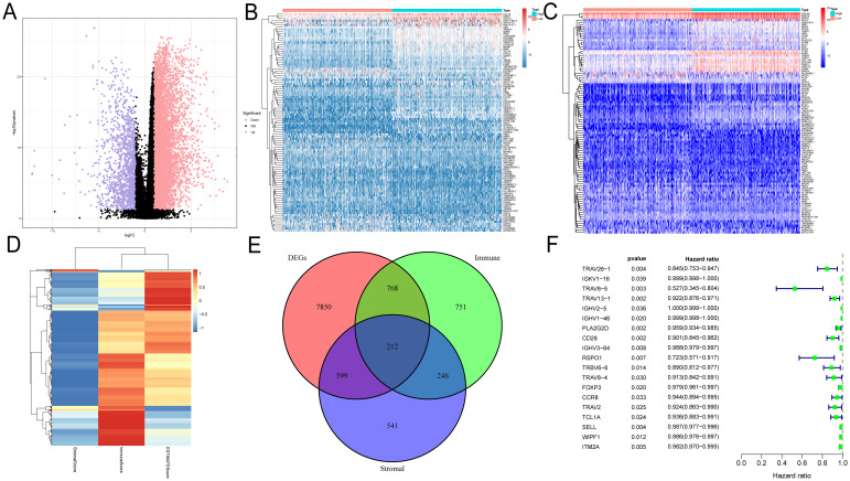
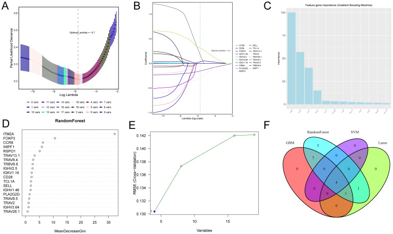
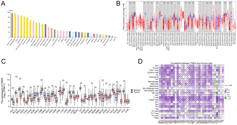
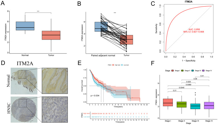
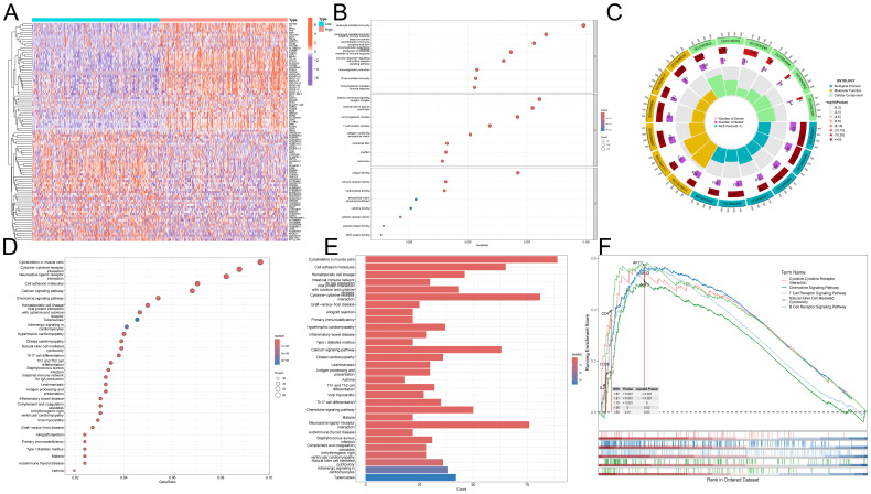
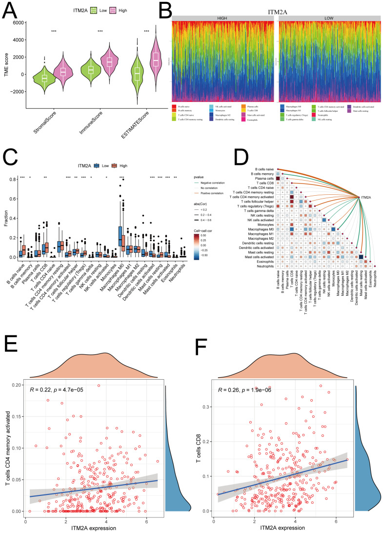
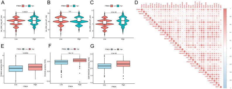
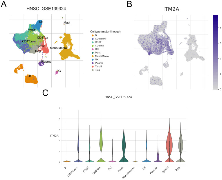
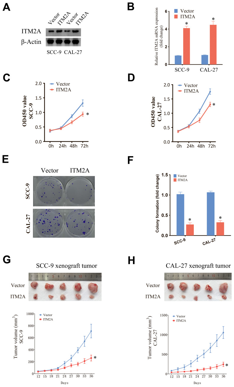
Head and neck squamous carcinoma (HNSC), characterized by a high degree of malignancy, develops in close association with the tumor immune microenvironment (TIME). Therefore, identifying effective targets related to HNSC and TIME is of paramount importance. Here, we employed the ESTIMATE algorithm to compute immune and stromal cell scores for HNSC samples from the TCGA database and identified differentially expressed genes (DEGs) based on these scores. Subsequently, we utilized four machine learning algorithms to identify four key genes: ITM2A, FOXP3, WIPF1, and RSPO1 from DEGs. Through a comprehensive pan-cancer analysis, our study identified aberrant expression of ITM2A across various tumor types, with a significant association with the TIME. Specifically, ITM2A expression was markedly reduced and correlated with poor prognosis in HNSC. Functional enrichment analysis revealed that ITM2A is implicated in multiple immune-related pathways, including immune-infiltrating cells, immune checkpoints, and immunotherapeutic responses. ITM2A expression was observed in various immune cell populations through single-cell analysis. Furthermore, we showed that ITM2A overexpression inhibited the growth of HNSC cells. Our results suggest that ITM2A may be a novel prognostic marker associated with TIME.

Keywords: HNSC; ITM2A; immune microenvironment; machine learning; prognostic.


Conflict of interest statement

The authors declare that the research was conducted in the absence of any commercial or financial relationships that could be construed as a potential conflict of interest.

Figures

Figure 1
Figure 1
Identification of prognosis-related genes based on ImmuneScore, StromalScore and DEG. (A) Volcano plots were used to show the distribution of DEGs. (B–D) Heatmaps were used to show DEG expression in ImmuneScore and StromalScore, respectively. (E) Wayne plots showing intersecting genes in ImmuneScore, StromalScore and DEG. (F) Univariate Cox analysis was used to screen the prognosis-related genes among the intersection genes.
Figure 2
Figure 2
Screening of candidate genes associated with TIME and prognosis in HNSC using four machine learning algorithms. (A, B) Shows the optimal parameter lambda value selected in the LASSO model and the variation of different genes with the Lamdba value. (C) The top 10 most important genes in the gradient boosting model. (D) Top 18 most important genes in the random forest model. (E) Optimal values of RMSE in the SVM-RFE algorithm model. (F) Wayne plots for filtering important genes for four models, LASSO regression, Support Vector Machine - Recursive Feature Elimination, Random Forest and Gradient Booster.
Figure 3
Figure 3
Expression of ITM2A in different tissues and cancer types. (A) Expression levels of ITM2A in various normal tissues. (B) Expression level of ITM2A in pan-cancer in TCGA database. (C) Expression levels of ITM2A in pan-cancer in the TCGA combined GTEx database. (D) Heatmap of the correlation between ITM2A in pan-cancer and immune cell infiltration using the ssGSEA algorithm. *p<0.05, **p<0.01, ***p<0.001.
Figure 4
Figure 4
Expression level of ITM2A in HNSC and its clinical significance. (A) Expression levels of ITM2A in normal and HNSC tumor tissues. (B) Expression levels of ITM2A in paired normal and HNSC tumor tissues. (C) ROC curves of ITM2A expression for identification of HNSC tissues. (D) Representative image of ITM2A immunohistochemistry. (E) Kaplan-Meier curves of OS. (F) Tumors in different clinical stages (stages I-IV) Comparison of ITM2A expression levels. ***p<0.001.
Figure 5
Figure 5
Functional role of ITM2A in HNSC. (A) Heatmap showing DEGs associated with ITM2A expression. (B, C) GO enrichment analysis. (D, E) KEGG enrichment analysis. (F) GSEA enrichment analysis of KEGG.
Figure 6
Figure 6
Relationship between ITM2A and tumor immune microenvironment. (A) Significant correlation of ITM2A with ImmuneScore, StromalScore and ESTIMATEScore. (B) Relative proportions of different immune cell types in HNSC samples. (C) Distribution of different types of immune cells in high and low ITM2A subgroups. (D) Correlation analysis of the level of infiltration of ITM2A and each type of immune cells. (E) Correlation analysis of ITM2A and CD4+ memory T cells. (F) Correlation analysis of ITM2A and CD8+ T cells. *p<0.05, **p<0.01, ***p<0.001.
Figure 7
Figure 7
Correlation between ITM2A expression and immunotherapy and chemotherapy in HNSC. (A–C) Violin plots demonstrating whether there is a difference between HNSC patients in the high and low ITM2A expression groups in response to treatment with PD-1 and or CTLA_4 inhibitors. (D) Association between ITM2A expression and immune checkpoints. (E–G) Semi-inhibitory concentration values of all three chemotherapeutic agents were elevated in patients with high ITM2A expression in HNSC.
Figure 8
Figure 8
Expression of ITM2A in different cell types of HNSC in single cells. (A) UMAP plots showing the distribution of different clusters as well as annotations. (B, C) Distribution of ITM2A in different cell types.
Figure 9
Figure 9
Upregulation of ITM2A inhibited the proliferation of HNSC cells. (A, B) Protein and mRNA levels were detected after transfection of empty vector or OE-ITM2A. (C, D) CCK-8 assay showing the proliferative capacity of HNSC cell lines with ITM2A overexpression. (E, F) Clone formation assay showing the clone formation ability of HNSC cell lines with ITM2A overexpression. (G, H) Xenograft tumor models were established using empty vector or ITM2A overexpression HNSC cells and tumor volumes were measured. The data represent the mean ± SEM. *p<0.05, versus vector.

Similar articles

References

    1. Solomon B, Young RJ, Rischin D. Head and neck squamous cell carcinoma: Genomics and emerging biomarkers for immunomodulatory cancer treatments. Semin Cancer Biol. (2018) 52:228–40. doi: 10.1016/j.semcancer.2018.01.008 - DOI - PubMed
    1. Johnson DE, Burtness B, Leemans CR, Lui V, Bauman JE, Grandis JR. Head and neck squamous cell carcinoma. Nat Rev Dis Primers. (2020) 6:92. doi: 10.1038/s41572-020-00224-3 - DOI - PMC - PubMed
    1. Vos JL, Elbers J, Krijgsman O, Traets J, Qiao X, van der Leun AM, et al. . Neoadjuvant immunotherapy with nivolumab and ipilimumab induces major pathological responses in patients with head and neck squamous cell carcinoma. Nat Commun. (2021) 12:7348. doi: 10.1038/s41467-021-26472-9 - DOI - PMC - PubMed
    1. Mu Q, Najafi M. Modulation of the tumor microenvironment (TME) by melatonin. Eur J Pharmacol. (2021) 907:174365. doi: 10.1016/j.ejphar.2021.174365 - DOI - PubMed
    1. Wang L, Ma L, Song Z, Zhou L, Chen K, Wang X, et al. . Single-cell transcriptome analysis profiling lymphatic invasion-related TME in colorectal cancer. Sci Rep. (2024) 14:8911. doi: 10.1038/s41598-024-59656-6 - DOI - PMC - PubMed

MeSH terms

Substances

LinkOut - more resources