. 2024 Dec 15;45(18):e70121.
doi: 10.1002/hbm.70121.

Understanding Cognitive Aging Through White Matter: A Fixel-Based Analysis

Affiliations

Understanding Cognitive Aging Through White Matter: A Fixel-Based Analysis

Emma M Tinney et al. Hum Brain Mapp. .

Abstract

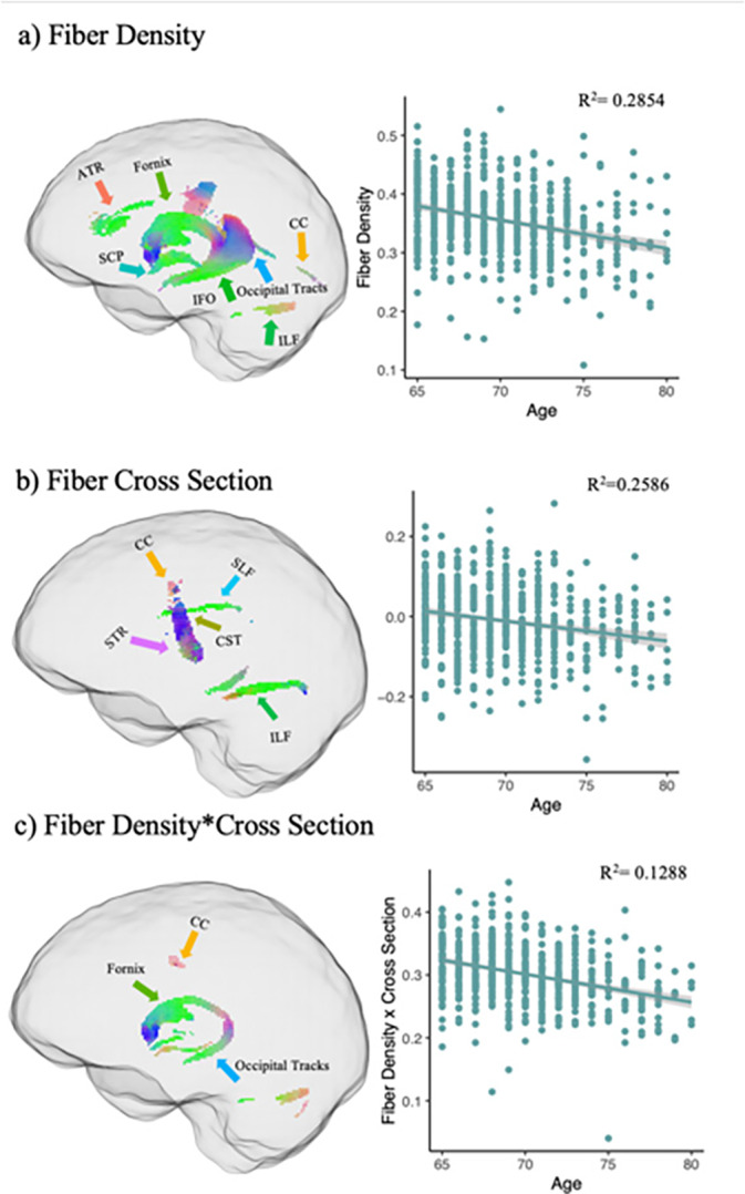
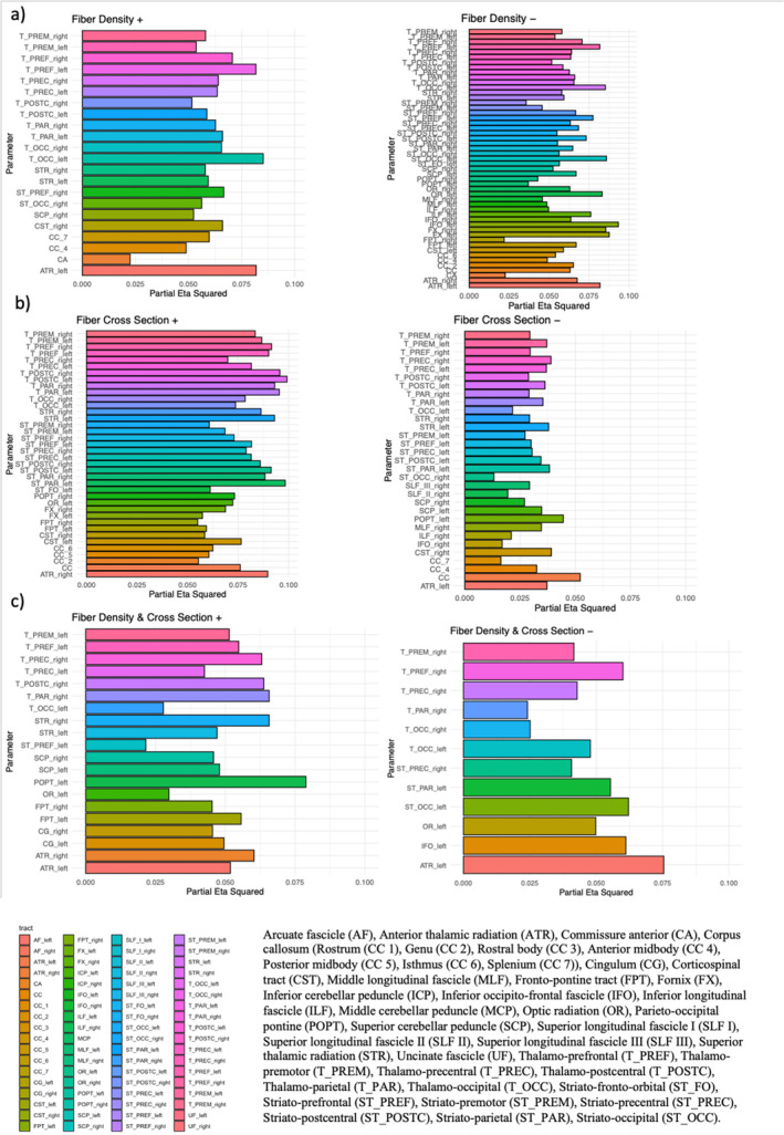
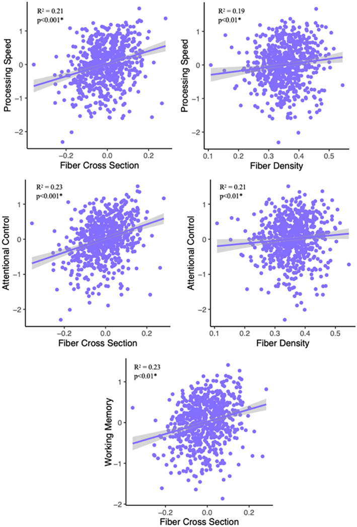
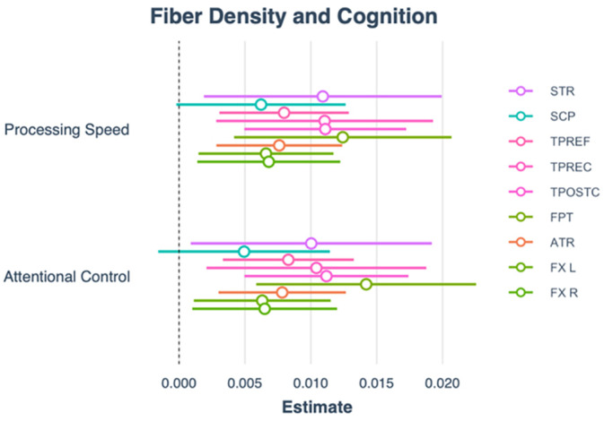
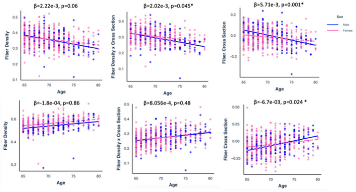
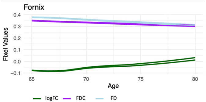
Diffusion-weighted imaging (DWI) has been frequently used to examine age-related deterioration of white matter microstructure and its relationship to cognitive decline. However, typical tensor-based analytical approaches are often difficult to interpret due to the challenge of decomposing and (mis)interpreting the impact of crossing fibers within a voxel. We hypothesized that a novel analytical approach capable of resolving fiber-specific changes within each voxel (i.e., fixel-based analysis [FBA])-would show greater sensitivity relative to the traditional tensor-based approach for assessing relationships between white matter microstructure, age, and cognitive performance. To test our hypothesis, we studied 636 cognitively normal adults aged 65-80 years (mean age = 69.8 years; 71% female) using diffusion-weighted MRI. We analyzed fixels (i.e., fiber-bundle elements) to test our hypotheses. A fixel provides insight into the structural integrity of individual fiber populations in each voxel in the presence of multiple crossing fiber pathways, allowing for potentially increased specificity over other diffusion measures. Linear regression was used to investigate associations between each of three fixel metrics (fiber density, cross-section, and density × cross-section) with age and cognitive performance. We then compared and contrasted the FBA results to a traditional tensor-based approach examining voxel-wise fractional anisotropy. In a whole-brain analysis, significant associations were found between fixel-based metrics and age after adjustments for sex, education, total brain volume, site, and race. We found that increasing age was associated with decreased fiber density and cross-section, namely in the fornix, striatal, and thalamic pathways. Further analysis revealed that lower fiber density and cross-section were associated with poorer performance in measuring processing speed and attentional control. In contrast, the tensor-based analysis failed to detect any white matter tracts significantly associated with age or cognition. Taken together, these results suggest that FBAs of DWI data may be more sensitive for detecting age-related white matter changes in an older adult population and can uncover potentially clinically important associations with cognitive performance.

Keywords: DWI; aging; cognitive decline; fixel‐based analysis; white matter.


Conflict of interest statement

The authors declare no conflicts of interest.

Figures

FIGURE 1
FIGURE 1
Negative relationships between fixel‐based metrics and age. Fixel‐wise regression analysis between age and baseline fixel‐based metrics, exhibiting significant negative associations with age are shown and color coded by direction (blue = superior–inferior; green = anterior–posterior; red = medial–lateral). (a) FBA showing FD × aging. (b) FBA showing FC × aging. (c) FBA showing FDC × aging. Anterior thalamic radiation (ATR). Corpus callosum (CC), Corticospinal tract (CST), Inferior occipito‐frontal fascicle (IFO), Inferior longitudinal fascicle (ILF), superior cerebellar peduncle (SCP), superior longitudinal fascicle (SLF), superior thalamic radiation (STR).
FIGURE 2
FIGURE 2
Positive relationship between fixel‐based metrics and age. Fixel‐wise regression analysis between age and baseline fixel‐based metrics, exhibiting significant positive associations with age are shown and color coded by direction (blue = superior–inferior; green = anterior–posterior; red = medial–lateral). (a) FBA showing FD × aging. (b) FBA showing FC × aging. (c) FBA showing FDC × aging. Anterior thalamic radiation (ATR), corpus callosum (CC), corticospinal tract (CST), fronto‐pontine tract (FPT), thalamo‐prefrontal (TPREF), thalamo‐premotor (TPREM), superior cerebellar peduncle (SCP), superior thalamic radiation (STR).
FIGURE 3
FIGURE 3
Effect sizes of each metric and associated tract. The color of each bar represents each tract. For FD+, left anterior thalamic radiation and left thalamic occipital tract had the largest effect of age. For FD−, the fornix had the largest effect. For FC+, right thalamo‐postcentral had the largest effect. For FC−, the corpus callosum had the largest effect. For FDC+, the left parieto‐occipital pontine had the largest effect. For FDC−, left anterior thalamic radiation had the largest effect.
FIGURE 4
FIGURE 4
Regression graphs demonstrating the significant positive relationships with age‐related FC and FD and processing speed, attentional control, and working memory.
FIGURE 5
FIGURE 5
Plot demonstrating beta coefficients and confidence intervals for each tract and processing speed and attentional control.
FIGURE 6
FIGURE 6
Scatter plots showing a significant positive association between age (x‐axis) and FBA metric (on the y‐axis) in males (blue) and females (pink).
FIGURE 7
FIGURE 7
Line graph demonstrating the relationship between FD, FC, and FDC in the FX (right and left) with age.

Similar articles

Cited by

References

    1. Ai, M. , Morris T. P., Noriega de la Colina A., et al. 2024. “Midlife Physical Activity Engagement Is Associated With Later‐Life Brain Health.” Neurobiology of Aging 134: 146–159. 10.1016/j.neurobiolaging.2023.11.004. - DOI - PubMed
    1. Alberton, B. A. V. , Nichols T. E., Gamba H. R., and Winkler A. M.. 2020. “Multiple Testing Correction Over Contrasts for Brain Imaging.” NeuroImage 216: 116760. 10.1016/j.neuroimage.2020.116760. - DOI - PMC - PubMed
    1. Aldehri, M. , Temel Y., Alnaami I., Jahanshahi A., and Hescham S.. 2018. “Deep Brain Stimulation for Alzheimer's Disease: An Update.” Surgical Neurology International 9: 58. 10.4103/sni.sni_342_17. - DOI - PMC - PubMed
    1. Andersson, J. L. R. , Graham M. S., Zsoldos E., and Sotiropoulos S. N.. 2016. “Incorporating Outlier Detection and Replacement Into a Non‐Parametric Framework for Movement and Distortion Correction of Diffusion MR Images.” NeuroImage 141: 556–572. 10.1016/j.neuroimage.2016.06.058. - DOI - PubMed
    1. Andersson, J. L. R. , Skare S., and Ashburner J.. 2003. “How to Correct Susceptibility Distortions in Spin‐Echo Echo‐Planar Images: Application to Diffusion Tensor Imaging.” NeuroImage 20, no. 2: 870–888. 10.1016/S1053-8119(03)00336-7. - DOI - PubMed

LinkOut - more resources