

Thin flap sulcus-deepening trochleoplasty in patellar instability yields good functional outcomes without progressive cartilage deterioration in the short-term follow-up-A retrospective single-surgeon cohort study
- PMID: 39720939
- PMCID: PMC12392387
- DOI: 10.1002/ksa.12566
Thin flap sulcus-deepening trochleoplasty in patellar instability yields good functional outcomes without progressive cartilage deterioration in the short-term follow-up-A retrospective single-surgeon cohort study
Abstract
Purpose: Sulcus-deepening trochleoplasty (TP) effectively treats patellofemoral (PF) instability (PFI) caused by high-grade trochlear dysplasia (TD), but current evidence is based on small case series. We hypothesised, that TP would result in significant functional improvements and a low re-dislocation rate but would not accelerate the progression of PF cartilage deterioration.
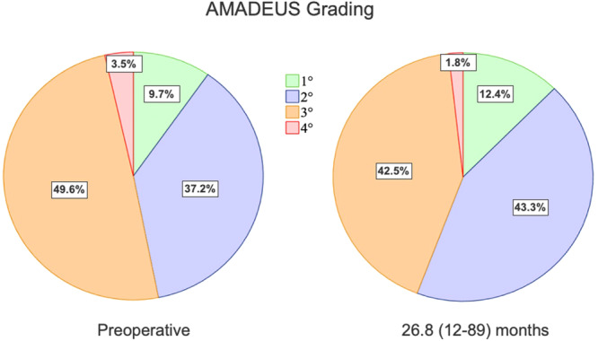
Methods: We retrospectively reviewed all TP cases performed by a single surgeon between 2015 and 2021. Inclusion criteria were postoperative Magnetic resonance imaging (MRI) >6 and >12 months and a clinical follow-up >12 months. Patients with simultaneous cartilage repair, open physes or incomplete records were excluded. Clinical outcomes were assessed using pre- and postoperative scores, postoperative Banff Patellofemoral Instability Instrument (BPII) 2.0 and Knee Injury and Osteoarthritis Outcome Score (KOOS), re-dislocation rate and patient satisfaction. PF cartilage was evaluated via Area Measurement and Depth & Underlying Structures (AMADEUS) scores preoperatively, at 6 months and at the final follow-up.
Results: We included 113 patients (25.8 ± 8.3 years) with high-grade TD (Dejour B-D; mean lateral inclination angle: -2.9 ± 9.1°), 85% of whom had advanced cartilage lesions. All underwent TP, lateral retinacular lengthening (LRL) and medial patellofemoral ligament reconstruction (MPFL-R). After 34.8 ± 20.9 months, function, pain levels and Tegner scores improved significantly (p < 0.001). KOOS dimensions were: symptoms 79.9 ± 13.5, pain 86.4 ± 12.1, activity 91.9 ± 8.3, sports 71.7 ± 22.2 and quality-of-life 58.1 ± 23.8. BPII 2.0 was 64.3 ± 31.4. Preoperative AMADEUS scores (55.2 ± 17.4) remained stable at 6 months (p = 0.343) but improved to 58.4 ± 16.0 at 28.6 (12-89) months (p = 0.004). Complication and re-dislocation rates were 5.3% and 1.8%, with 93% patient satisfaction.
Conclusion: Sulcus-deepening TP with MPFL-R and LRL yields good to excellent short-term results without progressive chondral deterioration, enabling patients to return to their prior or higher activity levels despite advanced preoperative chondral lesions. TP can be considered a safe, joint-preserving technique for PF stabilisation.
Level of evidence: Level III, retrospective cohort study.
Keywords: cartilage deterioration; maltracking; patellofemoral instability; trochlear dysplasia; trochleoplasty.
© 2024 The Author(s). Knee Surgery, Sports Traumatology, Arthroscopy published by John Wiley & Sons Ltd on behalf of European Society of Sports Traumatology, Knee Surgery and Arthroscopy.
Conflict of interest statement
Karl‐Heinz Frosch receives royalties from Arthrex (Naples, FL, USA). Karl‐Heinz Frosch and Matthias Krause receive honoraria for lectures from Arthrex (Naples, FL, USA). Andreas Weiler receives royalties from Medacta International (Castel San Pietro, Switzerland) and honoraria for lectures from Enovis (Freiburg, Germany). Arno Schmeling receives payments from a consulting contract with Arthrex (Naples, FL, USA) and honoraria for presentations from Conmed (Utica, NY, USA). He is head of the Patellofemoral Committee of the German Society for Knee Surgery. Jannik Frings and Eva Janssen declare no conflicts of interest.
Figures


Similar articles
-
MRI analysis of the chondral surface following deepening trochleoplasty for patellofemoral instability.Orthop Traumatol Surg Res. 2025 Jun 19:104322. doi: 10.1016/j.otsr.2025.104322. Online ahead of print. Orthop Traumatol Surg Res. 2025. PMID: 40543645
-
Results of medial patellofemoral ligament reconstruction compared with trochleoplasty plus individual extensor apparatus balancing in patellar instability caused by severe trochlear dysplasia: a systematic review and meta-analysis.Knee Surg Sports Traumatol Arthrosc. 2017 Dec;25(12):3869-3877. doi: 10.1007/s00167-016-4365-x. Epub 2016 Oct 27. Knee Surg Sports Traumatol Arthrosc. 2017. PMID: 27796419
-
Outcomes After Isolated Medial Patellofemoral Ligament Reconstruction for Recurrent Patellar Instability: Influence of Persistent Postoperative Apprehension and J-Sign.Am J Sports Med. 2025 Jul;53(8):1931-1939. doi: 10.1177/03635465251339822. Epub 2025 Jun 6. Am J Sports Med. 2025. PMID: 40478223
-
MPFL reconstruction vs. Insall procedure for adolescent patellar instability: nine-year follow-up on osteoarthritis, redislocations, and return to sports.BMC Musculoskelet Disord. 2025 Aug 4;26(1):749. doi: 10.1186/s12891-025-08992-3. BMC Musculoskelet Disord. 2025. PMID: 40759934 Free PMC article.
-
Medial patellofemoral ligament reconstruction with and without trochleoplasty for patients with patella instability-correlation of trochlear dysplasia and patient outcome, classification and outcome measure in the past decade-a systematic review.Eur J Orthop Surg Traumatol. 2022 May;32(4):595-607. doi: 10.1007/s00590-021-03030-z. Epub 2021 Jun 12. Eur J Orthop Surg Traumatol. 2022. PMID: 34120236
Cited by
-
[Why does the patellofemoral cartilage collapse? : A critical analysis of epidemiology, etiology and prevention].Orthopadie (Heidelb). 2025 Jun;54(6):425-435. doi: 10.1007/s00132-025-04649-0. Epub 2025 May 5. Orthopadie (Heidelb). 2025. PMID: 40323410 Review. German.
References
-
- Askenberger, M. , Janarv, P.M. , Finnbogason, T. & Arendt, E.A. (2017) Morphology and anatomic patellar instability risk factors in first‐time traumatic lateral patellar dislocations: a prospective magnetic resonance imaging study in skeletally immature children. The American Journal of Sports Medicine, 45, 50–58. Available from: 10.1177/0363546516663498 - DOI - PubMed
-
- Balcarek, P. , Ammon, J. , Frosch, S. , Walde, T.A. , Schüttrumpf, J.P. , Ferlemann, K.G. et al. (2010) Magnetic resonance imaging characteristics of the medial patellofemoral ligament lesion in acute lateral patellar dislocations considering trochlear dysplasia, patella alta, and tibial tuberosity‐trochlear groove distance. Arthroscopy: The Journal of Arthroscopic & Related Surgery, 26, 926–935. Available from: 10.1016/j.arthro.2009.11.004 - DOI - PubMed
MeSH terms
Grants and funding
LinkOut - more resources
Full Text Sources
Research Materials
