. 2024 Dec 25;30(1):274.
doi: 10.1186/s10020-024-01051-y.

Crosstalk between cancer-associated fibroblasts and non-neuroendocrine tumor cells in small cell lung cancer involves in glycolysis and antigen-presenting features

Affiliations

Crosstalk between cancer-associated fibroblasts and non-neuroendocrine tumor cells in small cell lung cancer involves in glycolysis and antigen-presenting features

Yuanhua Lu et al. Mol Med. .

Abstract

Background: Small cell lung cancer (SCLC) is a highly fatal malignancy, the complex tumor microenvironment (TME) is a critical factor affecting SCLC progression. Cancer-associated fibroblasts (CAFs) are crucial components of TME, yet their role in SCLC and the underlying mechanisms during their interaction with SCLC cells remain to be determined.

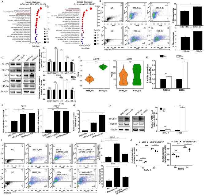
Methods: Microenvironmental cell components were estimated using transcriptome data from SCLC tissue available in public databases, analyzed with bioinformatic algorithms. A co-culture system comprising MRC5 fibroblasts and SCLC cell lines was constructed. RNA sequencing (RNA-seq) was performed on co-cultured and separately cultured MRC5 and H196 cells to identify differentially expressed genes (DEGs) and enriched signaling pathways. Glycolysis and STING signaling in SCLC cells were assessed using glucose uptake assays, qRT-PCR, and Western blot analysis. Immunohistochemical staining of SCLC tissue arrays quantified α-SMA, HLA-DRA and CD8 expression.

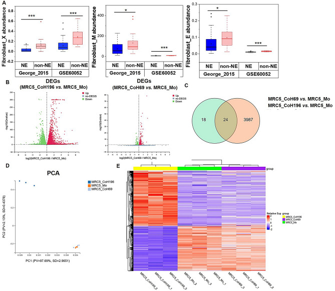
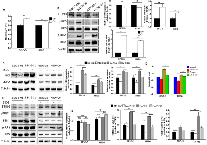
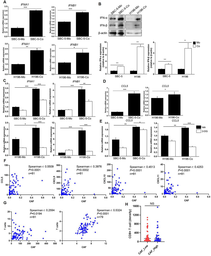
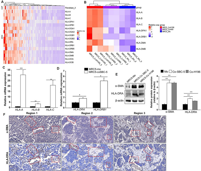
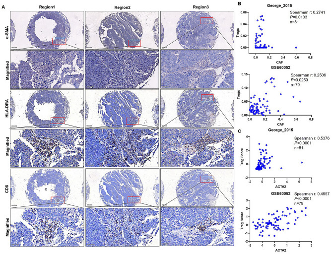
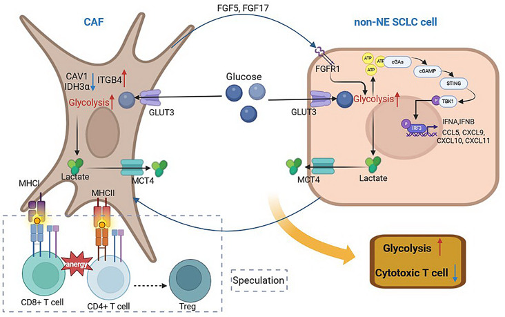
Results: Non-neuroendocrine (non-NE) SCLC-derived CAFs exhibited more abundance and DEGs than NE SCLC-derived CAFs did, which interact with non-NE SCLC cells can induce the enrichment of glycolysis-related genes, increasement of glucose uptake, upregulation of glycolytic signaling proteins in non-NE SCLC cells and accumulation of lactate in the extracellular environment, confirming CAF-mediated glycolysis promotion. Additionally, glycolysis-induced ATP production activated STING signaling in non-NE SCLC cells, which upregulated T cell chemo-attractants. However, CAF abundance did not correlate with CD8 + T cell numbers in SCLC tissues. Additionally, non-NE SCLC cell-educated CAFs exhibited features of antigen-presenting CAFs (apCAFs), as indicated by the expression of major histocompatibility complex (MHC) molecules. Co-localization of HLA-DRA and α-SMA signals in SCLC tissues confirmed apCAF presence. The apCAFs and CD8 + T cells were co-located in the SCLC stroma, and there was a positive correlation between CAFs and regulatory T cell (Treg) abundance.

Conclusion: Our findings suggest that crosstalk between CAFs and non-NE SCLC cells promotes glycolysis in non-NE SCLC cells, thereby increase T cell chemo-attractant expression via activating STING signaling. On the other hand, it promotes the presence of apCAFs, which probably contributes to CD8 + T cell trapping and Treg differentiation. This study emphasizes the pro-tumor function of CAFs in SCLC by promoting glycolysis and impairing T cell function, providing direction for the development of novel therapeutic approaches targeting CAF in SCLC.

Keywords: Antigen presentation; CAF; Glycolysis; SCLC; STING signaling.


Conflict of interest statement

Declarations. Ethics approval and consent to participate: The tissue arrays were obtained from ZK bioaitech company, informed patient consent was obtained prior to collection. The study was approved by Ethics Committee of Changsha Yaxiang Biotechnology Co., Ltd (Csyayj20231120). All methods were performed as per the relevant regulations and the Declaration of Helsinki. Consent for publication: Not applicable. Competing interests: The authors declare no competing interests.

Figures

Fig. 1
Fig. 1
CAF abundance and expression profile heterogeneity in SCLC. (A) The comparison of CAF abundance from George’s cohort and GSE60052 dataset estimated by xCell (X), MCP-counter (M) and EPIC (E) algorithms between NE and non-NE subtypes. Mann–Whitney U test was used for determine significant difference, *P < 0.05, **P < 0.01, ***P < 0.001. (B) Volcano plot of DEGs between MRC5_coH196 and MRC5_Mo cells and DEGs between MRC5_coH69 and MRC5_Mo cells. (C) Venn diagram of DEGs between MRC5_coH196 and MRC5_Mo cells and DEGs between MRC5_coH69 and MRC5_Mo cells (Log2 Fold Change > 0.58). (D) PCA analysis for samples of MRC5_Mo, MRC5_coH69 and MRC5_coH196 cells. (E) Heatmap presents gene expression of MRC5_Mo, MRC5_coH69 and MRC5_coH196 cells
Fig. 2
Fig. 2
Glucose metabolic reprogramming during crosstalk between CAFs and non-NE SCLC cells. (A) MSigDB_Hallmark enrichment analysis was performed in upregulated DEGs of MRC5coH196 vs. MRC5_Mo cells and upregulated DEGs of H196_Co vs. H196_Mo cells. (B) SBC-5 and H196 cells co-cultured with MRC5 cells or individually cultured for 5 days, the uptake of 2-NBDG in SBC-5 and H196 cells was detected by flow cytometry. (C) The expression of glycolytic proteins in co-cultured and individually cultured SBC-5 and H196 cells was determined using western blot analysis, quantitative analyses of blots were performed based on triple repeated experiments. (D) The FPKM expression level of MCT4 and MCT1 in H196_Mo and H196_Co cells. (E) Lactate concentration in culture media of individually cultured (MRC5 + H196 or SBC-5) or co-cultured (SBC-5-CoMRC5 or H196-CoMRC5) cells was determined and normalized to the Mo group. (F) Expression of FGF5 and FGF17 in co-cultured and individually cultured MRC5 cells was determined by qRT-PCR. (G) FGF17 level in culture media of co-cultured and individually cultured MRC5 cells was determined by ELISA. (H) The phosphorylated FGFR1 and expression of FGFR1 in co-cultured and individually cultured SBC-5 and H196 cells was determined using western blot analysis. Quantitative analyses of blots were performed based on triple repeated experiments. (I) The uptake of 2-NBDG in SBC-5 and H196 cells which co-cultured with MRC5 cells transfecting with siRNAs for FGF5 and FGF17 or scramble controls, and individually cultured SBC-5 and H196 cells was determined. (J) MRC5 cells transfected with siRNAs for FGF5 and FGF17, then co-cultured or individually cultured with SBC-5 or H196 cells. Lactate concentration in culture media of individually cultured or co-cultured cells was determined and normalized to the Mo-scramble group. *P < 0.05, **P < 0.01, ***P < 0.001
Fig. 3
Fig. 3
Glycolysis mediates STING signaling activation during crosstalk between CAFs and non-NE SCLC cells. (A) ATP production in co-cultured and individually cultured SBC-5 and H196 cells was detected using ATP Luminescent reagent. (B) Total and phosphorylated proteins of STING signaling was determined by western blot analysis. (C-E) Co-cultured and individually cultured SBC-5 and H196 cells were treated with 2-DG, the expression of glycolytic proteins (C), ATP production (D) and STING signaling in SBC-5 and H196 cells were determined. *P < 0.05, **P < 0.01, ***P < 0.001
Fig. 4
Fig. 4
CAFs promoted T cell chemo-attractants expression. (A, D) Expression of IFN I (A) and CCL5 (D) in co-cultured and individually cultured SBC-5 and H196 cells was determined by qRT-PCR. (B) The protein levels of IFN-α and IFN-β in co-cultured and individually cultured SBC-5 and H196 cells were determined by western blot. (C, E) After treatment with 2-DG, expression of IFN I (C) and CCL5 (E) in co-cultured and individually cultured SBC-5 and H196 cells was measured using qRT-PCR. (F) Spearman correlation was performed to measure the correlation between expression of T cell chemo-attractants and CAF abundance in George’s cohort. (G) Correlation between T cell abundance and CAF abundance estimated by MCP-counter was analyzed using Spearman correlation in George’s cohort and GSE60052 dataset. (H) SCLC tissue samples in tissue array (n = 74) were divided into CAF-High and CAF-Low groups based on the median value of α-SMA IHC score, the density of CD8 + T cells was compared between CAF-High and CAF-Low groups. *P < 0.05, **P < 0.01, ***P < 0.001
Fig. 5
Fig. 5
The apCAF exists in SCLC. (A) Heatmap presents the expression of HLA genes and fibroblast abundance in George’s cohort. (B) Differences in the expression of HLA-genes among MRC5_Mo cells, MRC5_coH69 cells and MRC5_coH196 cells. (C-D) The expression of HLA genes in MRC5_coSBC-5 cells and MRC5_Mo cells was determined by qRT-CPR. (E) The expression of α-SMA and HLA-DRA proteins in co-cultured and individually cultured MRC5 cells was detected by western blot. (F) CAF marker α-SMA and HLA-DRA were stained in SCLC tissue arrays by IHC, the regions where positive signals for α-SMA and HLA-DRA presented in the identical areas of consecutive slices were marked with red boxes. Scale bar: 50 μm. *P < 0.05, **P < 0.01, ***P < 0.001
Fig. 6
Fig. 6
CAFs involved in trapping CD8 + cells and Treg differentiation. (A) α-SMA, HLA-DRA and CD8 were detected in SCLC tissue arrays using IHC, the regions with presence of positive signals for α-SMA, HLA-DRA and CD8 were marked with red boxes. Scale bar: 200 μm and 50 μm (Magnified). (B) Correlation between CAF abundance and Treg abundance estimated by xCell algorithm from George’s cohort and GSE60052 dataset was performed using Spearman correlation. (C) Correlation between the expression of ACTA2 and Treg score, which calculated as the geometric mean of CD25 and FOXP3 expression, was analyzed by Spearman correlation
Fig. 7
Fig. 7
Schematic model on the crosstalk between CAF and non-NE SCLC cell and its’ impact on glycolysis and T cell function

Similar articles

Cited by

References

    1. Apostolova P, Pearce EL. Lactic acid and lactate: revisiting the physiological roles in the tumor microenvironment. Trends Immunol. 2022;43(12):969–77. 10.1016/j.it.2022.10.005. - PMC - PubMed
    1. Becker LM, O’Connell JT, Vo AP, et al. Epigenetic reprogramming of Cancer-Associated fibroblasts deregulates glucose metabolism and facilitates progression of breast Cancer. Cell Rep. 2020;31(9):107701. 10.1016/j.celrep.2020.107701. - PMC - PubMed
    1. Benonisson H, Altıntaş I, Sluijter M, et al. CD3-Bispecific antibody therapy turns solid tumors into Inflammatory sites but does not install protective memory. Mol Cancer Ther. 2019;18(2):312–22. 10.1158/1535-7163.mct-18-0679. - PubMed
    1. Bonuccelli G, Whitaker-Menezes D, Castello-Cros R, et al. The reverse Warburg effect: glycolysis inhibitors prevent the tumor promoting effects of caveolin-1 deficient cancer associated fibroblasts. Cell Cycle. 2010;9(10):1960–71. 10.4161/cc.9.10.11601. - PubMed
    1. Chen Y, McAndrews KM, Kalluri R. Clinical and therapeutic relevance of cancer-associated fibroblasts. Nat Reviews Clin Oncol. 2021;18(12):792–804. 10.1038/s41571-021-00546-5. - PMC - PubMed

MeSH terms