. 2024 Dec 26;24(1):1249.
doi: 10.1186/s12870-024-05967-9.

The effect of silicon supplementation and drought stress on the deposition of callose and chemical components in the cell walls of the Brassica napus roots

Affiliations

The effect of silicon supplementation and drought stress on the deposition of callose and chemical components in the cell walls of the Brassica napus roots

Saja-Garbarz Diana et al. BMC Plant Biol. .

Abstract

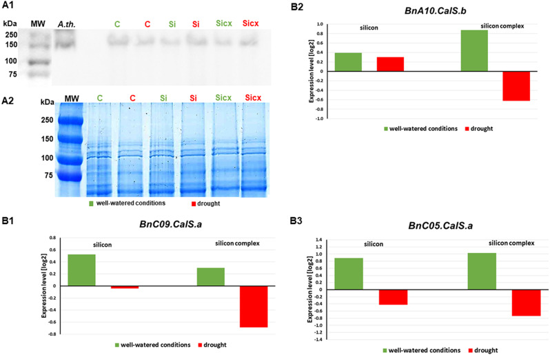
Background: Silicon has an important role in regulating water management in plants. It is deposited in cell walls and creates a mechanical barrier against external factors. The aim of this study was to determine the role of silicon supplementation in the synthesis and distribution of callose in oilseed rape roots and to characterize the modifications of cell wall structure of these organs after exposure to drought stress. Histological and ultrastructural analyses were performed to determine the changes in the distribution of arabinogalactan proteins, pectins, and extensin in roots of Brassica napus growing under drought and supplemented with silicon. Callose deposition and the accumulation of callose synthase protein were assessed, followed by transcriptional analysis of callose synthase genes.

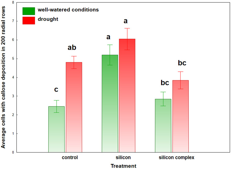
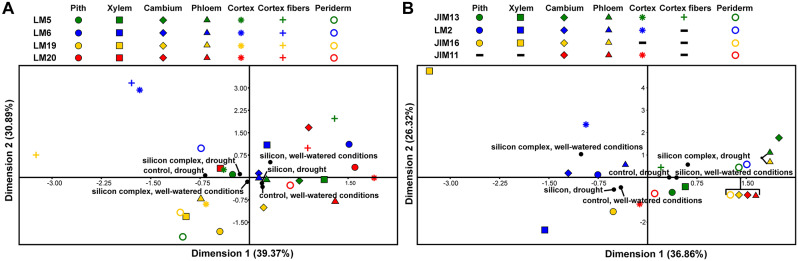
Results: The results showed that silicon supplementation under drought conditions alter the direction of cortex cell differentiation, promoting fiber formation and proliferation of callose-depositing cells in the roots of the tested plants. This was reflected in an increase in the level of callose synthase and a decrease in the transcriptional activity of the gene encoding this enzyme, indicating regulation based on negative feedback under drought stress. The changes in abundance and distribution of investigated arabinogalactan proteins, pectins and extensin in roots of Si supplemented plants growing under drought stress were observed, indicating cell walls remodeling.

Conclusion: Silicon supplementation in oilseed rape roots induced significant changes in cell wall composition, including increased callose deposition and altered pectins and arabinogalactan proteins distribution. These modifications, along with the formation of fibres in the root cortex, likely contribute to enhanced cell wall strength providing a physical barrier against water loss and mechanical stress, as a probable defence mechanism induced during drought stress.

Keywords: Brassica napus var napus L.; Callose synthase accumulation; Cells walls; Drought stress; Roots structure; Roots tissues; Silicon.


Conflict of interest statement

Declarations. Ethics approval and consent to participate: Not applicable. Consent for publication: Not applicable. Competing interests: The authors declare no competing interests.

Figures

Fig. 1
Fig. 1
Experimental design. In the experiment seeds of oilseed rape which were not subjected to sterilization and stratification were used. Samples were taken from the middle part of the root (root hair zone and lateral root zone). The roots were washed several times with distilled water and excess soil was removed with a paintbrush
Fig. 2
Fig. 2
Presence of fibres on cross sections in roots growing under different conditions. A, B – control roots, A - well-watered, B – under drought; C, D – roots treated with silicic acid, C - well-watered, D – under drought; E, F – roots treated with silicon complex, E - well-watered, F – under drought (arrows point to fibres; x- xylem; c – cambium; ph – phloem; cor – cortex; the analysis was carried out using a fluorescence microscope and the ability of the lignified walls to autofluorescence; scale bars = 50 μm)
Fig. 3
Fig. 3
Number of cells with callose deposition. Values which do not differ significantly are marked with the same letter (Duncan’s multiple range test at 0.05 probability level)
Fig. 4
Fig. 4
Histology and ultrastructure of callose deposition in control roots. A, B, C, D, E– well-watered; F, G, H, I – under drought; (A, F – cross-sections; B, C, D, E, G, H, I – TEM images). AE Control roots growing in well-watered conditions. A – Callose deposition in root cross-section (aniline blue staining). B – Organization of phloem cells. Two sieve elements are linked by a sieve plate and associated with companion cell and phloem parenchyma cell. C – Developing sieve plate with callose deposits. D – Branched plasmodesma (arrows) between a differentiating sieve element and a companion cell. The callose is visible on the sieve element side. E – Accumulation of callose in the cell wall between the sieve element and the phloem parenchyma cell. FI Control roots growing in drought. F– Callose deposition in root cross-section (aniline blue staining). G – Mature sieve plate with electron-lucent callose deposits. The sieve plate pores are plugged with P-protein. H – Callose deposition at the plasmodesma. I – Aggregates of callose in the cell wall of phloem parenchyma cell and sieve plate. Abbreviations: white arrows – callose deposits, c – cambium, Cal – callose, CC – companion cell, cor – cortex, cw – cell wall, Mi – middle lamella, Pd – plasmodesma, ph – phloem, Pp – P-protein filaments, Po – sieve pore, PC – phloem parenchyma cell, Pw – primary wall, SE – sieve element, Sp – sieve plate, x – xylem; the color of the walls of xylem cells and fibres is their autofluorescence; scale bars = 50 μm (A, F), 2 μm (B), 1 μm (C, G, I), 0.4 μm (D, E, H)
Fig. 5
Fig. 5
Histology and ultrastructure of callose deposition in roots treated with silicic acid. A, B, C, D, E – well-watered; F, G, H, I, J – under drought; (A, F cross-sections; B, C, D, E, G, H, I, J – TEM images). A – E Silicon treated roots growing in well-watered conditions. A – Callose deposition in root cross-section (aniline blue staining). B – Organization of phloem cells. Two sieve elements are connected by a sieve plate and associated with companion cells and phloem parenchyma cell. C – Callose deposits in the developing sieve plate. D – Callose is associated with the branched plasmodesma (arrows) on the sieve element side. E – Callose deposits in the cell wall between the parenchyma cell and sieve element. FJ Silicon treated roots growing in drought. G – Mature sieve plate with extensive electron-lucent deposits of callose around the pores and network of P-protein in the sieve elements lumen. H – High magnification of sieve plate with callose and the accumulation of dispersive P-proteins. The sieve plate pores are plugged with P-protein. I – Plasmodesma between a differentiating sieve element and a companion cell. The callose is visible on the sieve element side. J – Callose accumulation in the cell wall between the parenchyma cell and sieve element. Abbreviations: white arrows – callose deposits; c – cambium, Cal – callose, CC – companion cell, cor – cortex, cw– cell wall, Pd – plasmodesma, ph – phloem, Pp – P-protein filaments, PC– phloem parenchyma cell, SE – sieve element, Sp – sieve plate, x – xylem; the color of the walls of xylem cells and fibres is their autofluorescence; scale bars = 50 μm (A, F), 1 μm (B, G, J), 0.4 μm (C, D, E, H, I)
Fig. 6
Fig. 6
Histology and ultrastructure of callose deposition in roots treated with silicon complex. A, B, C, D, E – well-watered; F, G, H, I, J – under drought; (A, F cross-sections; B, C, D, E, G, H, I, J – TEM images). AE Silicon complex treated roots growing in drought. A – Callose deposition in root cross-section (aniline blue staining). B – Organization of phloem cells. Sieve elements are linked by a sieve plate and associated with companion cells. C – Developing a sieve plate with callose deposits around the plasmodesmata. D – Branched plasmodesma (arrows) with callose deposit on the sieve element side. E – Abundant electron-lucent aggregates of callose in the cell wall thickenings of phloem parenchyma cells. FJ Silicon complex treated roots growing in drought. F – Callose deposition in root cross-section (aniline blue staining). G – Organization of phloem cells. H – Developing a sieve plate with callose deposits around the plasmodesmata. I – Callose deposits at the branched plasmodesmata (arrows) between companion cell and sieve element. J – Callose accumulation in the cell wall between the parenchyma cell and sieve element. Abbreviations: white arrows – callose deposits; Cal – callose, CC – companion cell, cw– cell wall, Pd – plasmodesma, PC– phloem parenchyma cell, SE – sieve element, Sp – sieve plate, x – xylem; the color of the walls of xylem cells and fibres is their autofluorescence; scale bars = 50 μm (A, F), 2 μm (B), 1 μm (E, G), 0.4 μm (C, D, H, I, J)
Fig. 7
Fig. 7
The effect of silicon supplementation (silicon – Si or silicon complex – Si cx) and watered with water (control – C) on accumulation level of CalS12 protein (A1). Immunoblot analysis of the CalS12 protein in the roots of oilseed rape plants growing in different conditions using CALS12/PMR4/Callose synthase polyclonal antibody; MW – molecular weight marker (Thermo Scientific PageRuler Prestained Protein Ladder), A. th. – Arabidopsis thaliana. Loading control (A2), gel stained with Coomassie Brilliant Blue solution after electrophoretic separation of proteins isolated from studied plant material (5.5 µg total protein isolated from investigated samples was loaded on each lane). The expression of genes involved in callose synthase synthesis (B1-B3). The expression profiles of BnC09.CalS.a, BnC10.CalS.b and BnC05.CalS.a genes in roots of oilseed rape under well-watered conditions (green) and drought (red). The expression level of each examined gene was relative to non-treated plants in well-watered (green bars) and drought (red bars) conditions. Genes transcript accumulation is rendered as the log2 fold change in the expression of a specific gene in a particular sample in contrast to the endogenous reference genes – Actin and GAPDH
Fig. 8
Fig. 8
Scatter diagram presenting the results of multiple correspondence analysis (MCA) for particular experimental groups concerning pectic epitopes (A) and for particular experimental groups concerning AGP and extensin epitopes in different root tissues (B). Colors indicate epitopes and shapes indicate plant tissues. Experimental groups are marked with black circles

References

    1. Coskun D, Britto DT, Huynh WQ, Kronzucker HJ. The role of silicon in higher plants under salinity and drought stress. Front Plant Sci. 2016;7:1072. - PMC - PubMed
    1. Guntzer F, Keller C, Meunier JD. Benefits of plant silicon for crops: a review. Agron Sustain Dev. 2012;32:201–13.
    1. Chen DQ, Wang SW, Yin L, Deng X. How does silicon mediate plant water uptake and loss under water deficiency? Front Plant Sci. 2018;9:281. - PMC - PubMed
    1. Saja-Garbarz D, Ostrowska A, Kaczanowska K, Janowiak F. Accumulation of silicon and changes in water balance under drought stress in Brassica napus var. napus L. Plants-Basel. 2021;10:280. - PMC - PubMed
    1. Saja-Garbarz D, Libik-Konieczny M, Fellner M, Jurczyk B, Janowiak F. Silicon-induced alterations in the expression of aquaporins and antioxidant system activity in well-watered and drought-stressed oilseed rape. Plant Physiol Biochem. 2022;174:73–86. - PubMed

MeSH terms

LinkOut - more resources