

Image-enhanced endoscopy in upper gastrointestinal disease: focusing on texture and color enhancement imaging and red dichromatic imaging
- PMID: 39722144
- PMCID: PMC11983135
- DOI: 10.5946/ce.2024.159
Image-enhanced endoscopy in upper gastrointestinal disease: focusing on texture and color enhancement imaging and red dichromatic imaging
Abstract
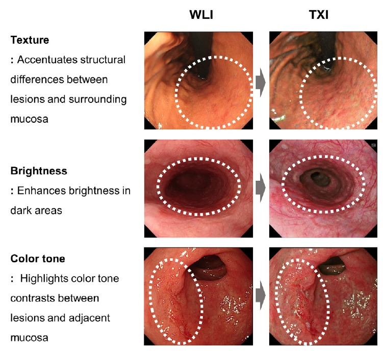
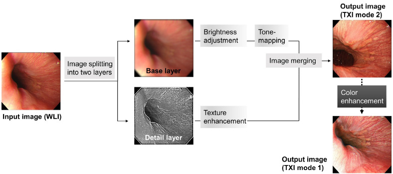
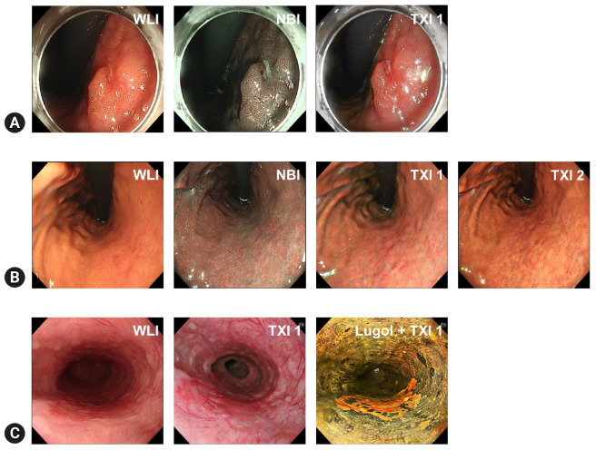
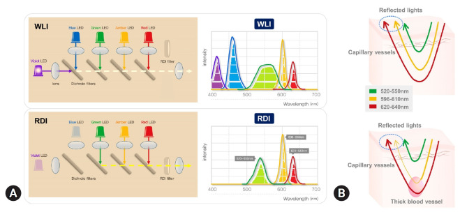
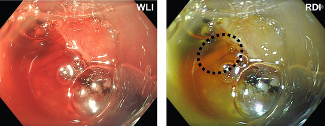
Endoscopic examination plays a crucial role in the diagnosis of upper gastrointestinal (UGI) tract diseases. Despite advancements in endoscopic imaging, the detection of subtle early cancers and premalignant lesions using white-light imaging alone remains challenging. This review discusses two novel image-enhanced endoscopy (IEE) techniques-texture and color enhancement imaging (TXI) and red dichromatic imaging (RDI)-and their potential applications in UGI diseases. TXI enhances texture, brightness, and color tone, which improves the visibility of mucosal irregularities and facilitates earlier detection of neoplastic lesions. Studies have suggested that TXI enhances the color differences between lesions and the surrounding mucosa and improves the visibility of the lesion. TXI aids in the diagnosis of various UGI diseases, including early gastric cancer, esophageal cancer, premalignant conditions such as atrophic gastritis and Barrett's esophagus, and duodenal tumors. RDI utilizes specific wavelengths to enhance the visualization of deep blood vessels or bleeding points, aiding in the rapid and accurate identification of bleeding sources during endoscopic procedures. Although promising, TXI and RDI require further large-scale studies across diverse populations to establish their clinical utility, diagnostic performance, and cost-effectiveness before integration into the guidelines. Standardized training is also required for effective utilization. Overall, these IEE techniques has the potential to improve the diagnosis and management of UGI.
Keywords: Diagnosis; Endoscopy; Gastric cancer; Image enhancement; Upper gastrointestinal tract.
Conflict of interest statement
Jae Yong Park is currently serving as a publication committee member in
Figures
Similar articles
-
Using texture and colour enhancement imaging to evaluate gastrointestinal diseases in clinical practice: a review.Ann Med. 2022 Dec;54(1):3315-3332. doi: 10.1080/07853890.2022.2147992. Ann Med. 2022. PMID: 36420822 Free PMC article. Review.
-
Third-Generation High-Vision Ultrathin Endoscopy Using Texture and Color Enhancement Imaging and Narrow-Band Imaging to Evaluate Barrett's Esophagus.Diagnostics (Basel). 2022 Dec 13;12(12):3149. doi: 10.3390/diagnostics12123149. Diagnostics (Basel). 2022. PMID: 36553156 Free PMC article.
-
Recent advancements in image-enhanced endoscopy in the pancreatobiliary field.DEN Open. 2024 May 14;5(1):e382. doi: 10.1002/deo2.382. eCollection 2025 Apr. DEN Open. 2024. PMID: 38746904 Free PMC article. Review.
-
Utility of texture and color enhancement and red dichromatic imaging during endoscopic resection.VideoGIE. 2025 Apr 23;10(8):439-442. doi: 10.1016/j.vgie.2025.04.004. eCollection 2025 Aug. VideoGIE. 2025. PMID: 40704121 Free PMC article. Review.
-
Texture and Color Enhancement Imaging Increases Color Changes and Improves Visibility for Squamous Cell Carcinoma Suspicious Lesions in the Pharynx and Esophagus.Diagnostics (Basel). 2021 Oct 23;11(11):1971. doi: 10.3390/diagnostics11111971. Diagnostics (Basel). 2021. PMID: 34829318 Free PMC article.
Cited by
-
Navigating the Treatment Landscape for Widespread Superficial Esophageal Squamous Cell Neoplasia.Gut Liver. 2025 May 15;19(3):305-306. doi: 10.5009/gnl250084. Gut Liver. 2025. PMID: 40356327 Free PMC article. No abstract available.
-
Classification of image-enhanced endoscopy in colon tumors.Clin Endosc. 2025 May;58(3):337-351. doi: 10.5946/ce.2024.263. Epub 2025 May 8. Clin Endosc. 2025. PMID: 40336268 Free PMC article. Review.
References
-
- Hahn KY, Park CH, Lee YK, et al. Comparative study between endoscopic submucosal dissection and surgery in patients with early gastric cancer. Surg Endosc. 2018;32:73–86. - PubMed
-
- Jun JK, Choi KS, Lee HY, et al. Effectiveness of the Korean national cancer screening program in reducing gastric cancer mortality. Gastroenterology. 2017;152:1319–1328. - PubMed
Publication types
LinkOut - more resources
Full Text Sources
