. 2025 Feb;14(4):e2403264.
doi: 10.1002/adhm.202403264. Epub 2024 Dec 25.

Microfluidics-Driven Manufacturing and Multiscale Analytical Characterization of Nanoparticle-Vesicle Hybrids

Affiliations

Microfluidics-Driven Manufacturing and Multiscale Analytical Characterization of Nanoparticle-Vesicle Hybrids

Jacopo Cardellini et al. Adv Healthc Mater. 2025 Feb.

Abstract

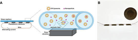
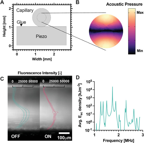
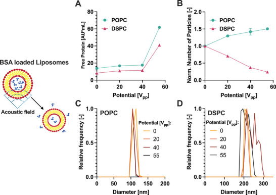
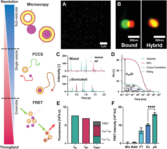
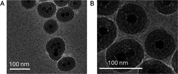
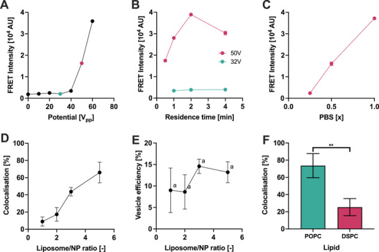
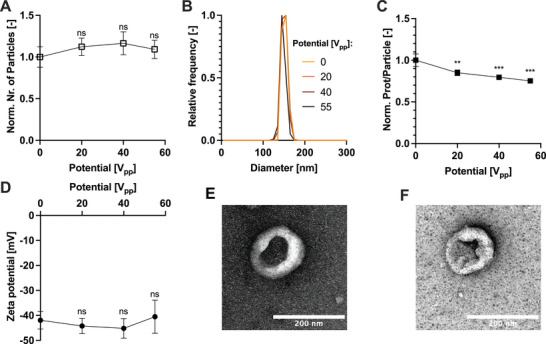
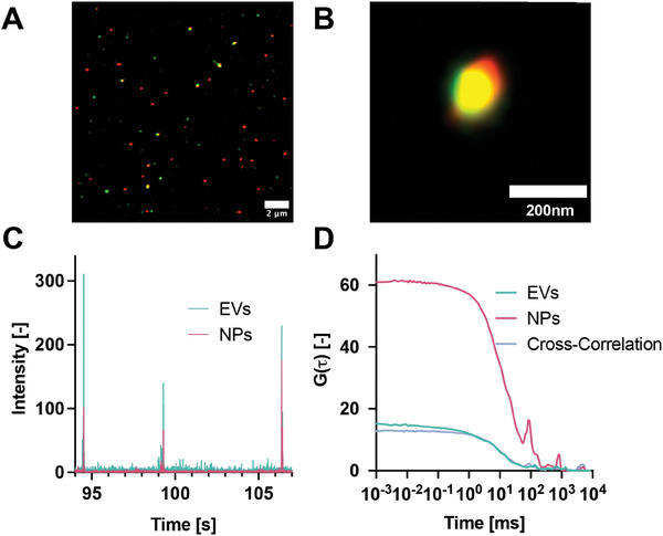
Coating synthetic nanoparticles (NPs) with lipid membranes is a promising approach to enhance the performance of nanomaterials in various biological applications, including therapeutic delivery to target organs. Current methods for achieving this coating often rely on bulk approaches which can result in low efficiency and poor reproducibility. Continuous processes coupled with quality control represent an attractive strategy to manufacture products with consistent attributes and high yields. Here, this concept is implemented by developing an acoustic microfluidic device together with an analytical platform to prepare nanoparticle-vesicle hybrids and quantitatively characterize the nanoparticle coverage using fluorescence-based techniques at different levels of resolution. With this approach polymethyl methacrylate (PMMA) nanoparticles are successfully coated with liposomes and extracellular vesicles (EVs), achieving a high encapsulation efficiency of 70%. Moreover, the approach enables the identification of design rules to control the efficiency of encapsulation by tuning various operational parameters and material properties, including buffer composition, nanoparticle/vesicle ratio, and vesicle rigidity.

Keywords: acoustofluidics; lipid vesicles; microfluidics; nanoparticles; nanoparticle‐vesicle hybrids.


Conflict of interest statement

The authors declare no conflict of interest.

Figures

Figure 1
Figure 1
A) Illustration of the µSonicator. An alternating current at a defined frequency is applied to the piezoelectric devices, causing them to vibrate and generate an acoustic field inside the capillary, leading to the formation of vesicle‐NP hybrids. B) Image of the µSonicator. For scale a coin with a diameter of 27.40 mm.
Figure 2
Figure 2
A) Model of the µSonicator cross‐section. B) Simulated acoustic pressure distribution in the glass capillary cross‐section at the 1.75 MHz excitation frequency. C) Distribution of 100 nm polystyrene particles in the microfluidic channel with the acoustic field OFF and ON. The particles migrate across the capillary due to the acoustic field. Overlay ‐ Fluorescence intensity distribution across the channel. The lowest value was normalized to 0. The dotted lines represent the standard deviation of technical triplicates. D) Simulated average acoustic energy densities (E ac ) at different excitation frequencies.
Figure 3
Figure 3
Effect of the microfluidic sonication on liposomes. A) Free protein released from the liposomes as measured by SEC‐FLD; B) Normalised particle concentration measured by SEC‐MALS as a function of the applied voltage. The data are normalized based on the concentration measured at 0 V. C,D) Size distribution of POPC (C) and DSPC (D) liposomes measured by SEC‐MALS. Error bars represent the standard deviation of technical triplicates.
Figure 4
Figure 4
Characterisation of hybrid liposome‐NPs by fluorescence‐based methods at different resolutions and throughputs. A) TIRF microscopy image after microfluidic sonication. Green ‐ DiO‐Liposomes, Red ‐ RhodamineB‐PMMA‐NPs. B) The high resolution of TIRF microscopy can distinguish between liposomes bound to NPs (partial overlap) and hybrids (full overlap). C) Fluorescence intensity traces measured by FCCS. After µSonication the fluorescence peaks are overlapping. Top ‐ after mixing, Bottom ‐ after µSonication at 55V pp . Green ‐ DiO‐Liposomes, Red ‐ RhodamineB‐PMMA‐NPs. D) FCS correlation functions of vesicles, PMMA‐NPs, and Cross‐Correlation. The colocalization is calculated by dividing the amplitude of the cross‐correlation by the amplitude autocorrelation of vesicles. Black curves represent fits of autocorrelation functions of vesicles and PMMA‐NPs; E) Three fluorescence intensities were measured: fluorescence intensity in the vesicle channel I Ve , fluorescence intensity in the nanoparticle channel I NP and the fluorescence intensity in the FRET channel I FRET . The FRET signal was calculated from the measured I FRET subtracting the contributions from the direct fluorescence of the nanoparticles and vesicles (c x × I x ). See the Methods section for further details. F)FRET intensity after different loading methods. Bath ‐ Bath sonication, FT ‐ Freeze‐Thaw, Ex ‐ Coextrusion, µS ‐ µSonication. Error bars represent the standard deviation of technical triplicates.**** ‐ P < 0.0001, one‐way ordinary ANOVA post‐hoc analysis.
Figure 5
Figure 5
Cryo‐EM images of hybrids composed of core‐shell iron oxide NPs coated by lipid bilayers generated with the acoustic microfluidic device.
Figure 6
Figure 6
Parameters affecting the formation of hybrid liposome‐nanoparticles. A) Effect of the applied electrical potential on the FRET signal. B) Effect of residence time in the device on FRET signal at 32 and 50 V pp . C) Effect of the PBS concentration on hybrid formation measured by FRET signal. D) The percentage of colocalization measured by FCCS after sonicating different liposome/NP ratios in the µSonicator. E) Calculated percentage of vesicles converted into liposome‐NP hybrids after sonicating different liposome/NP ratios in the µSonicator. The values are statistically not significantly different from another (a ‐ P > 0.05, one way ANOVA multiple comparisons between every column pair). F) Effect of POPC and DSPC liposome rigidity on the percentage of colocalization calculated by cross‐correlation measured by FCCS.Error bars represent the standard deviation of technical triplicates. ** ‐ P < 0.01, unpaired t‐test post‐hoc analysis.
Figure 7
Figure 7
Effect of the sonication on EVs. Both the A) number of particles and B) size distribution measured by MALS is not affected by the acoustic field. However, C) the protein per particle decreases with the increased acoustic field strength. D) The zeta potential measured by NTA remains constant despite an increasing acoustic field strength. The morphology of the HEK‐EVs E) before and F) after µSonication at 55 V pp imaged by negative staining TEM. Error bars represent the standard deviation of technical triplicates. ns ‐ not significant; ** ‐ P < 0.01; *** ‐ P < 0.001, one‐way ordinary ANOVA post‐hoc analysis. Each measurement was compared to the control value (0V) for statistical significance.
Figure 8
Figure 8
Characterisation of HEK‐EV membrane‐covered PMMA‐NP hybrids. A) dSTORM images of HEK‐EVs‐PMMA show the colocalization of the surface marker CD81 and PMMA‐NPs. B) Zoomed image of the formed hybrid. C) Intensity traces in two channels (EV Channel: λ ex = 488 and λ em = 520 nm; NPs channel: λ ex = 561 and λ em = 650 nm). D) Autocorrelation functions and cross‐correlation of HEK‐EVs and PMMA‐NPs after sonication.

Similar articles

Cited by

References

    1. Jiao M., Zhang P., Meng J., Li Y., Liu C., Luo X., Gao M., Biomater. Sci. 2018, 6, 726. - PubMed
    1. Shi J., Kantoff P. W., Wooster R., Farokhzad O. C., Nat. Rev. Cancer 2017, 17, 20. - PMC - PubMed
    1. Youn Y. S., Bae Y. H., Adv. Drug Delivery Rev. 2018, 130, 3. - PubMed
    1. Aftab S., Shah A., Nadhman A., Kurbanoglu S., Aysıl Ozkan S., Dionysiou D. D., Shukla S. S., Aminabhavi T. M., Int. J. Pharm. 2018, 540, 132. - PubMed
    1. Dallari C., Lenci E., Trabocchi A., Bessi V., Bagnoli S., Nacmias B., Credi C., Pavone F. S., ACS Sens. 2023, 8, 3693. - PMC - PubMed

MeSH terms

Grants and funding

LinkOut - more resources