. 2024 Dec;30(12):e70188.
doi: 10.1111/cns.70188.

Clinical Insights Into Default Mode Network Abnormalities in Mild Traumatic Brain Injury: Unraveling Axonal Injury Through Functional, Structural, and Molecular Analyses

Affiliations

Clinical Insights Into Default Mode Network Abnormalities in Mild Traumatic Brain Injury: Unraveling Axonal Injury Through Functional, Structural, and Molecular Analyses

Dewen Ru et al. CNS Neurosci Ther. 2024 Dec.

Abstract

Background: Mild traumatic brain injury (mTBI) frequently results in persistent cognitive, emotional, and functional impairments, closely linked to disruptions in the default mode network (DMN). Understanding the mechanisms driving these network abnormalities is critical for advancing diagnostic and therapeutic strategies.

Methods: This study adopted a multimodal approach, combining functional connectivity (FC) analysis, diffusion tensor imaging (DTI), and gene expression profiling to investigate DMN disruptions in mTBI. A primary focus was placed on the middle cingulate cortex (MCC), a region consistently identified with increased connectivity. We explored the structural and molecular changes underlying this phenomenon. Receiver operating characteristic (ROC) curve analysis was utilized to assess the diagnostic potential of DTI-derived metrics, while white matter tractography was employed to explore structural connectivity between the MCC and the dorsolateral prefrontal cortex (DLPFC).

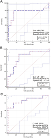
Results: Our findings revealed significant disruptions in DMN connectivity, with the MCC prominently involved in mTBI pathology. DTI analyses identified pronounced axonal injury in the MCC, characterized by decreased fractional anisotropy (FA) and axial diffusivity (AD), alongside increased isotropy (ISO), indicating compromised white matter integrity and diffuse axonal injury. Gene expression profiling revealed the upregulation of pathways related to synaptic transmission, ion channel regulation, and axonal injury response. ROC analysis demonstrated that ISO serves as a particularly effective biomarker for mTBI, showing high diagnostic accuracy (AUC = 0.871). White matter tractography further confirmed strong structural connectivity between the MCC and the DLPFC, identifying potential therapeutic targets for neuromodulation.

Conclusion: This study provides robust evidence that diffuse axonal injury plays a pivotal role in DMN abnormalities observed in mTBI. The integration of FC, DTI, and gene expression profiling offers a comprehensive framework for understanding mTBI's impact on brain networks. Our findings also highlight the DLPFC as a promising target for therapeutic interventions aimed at addressing cognitive and emotional deficits associated with mTBI.

Keywords: axonal injury; default mode network; diffusion tensor imaging; functional connectivity; mild traumatic brain injury.


Conflict of interest statement

The authors declare no conflicts of interest.

Figures

FIGURE 1
FIGURE 1
Flowchart of studies selection process for meta‐analysis on DMN abnormalities in mTBI patients. As is shown above, 10 studies met all inclusion criteria and were incorporated into the final ALE meta‐analysis, which aimed to investigate DMN abnormalities associated with mTBI. The use of these criteria ensured a rigorous and focused analysis of the literature on DMN alterations in the context of mTBI. ALE, activation likelihood estimation; DMN, default mode network; mTBI, mild traumatic brain injury.
FIGURE 2
FIGURE 2
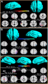
Changes in DMN connectivity in mTBI patients compared to healthy controls across acute and subacute phases. Acute Phase (within 7 days post‐injury): (A) Increased DMN connectivity. 3D surface renderings of the brain highlight regions where mTBI patients exhibited significantly increased DMN connectivity relative to healthy controls. Key regions include the Posterior Lobe, Declive, Temporal Lobe, Superior Temporal Gyrus, Parietal Lobe, Precuneus, Anterior Lobe, and Culmen. (B) Decreased DMN connectivity. 3D brain renderings show regions where mTBI patients exhibited significantly decreased DMN connectivity compared to healthy controls. Key regions include the Parietal Lobe and Angular Gyrus. Subacute Phase (beyond 7 days post‐injury): (C) The 3D surface renderings depict regions in both the left and right hemispheres where mTBI patients exhibited significantly increased DMN connectivity. Key regions include the Limbic Lobe, Cingulate Gyrus, Brodmann area 23, Insula, Brodmann area 13, Claustrum, Postcentral Gyrus, Brodmann area 7, Superior Frontal Gyrus, and Brodmann area 9. ALE, activation likelihood estimation; MNI, Montreal Neurological Institute.
FIGURE 3
FIGURE 3
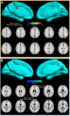
Increased DMN connectivity in all mTBI patients compared to healthy controls. (A) Increased DMN connectivity: The top row presents 3D surface renderings of the brain, highlighting regions in both hemispheres where mTBI patients exhibited significantly increased DMN connectivity. Key regions include the Limbic Lobe and Cingulate Gyrus. The bottom row shows axial slices at specific z‐coordinates (z = 43, z = 35.5), offering a more detailed view of the areas with enhanced connectivity. (B) Decreased DMN connectivity: This panel displays regions where mTBI patients showed significantly decreased DMN connectivity compared to healthy controls. Key regions include the Limbic Lobe, Posterior Cingulate, Occipital Lobe, and Precuneus. The top row provides 3D brain renderings, while the bottom row presents axial slices at various z‐coordinates (z = 28, z = 18) to illustrate these areas with decreased connectivity. ALE, activation likelihood estimation; MNI, Montreal Neurological Institute.
FIGURE 4
FIGURE 4
Integrated molecular and structural changes in the MCC of mTBI patients. (A) Location of the MCC: The top panel illustrates the location of the MCC as a region of interest in the brain, based on meta‐analysis findings. (B) Gene Enrichment Analysis: This panel displays the results of the gene enrichment analysis conducted on the MCC. The analysis identifies the most significantly enriched biological processes associated with the DEGs in this region, with particular emphasis on processes such as “response to axon injury”. (C) Gene Interaction Network: The comprehensive gene interaction network is depicted, where nodes represent individual genes and edges indicate the interactions between them. (D) The FA values for the MCC, comparing mTBI patients to healthy controls. Notice that a significant reduction in FA is observed in mTBI patients, indicating compromised white matter integrity in the MCC. (E) The AD values for the MCC, which reflect the magnitude of water diffusion along the principal axis of white matter fibers. Notice that in mTBI patients, AD is significantly reduced compared to healthy controls, further supporting the presence of axonal damage in the MCC. (F) The ISO values for the MCC, which measure the extent of isotropic water diffusion. Notice that in mTBI patients, ISO values are significantly higher than those in healthy controls. AD, axial diffusivity; Con, healthy control; FA, fractional anisotropy; ISO, iisotropy; mTBI, mild traumatic brain injury.
FIGURE 5
FIGURE 5
Functional enrichment analysis of the MCC as a common region of increased connectivity in mTBI patients. Key terms: Prominent terms such as “memory,” “control,” “inhibition,” “recognition,” and “social” are displayed in larger font sizes, indicating their strong relevance to the MCC's connectivity in mTBI patients. Additional relevant terms: Other notable terms, including “pain,” “anxiety,” “reward,” “cognition,” “emotional,” and “personality,” are also highlighted, reflecting the MCC's involvement in a wide range of emotional and cognitive processes. Interpretation: The word cloud effectively emphasizes the relationship between increased MCC connectivity and the diverse cognitive, emotional, and social processes affected in mTBI patients.
FIGURE 6
FIGURE 6
ROC curve analysis for identifying mTBI patients using DTI metrics in the MCC. (A) ROC curve for FA in the MCC. The analysis reveals that FA is a moderately effective biomarker for identifying mTBI patients. (B) ROC curve for AD in the MCC. Notice that AD is a reliable marker for distinguishing mTBI patients from healthy controls. (C) ROC curve for ISO in the MCC. Notice that ISO is a highly effective biomarker for mTBI. AUC, area under the curve.
FIGURE 7
FIGURE 7
Integrated functional and structural connectivity analysis between the MCC and dorsolateral prefrontal cortex (DLPFC). (A)Whole‐brain FC analysis using the MCC as the ROI in a cohort of 40 healthy controls. Central Panel: The central 3D cortical surface visualization shows the MCC's FC with the rest of the brain. Warm colors (yellow to red) indicate regions of strong positive connectivity with the MCC, while cooler colors (blue) represent areas of weaker connectivity. Surrounding Axial Slices: The axial slices surrounding the central panel provide a more detailed view of specific brain regions with significant FC to the MCC. (B) White matter tractography analysis in mTBI patients. The left panel visualizes the white matter fiber tracts connecting the MCC and DLPFC in mTBI patients, as identified through DTI tractography. The central panel provides a close‐up of these tracts, while the right panel highlights the role of the superior longitudinal fasciculus, a major white matter pathway mediating this structural connection.

Similar articles

Cited by

References

    1. Visser K., Koggel M., Blaauw J., van der Horn H. J., Jacobs B., and van der Naalt J., “Blood‐Based Biomarkers of Inflammation in Mild Traumatic Brain Injury: A Systematic Review,” Neuroscience and Biobehavioral Reviews 132 (2022): 154–168. - PubMed
    1. Kahriman A., Bouley J., Tuncali I., et al., “Repeated Mild Traumatic Brain Injury Triggers Pathology in Asymptomatic C9ORF72 Transgenic Mice,” Brain 146, no. 12 (2023): 5139–5152. - PMC - PubMed
    1. Niu X., Bai L., Sun Y., et al., “Mild Traumatic Brain Injury is Associated With Effect of Inflammation on Structural Changes of Default Mode Network in Those Developing Chronic Pain,” Journal of Headache and Pain 21, no. 1 (2020): 135. - PMC - PubMed
    1. Zhou Y., Milham M. P., Lui Y. W., et al., “Default‐Mode Network Disruption in Mild Traumatic Brain Injury,” Radiology 265, no. 3 (2012): 882–892. - PMC - PubMed
    1. Li F., Lu L., Sa S., et al., “Disrupted Functional Network Connectivity Predicts Cognitive Impairment After Acute Mild Traumatic Brain Injury,” CNS Neuroscience & Therapeutics 26, no. 10 (2020): 1083–1091. - PMC - PubMed

Publication types

MeSH terms

LinkOut - more resources