Review
. 2024 Dec 21:17:11419-11436.
doi: 10.2147/JIR.S497775. eCollection 2024.

Regulated Cell Death of Alveolar Macrophages in Acute Lung Inflammation: Current Knowledge and Perspectives

Affiliations
Review

Regulated Cell Death of Alveolar Macrophages in Acute Lung Inflammation: Current Knowledge and Perspectives

Siwei Xia et al. J Inflamm Res. .

Abstract

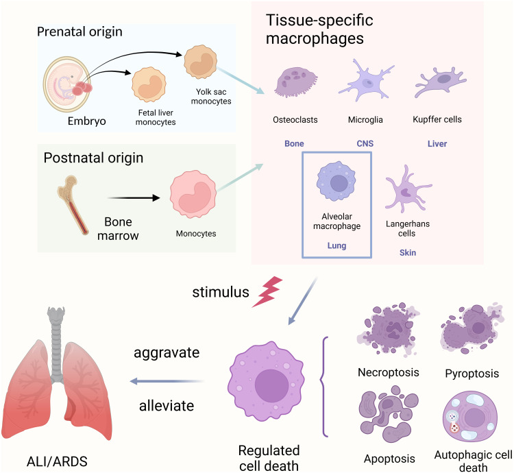
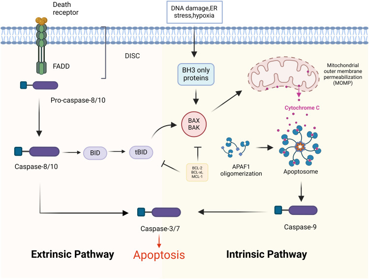
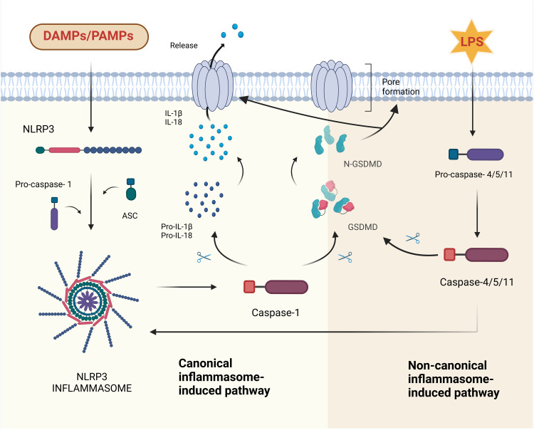
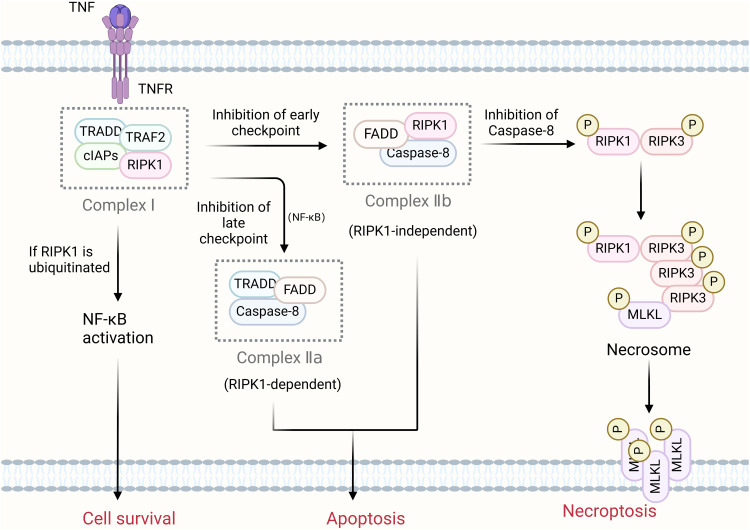
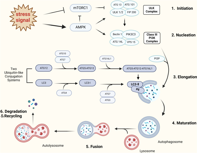
Acute lung injury/acute respiratory distress syndrome (ALI/ARDS) is a common and serious clinical lung disease characterized by extensive alveolar damage and inflammation leading to impaired gas exchange. Alveolar macrophages (AMs) maintain homeostatic properties and immune defenses in lung tissues. Several studies have reported that AMs are involved in and regulate ALI/ARDS onset and progression via different regulated cell death (RCD) programs, such as pyroptosis, apoptosis, autophagic cell death, and necroptosis. Notably, the effects of RCD in AMs in disease are complex and variable depending on the environment and stimuli. In this review, we provide a comprehensive perspective on how regulated AMs death impacts on ALI/ARDS and assess its potential in new therapeutic development. Additionally, we describe the crosstalk between different RCD types in ALI, and provide new perspectives for the treatment of ALI/ARDS and other severe lung diseases.

Keywords: acute lung injury; alveolar macrophage; apoptosis; autophagic cell death; necroptosis; pyroptosis.


Conflict of interest statement

The authors report no conflicts of interest in this work.

Figures

Figure 1
Figure 1
Alveolar macrophages. Macrophages resident in different organs can be derived from embryonic monocyte progenitors that originate in the yolk sac or fetal liver during embryonic development and then migrate to different tissues. Circulating monocytes that develop from haematopoietic stem cells in the bone marrow can also differentiate into tissue-resident macrophages. They exhibit diverse functions. When resident alveolar macrophages are exposed to external stimuli (such as environmental factors, drugs, genetic factors, etc), they undergo regulated cell death, which affects their phenotype and function, thereby either exacerbating or alleviating pulmonary inflammation. Created in BioRender. Xia, S. (2024) https://BioRender.com/e15t922.
Figure 2
Figure 2
The extrinsic and intrinsic apoptotic pathways. The extrinsic apoptotic pathway is activated when the ligand activates the death receptor, recruiting FADD and Caspase-8/10 to form the death-inducing signalling complex (DISC). Subsequent activation of Caspase-8/10 and death effector Caspase-3/7 induces apoptosis. The intrinsic apoptotic pathway involves the activity of the BCL-2 family of proteins located in the outer mitochondrial membrane, and many stimuli lead to permeability of the mitochondrial outer membrane (MOMP) and further release of cytochrome C. The latter binds to APAF-1 to form the DISC. The latter binds APAF-1 to form apoptotic bodies, which then activate Caspase-9 and downstream Caspase-3/7. In some cells, Bid cleavage also activates BAK/BAX thereby leading to the release of cytochrome c from mitochondria and promoting apoptosis. Created in BioRender. Xia, S. (2024) https://BioRender.com/z40q507.
Figure 3
Figure 3
The canonical and non-canonical inflammasome pathways in pyroptosis. In the canonical pyroptosis pathway, active NLRP3 binds Pro-caspase-1 via ACS to form NLRP3 inflammasome, followed by activation of Caspase-1. Active Caspase-1 mediates the maturation and cleavage of IL-18 and IL-1β from the GSDMD, which forms membrane pores and releases cytoplasmic inflammatory contents, leading to cellular pyroptosis. In the non-canonical pyroptosis pathway, lipopolysaccharide (LPS) derived from Gram-negative bacteria directly activates Caspase-4/5/11. Activated Caspase-4/5/11 cleaves the GSDMD to induce pore formation, while activating the NLRP3 inflammasome through K efflux, leading to pyroptosis. Created in BioRender. Xia, S. (2024) https://BioRender.com/y83o292.
Figure 4
Figure 4
Necroptosis pathway. TNFα is the predominant upstream signalling component of necroptosis apoptosis.TNF-α activates TNFR1, which recruits scaffolding proteins TRADD, TRAF2, RIPK1 and cIAP1/2 to form plasma membrane-associated complex I. Linear ubiquitination stabilises complex I, which activates the NF-κB signalling pathway, leading to cell survival. When NF-κB is inhibited, complex IIa is activated, initiating apoptosis. Disruption of the signalling checkpoint early in cell survival leads to induction of complex IIb, which induces apoptosis via activated Caspase-8. When Caspase-8 is inhibited, phosphorylated RIPK1 and phosphorylated RIPK3 recruit and phosphorylate their substrate MLKL to form the necrosome complex. Activated MLKL oligomerises and migrates to the plasma membrane, thereby triggering necroptosis. Created in BioRender. Xia, S. (2024) https://BioRender.com/d66m064.
Figure 5
Figure 5
Classical autophagy pathway. In stress-induced macroautophagy, AMPK activation or inhibition of mTORC1 activity leads to the activation of the ULK complex and class III PI3K complex, resulting in the formation of phagocytic carriers. Two different ubiquitin-like coupling systems are involved in phagocytic carrier elongation: one involves ATG5-ATG12-ATG16L, and the other involves LC3-PE (LC3II), which helps to seal off double-membrane autophagosome formation. Eventually, autophagosomes fuse with lysosomes to form autophagic lysosomes for degradation. Created in BioRender. Xia, S. (2024) https://BioRender.com/i71l142.

Similar articles

Cited by

References

    1. Matuschak GM, Lechner AJ. Acute lung injury and the acute respiratory distress syndrome: pathophysiology and treatment. Mo Med. 2010;107(4):252–258. - PMC - PubMed
    1. Mokra D, Kosutova P. Biomarkers in acute lung injury. Respir Physiol Neurobiol. 2015;209:52–58. doi:10.1016/j.resp.2014.10.006 - DOI - PubMed
    1. Meyer NJ, Gattinoni L, Calfee CS. Acute respiratory distress syndrome. Lancet. 2021;398(10300):622–637. doi:10.1016/S0140-6736(21)00439-6 - DOI - PMC - PubMed
    1. Tang D, Kang R, Berghe TV, Vandenabeele P, Kroemer G. The molecular machinery of regulated cell death. Cell Res. 2019;29(5):347–364. doi:10.1038/s41422-019-0164-5 - DOI - PMC - PubMed
    1. Vitale I, Pietrocola F, Guilbaud E, et al. Apoptotic cell death in disease-current understanding of the NCCD 2023. Cell Death Differ. 2023;30(5):1097–1154. doi:10.1038/s41418-023-01153-w - DOI - PMC - PubMed

LinkOut - more resources