

Epithelial-Mesenchymal Transition Induced by a Metal Mixture in Liver Cells With Antioxidant Barrier Decreased
- PMID: 39722890
- PMCID: PMC11669431
- DOI: 10.1155/omcl/6983256
Epithelial-Mesenchymal Transition Induced by a Metal Mixture in Liver Cells With Antioxidant Barrier Decreased
Abstract
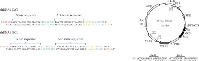
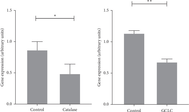
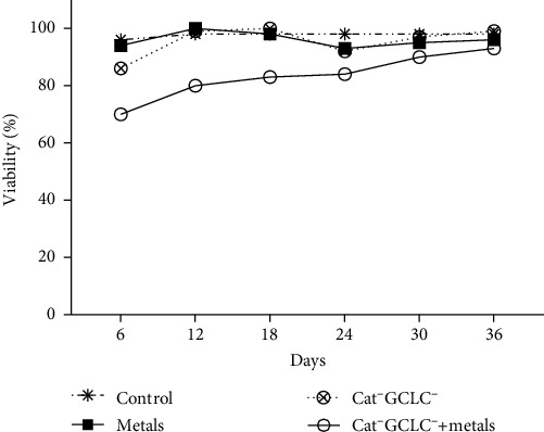
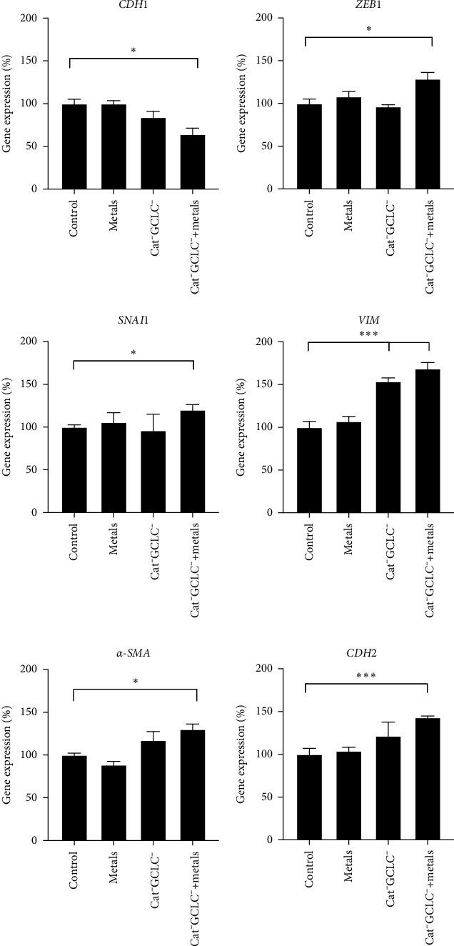
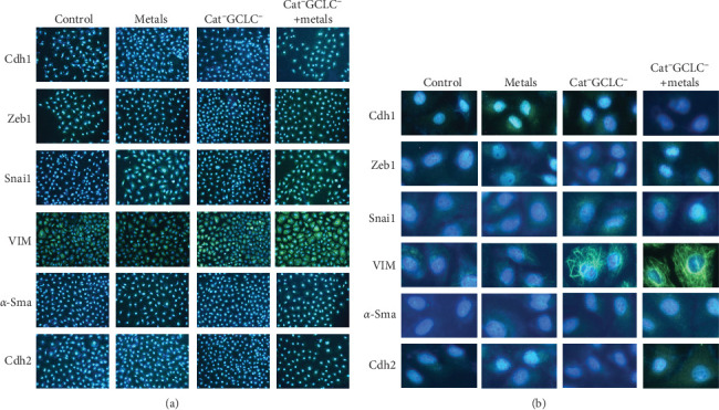
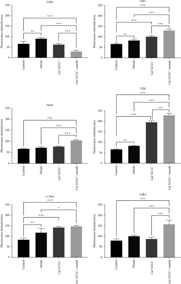
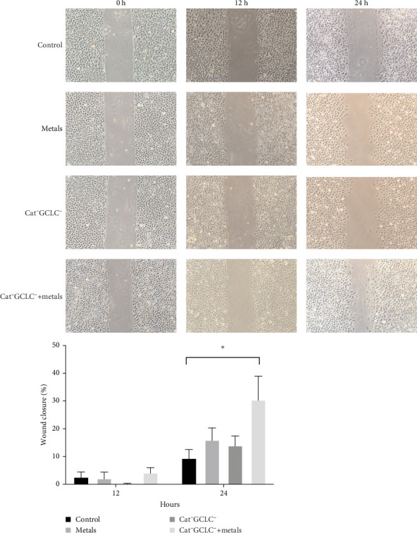
Occupational exposure to arsenic (As), cadmium (Cd), and lead (Pb) affects many sectors, necessitating research to understand their transformation mechanisms. In this study, we characterized the process of epithelial-mesenchymal transition (EMT) in a rat hepatic epithelial cell line with decreased expression of catalase and glutamate cysteine ligase catalytic (GCLC) subunit that was exposed to a mixture of As, Cd, and Pb at equimolar occupational exposure concentrations. We evaluated the expression of genes and proteins involved in EMT. Our findings revealed that cells with a decreased antioxidant barrier showed a decreased expression and abundance of epithelial genes when exposed to a mixture of metals. Additionally, we observed alterations in the expression of transcription factors (TFs) associated with EMT and an increase in the expression and abundance of mesenchymal genes. Specifically, we found that E-cadherin expression decreased by ~50% at both the gene and protein levels. In contrast, the expression of vimentin, α-smooth muscle actin, and N-cadherin genes increased by ~70%, whereas their corresponding protein levels increased by nearly 100%. Furthermore, the TFs zinc finger e-box binding homeobox 1 and snail family transcriptional repressor 1 showed a 30% increase in gene expression and an ~80% increase in protein expression. These changes enable the cells to acquire migratory capabilities. Our results confirmed that exposure to this mixture of As, Cd, and Pb can induce EMT in cells with a decreased antioxidant barrier.
Keywords: catalase and GSH abatement; epithelial–mesenchymal transition; metals mixture; wound healing.
Copyright © 2024 M. Valverde et al.
Conflict of interest statement
The authors declare no conflicts of interest.
Figures
Similar articles
-
Cadmium exposure upregulates SNAIL through miR-30 repression in human lung epithelial cells.Toxicol Appl Pharmacol. 2019 Jun 15;373:1-9. doi: 10.1016/j.taap.2019.04.011. Epub 2019 Apr 16. Toxicol Appl Pharmacol. 2019. PMID: 30998937 Free PMC article.
-
Epithelial to Mesenchymal Transition in Human Mesothelial Cells Exposed to Asbestos Fibers: Role of TGF-β as Mediator of Malignant Mesothelioma Development or Metastasis via EMT Event.Int J Mol Sci. 2019 Jan 3;20(1):150. doi: 10.3390/ijms20010150. Int J Mol Sci. 2019. PMID: 30609805 Free PMC article.
-
Targeting the Nuclear Cathepsin L CCAAT Displacement Protein/Cut Homeobox Transcription Factor-Epithelial Mesenchymal Transition Pathway in Prostate and Breast Cancer Cells with the Z-FY-CHO Inhibitor.Mol Cell Biol. 2017 Feb 15;37(5):e00297-16. doi: 10.1128/MCB.00297-16. Print 2017 Mar 1. Mol Cell Biol. 2017. PMID: 27956696 Free PMC article.
-
Epithelial-mesenchymal transition in breast epithelial cells treated with cadmium and the role of Snail.Toxicol Appl Pharmacol. 2018 Apr 1;344:46-55. doi: 10.1016/j.taap.2018.02.022. Epub 2018 Mar 6. Toxicol Appl Pharmacol. 2018. PMID: 29501589 Free PMC article.
-
The Post-Translational Regulation of Epithelial-Mesenchymal Transition-Inducing Transcription Factors in Cancer Metastasis.Int J Mol Sci. 2021 Mar 30;22(7):3591. doi: 10.3390/ijms22073591. Int J Mol Sci. 2021. PMID: 33808323 Free PMC article. Review.
References
MeSH terms
Substances
LinkOut - more resources
Full Text Sources
Medical
Research Materials
Miscellaneous
