. 2024 Dec 11:15:1525376.
doi: 10.3389/fimmu.2024.1525376. eCollection 2024.

Ring finger protein 5 mediates STING degradation through ubiquitinating K135 and K155 in a teleost fish

Affiliations

Ring finger protein 5 mediates STING degradation through ubiquitinating K135 and K155 in a teleost fish

Xiaowei Qin et al. Front Immunol. .

Abstract

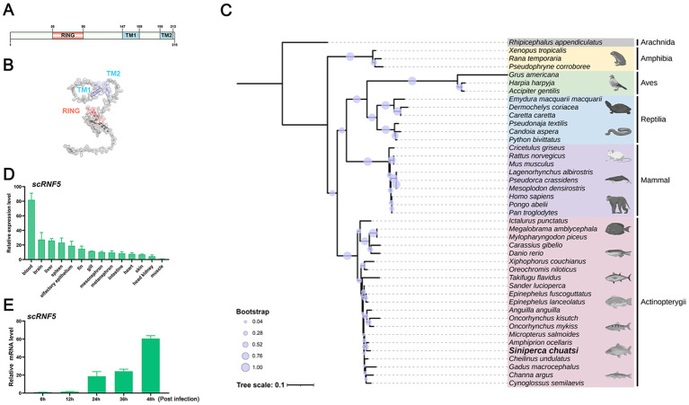
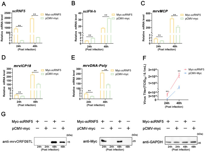
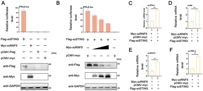
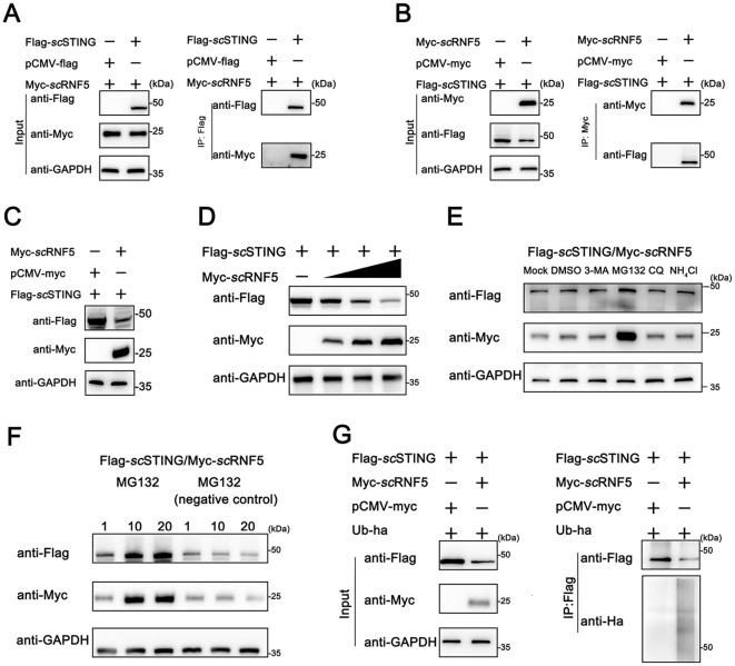
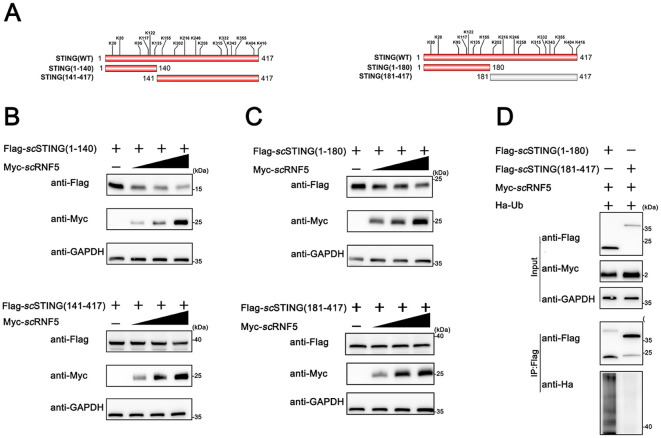
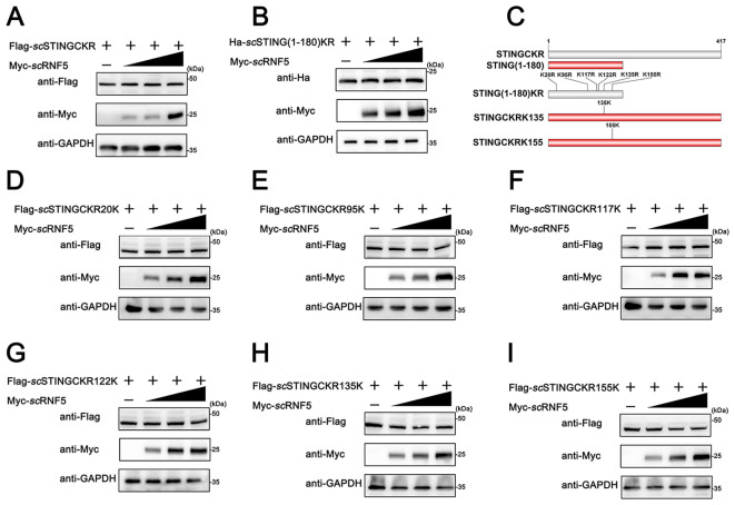
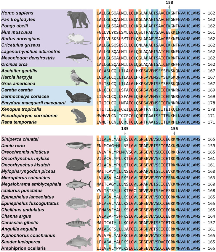
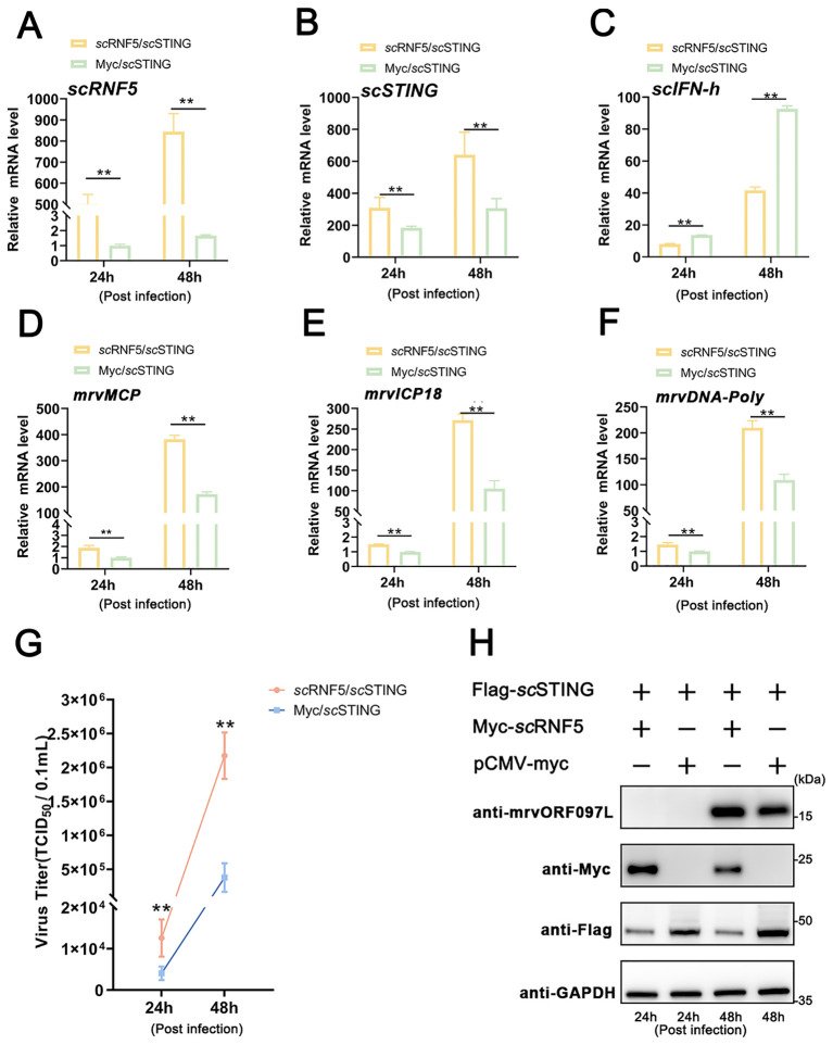
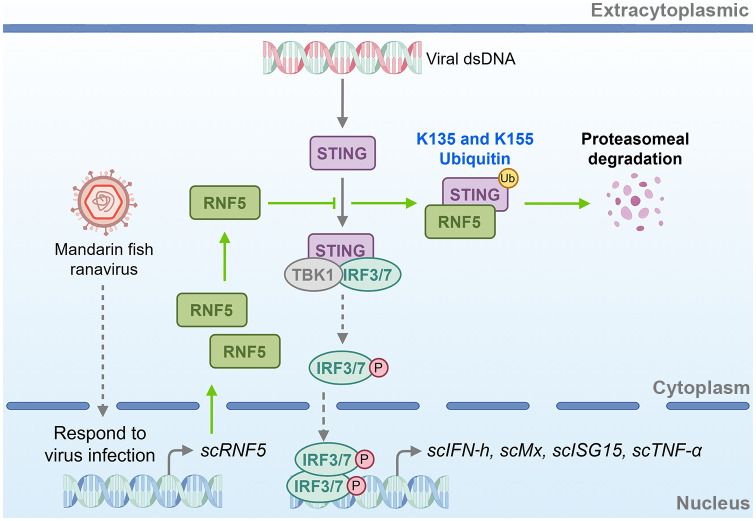
Stimulator of interferon genes (STING) is a key connector protein in interferon (IFN) signaling, crucial for IFN induction during the activation of antiviral innate immunity. In mammals, ring finger protein 5 (RNF5) functions as an E3 ubiquitin ligase, mediating STING regulation through K150 ubiquitylation to prevent excessive IFN production. However, the mechanisms underlying RNF5's regulation of STING in teleost fish remain unknown. This study investigated the regulatory role of the mandarin fish (Siniperca chuatsi) RNF5 (scRNF5) in the STING-mediated antiviral immune response and identified the specific regulatory sites on scSTING. Furthermore, an examination of scRNF5 expression patterns in virus-infected cells revealed its responsiveness to mandarin fish ranavirus (MRV) infection. The ectopic expression of scRNF5 suppressed scSTING-mediated IFN signaling and facilitated MRV replication. Co-immunoprecipitation experiments indicated an interaction between scRNF5 and scSTING. The further experiments demonstrated that scRNF5 exerted its inhibitory effect by promoting the degradation of scSTING, which was observed to be blocked by MG132 treatment. Ubiquitination assays with various scSTING mutants showed that scRNF5 catalyzed the ubiquitination of scSTING at K135 and K155 residues. Furthermore, we provided evidence that scRNF5 significantly attenuated scSTING-dependent antiviral immunity by targeting negative regulators within the scSTING signaling cascade. This study underscored that RNF5 negatively regulated the STING-mediated IFN signaling pathway in mandarin fish, attenuated STING's antiviral activity, and facilitated STING degradation via the ubiquitin-proteasome pathway at two novel lysine sites (K135 and K155). Our work offered valuable insights into the regulatory mechanisms of STING-mediated signaling in teleost fish, paving the way for further research.

Keywords: RNF5; STING; innate immunity; interferons; ubiquitination.


Conflict of interest statement

The authors declare that the research was conducted in the absence of any commercial or financial relationships that could be construed as a potential conflict of interest.

Figures

Figure 1
Figure 1
Molecular characteristics of scRNF5. (A, B) Domain and 3–D structure prediction of scRNF5. (C) A phylogenetic tree was generated using the MEGA v11.0.13 program, based on RNF5 sequences from various vertebrate species (GenBank accession numbers are provided in the Supplementary Material 2 ). (D) Tissue distributions of scRNF5 expressions in different tissues of mandarin fish. Samples were collected from the blood, brain, liver, spleen, fin, gill, intestine, heart, skin, head kidney, and muscle of three healthy mandarin fish, and the expression levels of the scRNF5 were detected by qRT-PCR. (E) Expression of scRNF5 in response to MRV infection. scRNF5 mRNA levels in MFF-1 cells were measured by qRT-PCR and normalized to the reference β-actin gene after infection with MRV at 0.1 MOI.
Figure 2
Figure 2
Overexpression of scRNF5 enhances MRV infection. Cells were transfected with scRNF5-myc or pCMV-myc for 24 h, then infected with MRV, and harvested at the indicated time points for qRT-PCR analysis. (A) Expression levels of scRNF5 in MRV-infected cells at indicated times. (B) Expression levels of scIFN-h genes in MRV-infected cells at various time points. (C–E) Expression levels of mrvMCP, mrvICP18, mrvDNA-Poly genes in MRV-infected cells at indicated times. For qRT-PCR analysis, the baseline was established as the group exhibiting the lowest relative expression among all groups, with this value set to 1. All groups represented different time points following viral infection. (F) Virus infection was measured on a 96-well cell culture plate via the finite dilution method. (G) Protein levels of mrvORF097L were detected via WB analysis. Vertical bars represent ± SD (n = 3). Statistical significance is indicated by asterisks, with ** referring to p < 0.01.
Figure 3
Figure 3
Suppression of scSTING-triggered IFN signaling by scRNF5. (A) Relative luciferase activities of IFN-β-luc were measured relative to controls after cells were co-transfected with scSTING-flag and scRNF5-myc. Cells transfected with pCMV-myc alone served as the negative control. (B) MFF-1 cells were co-transfected with the reporter plasmids pRL-TK and IFN-β-luc, as well as the indicated plasmids scRNF5 and scSTING in 24-well plates. A reporter assay was performed 36 h post-transfection. (C–F) Cells were collected 24 h post-transfection after co-transfection scRNF5 or vector alone alongside scSTING, and levels of expression levels of scIFN-h (C), scMx (D), scISG15 (E) and scTNF-α (F) were assessed. Data shown are representative of three independent experiments (n=3). Statistical analysis revealed significant differences (**p < 0.01).
Figure 4
Figure 4
Ubiquitination-mediated degradation of scSTING by scRNF5. (A, B) FHM cells were transfected with pCMV-Myc-scRNF5 or empty vector and pCMV-Flag-scSTING plasmids. Co-IP and WB analyses were performed for detection. IB: immunoblotting; IP: immunoprecipitation. (C) Western blotting analyses were used for detection. FHM cells were transfected with pCMV-Myc-scRNF5 or empty vector and pCMV-Flag-scSTING plasmids. (D) scRNF5 overexpression leads to dose-dependent STING degradation. FHM cells were seeded into 6-well plates, incubated overnight, and co-transfected with 2 μg Flag-scSTING and Myc-scRNF5 (0.5, 1, and 2 μg, respectively), After 24 h, lysates were immunoblotted with anti-Flag, anti-Myc, and anti-GAPDH antibodies. (E) The effects of MG132, 3-MA, CQ and NH4Cl on scRNF5-induced scSTING degradation were assessed. FHM cells in 6-well plates were transfected with Flag-scSTING (2 μg) and Myc-scRNF5 (2 μg). At 24 h post-transfection (hpt), cells were treated with DMSO or the respective inhibitors (MG132: 20 mM, 3-MA: 5 mM, CQ: 10 mM, NH4Cl: 20 mM). After 12 h, lysates were immunoblotted using anti-Flag, anti-Myc, and anti-GAPDH antibodies. (F) FHM cells were transfected and treated as in (E), but with MG132 concentrations of 1, 10, and 20 mM, and a negative control (MG-132). Lysates were immunoblotted as before. (G) Ubiquitination of scSTING was assessed in FHM cells co-expressing Flag-scSTING, ha-ubiquitin, and either Myc-scRNF5 (lane 2) or empty vector (lane 1). Flag-scSTING was immunoprecipitated with anti-Flag, and poly-ubiquitin chains were detected with anti-Ha.
Figure 5
Figure 5
scRNF5 ubiquitinated scSTING within its N-terminal 180 aas. (A) Lysine (K) residues positions in truncated STING variants. (B) FHM cells were seeded overnight in 6-well plates and co-transfected with 2 μg of either Flag-scSTING(1-140) or Flag-scSTING(141-417), along with Myc-scRNF5 at concentrations of 0.5, 1, and 2 μg. After 24 h, lysates were immunoblotted using anti-Flag, anti-Myc, and anti-GAPDH antibodies. (C) Similarly, FHM cells were seeded and co-transfected as in (B), but with Flag-scSTING(1-180) or Flag-scSTING(181-417). Lysates were processed for immunoblotting as before. (D) Ubiquitination of scSTING fragments was assessed. FHM cells were co-expressed with Myc-scRNF5, Ha-ubiquitin, and either Flag-scSTING(1-180) or Flag-scSTING(181-417). Flag-scSTING was immunoprecipitated using anti-Flag, and poly-ubiquitin chains were detected with anti-Ha.
Figure 6
Figure 6
scRNF5 targeted scSTING for ubiquitination at K135 and K155. A (A, B) FHM cells were co-transfected with 2 μg of Flag-scSTINGCKR/Ha-scSTING(1-180)KR and Myc-scRNF5 (0.5, 1 and 2 μg) for 24 h. Lysates were then IB with anti-Ha, anti-Myc, and anti-GAPDH Abs. (C) Schematic of scSTING mutants used in this study. (D–I) FHM cells were co-transfected with 2 μg of various Flag-tagged scSTING mutants (Flag-scSTINGKR20K, Flag-scSTINGKR95K, Flag-scSTINGKR117K, Flag-scSTINGKR122K, Flag-scSTINGKR135K or Flag-scSTINGKR155K) and Myc-scRNF5 (0.5, 1 and 2 μg) for 24 h. Lysates were then subjected to immunoblotted with anti-Flag, anti-Myc, and anti-GAPDH Abs.
Figure 7
Figure 7
Protein sequence alignment of STING. The protein amino acid alignment reveals the conserved aa sequences and lysine site distributions of STING across different species. Accession numbers for these sequences were provided in the Supplementary Material 2 . Arrows indicate lysine at the 150th position in hsSTING and lysine at the 135th and 155th positions in scSTING.
Figure 8
Figure 8
scRNF5 inhibited scSTING-mediated antiviral activity. (A) MFF-1 cells were infected with MRV and harvested for RT-qPCR, WB, and virus titers analysis independently at the indicated time points. Expression levels of scRNF5 (A), scSTING (B), scIFN-h (C), mrvMCP (D), mrvICP18 (E), mrvDNA-Poly (F) in MRV-infected cells at indicated times. For qRT-PCR analysis, the baseline was established as the group exhibiting the lowest relative expression among all groups, with this value set to 1. All groups represented different time points following viral infection. (G) Virus titers were measured on a 96-well cell culture plate using the finite dilution method. (H) WB analysis was performed to detect levels of viral protein mrvORF097L. The left two lanes indicate samples taken at 24 h post-infection, while the right two lanes indicate samples taken at 48 h post-infection. Vertical bars represent ± SD (n = 3). Statistical significance is indicated by asterisks, with ** referring to p < 0.01.
Figure 9
Figure 9
Schematic of RNF5-mediated negative regulation of STING signaling in mandarin fish. scRNF5 interacts with scSTING, ubiquitinating lysine residues at positions 135 and 155. This ubiquitination marks scSTING for proteasomal degradation, preventing IFN antiviral interferon response. By promoting scSTING degradation, scRNF5 suppresses the host’s antiviral immune response, thereby facilitating viral replication.

Similar articles

Cited by

References

    1. Ishikawa H, Barber GN. STING is an endoplasmic reticulum adaptor that facilitates innate immune signalling. Nature. (2008) 455:674–78. doi: 10.1038/nature07317 - DOI - PMC - PubMed
    1. Zhong B, Yang Y, Li S, Wang YY, Li Y, Diao F, et al. . The adaptor protein MITA links virus-sensing receptors to IRF3 transcription factor activation. Immunity. (2008) 29:538–50. doi: 10.1016/j.immuni.2008.09.003 - DOI - PubMed
    1. Ishikawa H, Ma Z, Barber GN. STING regulates intracellular DNA-mediated, type I interferon-dependent innate immunity. Nature. (2009) 461:788–92. doi: 10.1038/nature08476 - DOI - PMC - PubMed
    1. Phelan T, Little MA, Brady G. Targeting of the cGAS-STING system by DNA viruses. Biochem Pharmacol. (2020) 174:113831. doi: 10.1016/j.bcp.2020.113831 - DOI - PubMed
    1. Sun W, Li Y, Chen L, Chen H, You F, Zhou X, et al. . ERIS, an endoplasmic reticulum IFN stimulator, activates innate immune signaling through dimerization. Proc Natl Acad Sci U.S.A. (2009) 106:8653–58. doi: 10.1073/pnas.0900850106 - DOI - PMC - PubMed

MeSH terms

LinkOut - more resources