Review
. 2024 Dec 11:12:1500474.
doi: 10.3389/fcell.2024.1500474. eCollection 2024.

Multi-omics in exploring the pathophysiology of diabetic retinopathy

Affiliations
Review

Multi-omics in exploring the pathophysiology of diabetic retinopathy

Xinlu Li et al. Front Cell Dev Biol. .

Abstract

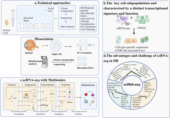
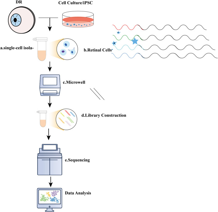
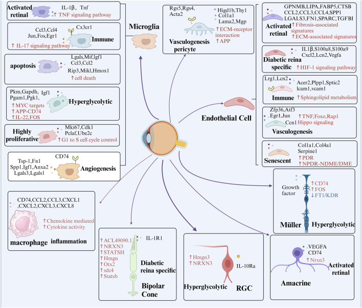
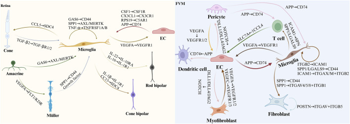
Diabetic retinopathy (DR) is a leading global cause of vision impairment, with its prevalence increasing alongside the rising rates of diabetes mellitus (DM). Despite the retina's complex structure, the underlying pathology of DR remains incompletely understood. Single-cell RNA sequencing (scRNA-seq) and recent advancements in multi-omics analyses have revolutionized molecular profiling, enabling high-throughput analysis and comprehensive characterization of complex biological systems. This review highlights the significant contributions of scRNA-seq, in conjunction with other multi-omics technologies, to DR research. Integrated scRNA-seq and transcriptomic analyses have revealed novel insights into DR pathogenesis, including alternative transcription start site events, fluctuations in cell populations, altered gene expression profiles, and critical signaling pathways within retinal cells. Furthermore, by integrating scRNA-seq with genetic association studies and multi-omics analyses, researchers have identified novel biomarkers, susceptibility genes, and potential therapeutic targets for DR, emphasizing the importance of specific retinal cell types in disease progression. The integration of scRNA-seq with metabolomics has also been instrumental in identifying specific metabolites and dysregulated pathways associated with DR. It is highly conceivable that the continued synergy between scRNA-seq and other multi-omics approaches will accelerate the discovery of underlying mechanisms and the development of novel therapeutic interventions for DR.

Keywords: diabetic retinopathy (DR); genomics; lipidomic; metabolomic; multi-omics; single-cell RNA sequencing(scRNA-seq); transcriptomics.


Conflict of interest statement

The authors declare that the research was conducted in the absence of any commercial or financial relationships that could be construed as a potential conflict of interest.

Figures

None
Overview of single-cell RNA sequencing (scRNA-seq) Applications in Diabetic Retinopathy Research. The application of single-cell RNA sequencing (scRNA-seq) methodologies in the study of diabetic retinopathy(DR) highlights four key areas: (A) The workflow of a typical scRNA-seq experiment; (B) The identification of key cell subpopulations, each characterized by a distinct transcriptional signature and function; (C) The integration of scRNA-seq with multi-omics approaches for DR research; (D) The advantages and challenges associated with scRNA-seq.
FIGURE 1
FIGURE 1
Illustration of single-cell RNA sequencing (scRNA-seq) experiments. (A) Typical single-cell RNA sequencing (scRNA-seq) workflow encompasses several key steps: a. single-cell isolation, which can be achieved through techniques such as micromanipulation, laser-capture microdissection, microfluidics, or fluorescence-activated cell sorting; (B). Cell lysis; (C) Reverse transcription of mRNA into complementary DNA (cDNA); (D) Amplification of cDNA and library preparation; (E) Sequencing.
FIGURE 2
FIGURE 2
Summary of Crucial Cellular Components and Their Features in DR Revealed by scRNA-seq. ScRNA-seq technology provides biological information at the single-cell level and has been extensively applied to DR research. This review concludes that DR-associated microenvironments are characterized by diverse, highly heterogeneous, and dynamic cell populations. scRNA-seq has been instrumental in identifying various temporal states of different cell types, each with a distinct transcriptional signature and function. As scRNA-seq technology matures, it offers new approaches to study DR, deepening our understanding of its pathogenesis and paving the way for innovative treatments. Key cellular components include endothelial cells (ECs), retinal pigment epithelium (RPE), and retinal ganglion cells (RGCs).
FIGURE 3
FIGURE 3
Representative strategy of single-cell RNA sequencing (scRNA-seq) and Multi-Omics. This figure summarizes the four key applications of single-cell RNA sequencing in DR research, each accompanied by a corresponding graph for better understanding. (A) ScRNA-seq combined with Transcriptomics; (B) ScRNA-seq combined with Genomics; (C) ScRNA-seq combined with lipidomic; (D) ScRNA-seq combined with Metabolomic; (E) ScRNA-seq combined with proteomics.
FIGURE 4
FIGURE 4
Presents the identification of key cell subpopulations in DR through multi-omics analysis. The figure highlights distinct cell subpopulations to emphasize the cellular heterogeneity observed during DR progression, offering insights into the specific roles of different cell clusters in the disease’s pathophysiology. Red arrows and text represent pathways that are significantly upregulated within these subpopulations, while blue arrows and text signify pathways that are downregulated. Additionally, black text denotes unique intracellular or secreted proteins, as well as membrane receptors, that are specifically expressed within each subpopulation.
FIGURE 5
FIGURE 5
Illustrates cell-cell communication in DR as uncovered by scRNA-seq. The figure depicts ligand-receptor interactions associated with DR, based on findings from previous scRNA-seq studies. In the diagram, the arrowhead points to the receptor protein and the corresponding cell expressing it, while the arrowtail indicates the ligand protein and the cell producing it. This representation sheds light on the disrupted communication networks in diabetic retinas and the intercellular signaling within FVM, which may contribute to the pathogenesis of DR.

Similar articles

Cited by

References

    1. Abbas A., Almaghrbi H., Giordo R., Zayed H., Pintus G. (2024). Pathogenic mechanisms, diagnostic, and therapeutic potential of microvesicles in diabetes and its complications. Archives Biochem. Biophysics 761, 110168. 10.1016/j.abb.2024.110168 - DOI - PubMed
    1. Ahmad A. F., Dwivedi G., O'Gara F., Caparros-Martin J., Ward N. C. (2019). The gut microbiome and cardiovascular disease: current knowledge and clinical potential. Am. J. Physiology. Heart Circulatory Physiology 317 (5), H923–H938. 10.1152/ajpheart.00376.2019 - DOI - PubMed
    1. Ancel P., Martin J. C., Doukbi E., Houssays M., Gascon P., Righini M., et al. (2023). Untargeted multiomics approach coupling lipidomics and metabolomics profiling reveals new insights in diabetic retinopathy. Int. J. Mol. Sci. 24 (15), 12053. 10.3390/ijms241512053 - DOI - PMC - PubMed
    1. Ashraf M., Sampani K., Rageh A., Silva P. S., Aiello L. P., Sun J. K. (2020). Interaction between the distribution of diabetic retinopathy lesions and the association of optical coherence tomography angiography scans with diabetic retinopathy severity. JAMA Ophthalmol. 138 (12), 1291–1297. 10.1001/jamaophthalmol.2020.4516 - DOI - PMC - PubMed
    1. Bawa G., Liu Z., Yu X., Qin A., Sun X. (2022). Single-cell RNA sequencing for plant research: insights and possible benefits. Int. J. Mol. Sci. 23 (9), 4497. 10.3390/ijms23094497 - DOI - PMC - PubMed

LinkOut - more resources