. 2024 Dec 11:12:1496244.
doi: 10.3389/fcell.2024.1496244. eCollection 2024.

Frataxin is essential for zebrafish embryogenesis and pronephros formation

Affiliations

Frataxin is essential for zebrafish embryogenesis and pronephros formation

Wesley S Ercanbrack et al. Front Cell Dev Biol. .

Abstract

Background and objectives: Friedreich's Ataxia (FRDA) is a genetic disease that affects a variety of different tissues. The disease is caused by a mutation in the frataxin gene (FXN) which is important for the synthesis of iron-sulfur clusters. The primary pathologies of FRDA are loss of motor control and cardiomyopathy. These occur due to the accumulation of reactive oxygen species (ROS) in the brain and the heart due to their high metabolic rates. Our research aims to understand how developmental processes and the kidney are impacted by a deficiency of FXN.

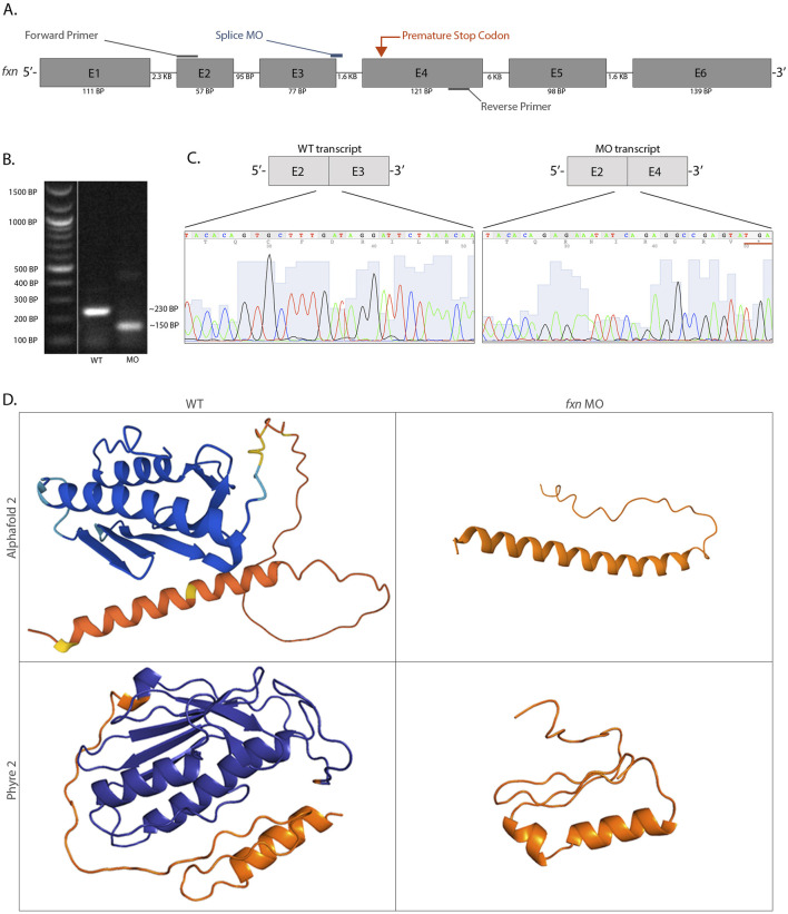
Methods: We utilized an antisense oligomer, or morpholino, to knockdown the frataxin gene (fxn) in zebrafish embryos. Knockdown was confirmed via RT-PCR, gel electrophoresis, and Sanger sequencing. To investigate phenotypes, we utilized several staining techniques including whole mount in situ hybridization, Alcian blue, and acridine orange, as well as dextran-FITC clearance assays.

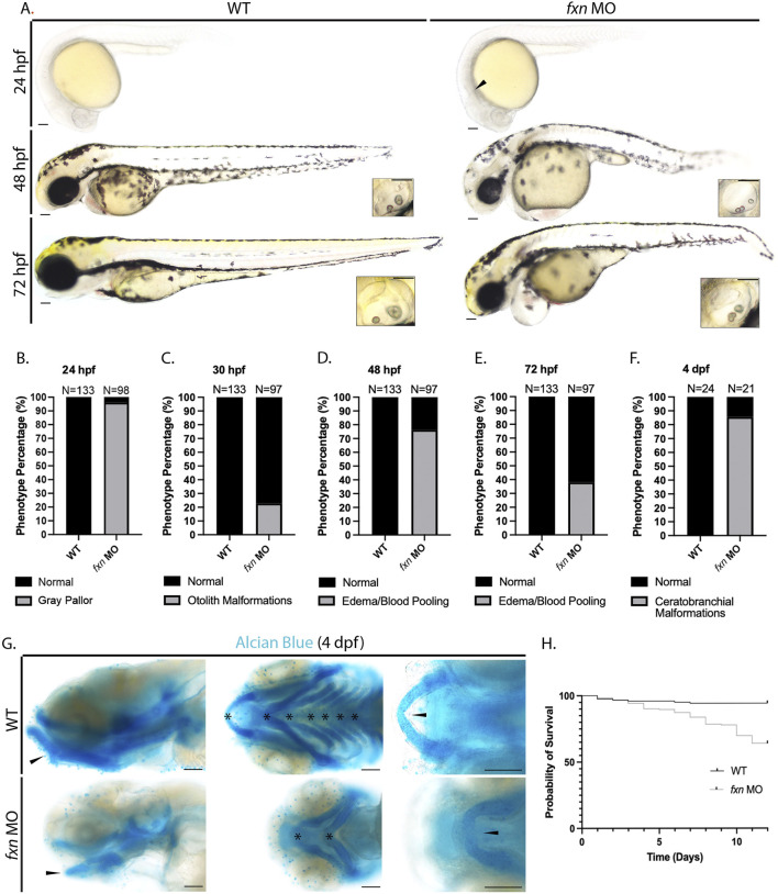
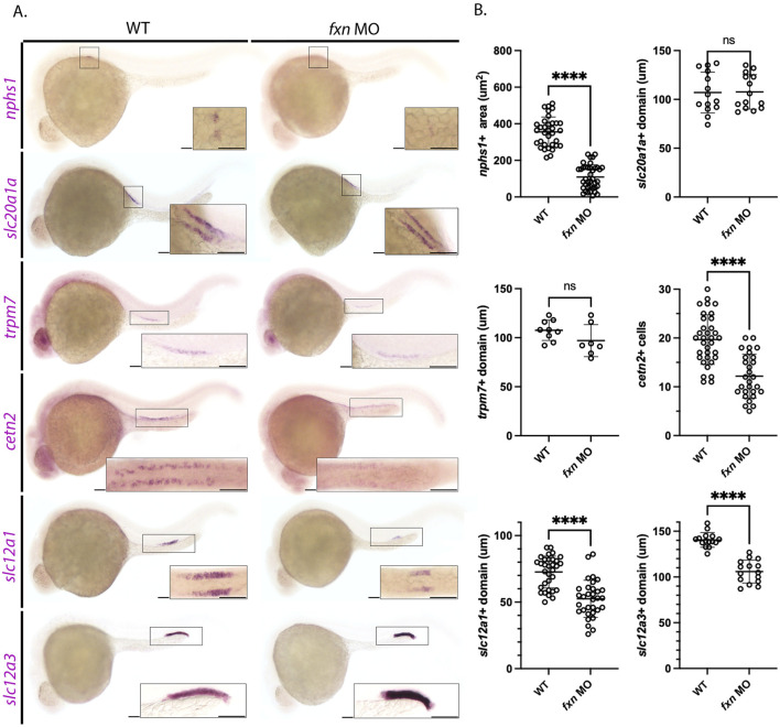
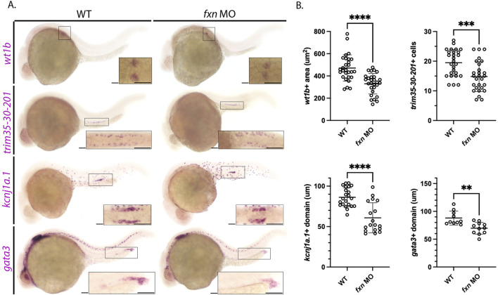
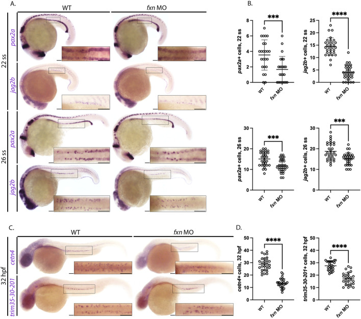
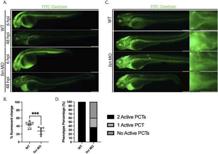
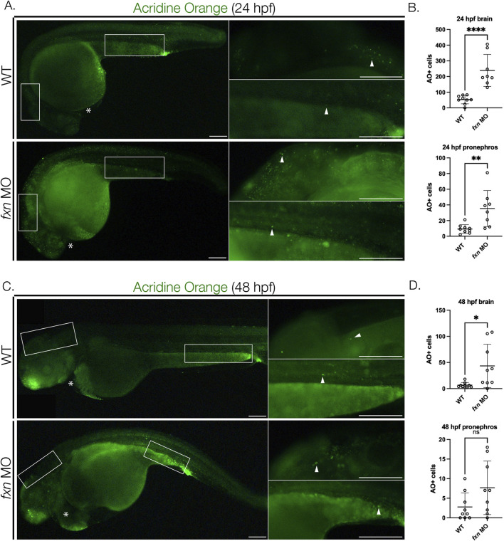
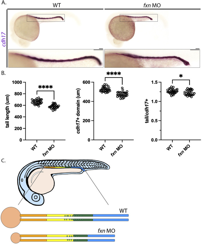
Results: fxn deficient animals displayed otolith malformations, edema, and reduced survival. Alcian blue staining revealed craniofacial defects in fxn deficient animals, and gene expression studies showed that the pronephros, or embryonic kidney, had several morphological defects. We investigated the function of the pronephros through clearance assays and found that the renal function is disrupted in fxn deficient animals in addition to proximal tubule endocytosis. Utilizing acridine orange staining, we found that cell death is a partial contributor to these phenotypes.

Discussion and conclusion: This work provides new insights about how fxn deficiency impacts development and kidney morphogenesis. Additionally, this work establishes an additional model system to study FRDA.

Keywords: development; frataxin; kidney; metabolism; nephron; zebrafish.


Conflict of interest statement

The authors declare that the research was conducted in the absence of any commercial or financial relationships that could be construed as a potential conflict of interest. The author(s) declared that they were an editorial board member of Frontiers, at the time of submission. This had no impact on the peer review process and the final decision.

Figures

FIGURE 1
FIGURE 1
An antisense morpholino oligomer targeting the exon 3 and exon 4 splice region is sufficient to completely knockdown fxn expression in zebrafish. (A) A schematic of the fxn gene with annotations showing the morpholino and primer locations. (B) After cDNA synthesis using RT-PCR, analysis of the products through gel electrophoresis revealed that morphant animals produced a shorter fxn transcript and a wild-type product was absent. (C) Sanger sequencing data revealed that the fxn morphant transcript had a complete deletion of exon 3, which encoded an in-frame premature stop codon (underlined). (D) Three-dimensional protein folding predictions suggested a highly disrupted Fxn protein structure based on the altered mRNA sequence caused by splice interference due to the fxn morpholino. The endogenous Fxn consists of three alpha helices and five beta sheets, and the altered Fxn consists of an alpha helix and an interdomain loop. The orange refers to the mitochondrial trafficking sequence, an area of the protein that has low confidence for the folding prediction because of the lack of conservation between species in this specific domain. These folding predictions strongly suggest that significant regions of the Fxn protein are abrogated in the morphants.
FIGURE 2
FIGURE 2
Morphological analysis of fxn-deficient zebrafish embryos reveal phenotypes indicative of several developmental malformations. (A) Brightfield microscopy revealed a diffuse gray pallor in the cranium of 24 hpf fxn morphants as well as blood pooling and pericardial edema at the 48 and 72 hpf stages. Insets show otolith malformations in the fxn morphants. Scale bars = 50 um. (B–F) Penetrance of observed phenotypes. (B) Gray pallor at 24 hpf. (C) Otolith malformations at 30 hpf. Edema/blood pooling at (D) 48 hpf and (E) 72 hpf. (F) Ceratobranchial malformations at 4 dpf. (G) Alcian blue staining revealed ceratobranchials 1-5 were missing in fxn morphants. Additionally, Meckel’s cartilage failed to fuse and the mandibular prominence did not curve ventrally as it does in wild-type animals. Scale bars = 50 um. (H) Kaplan-Meier survival curve documenting survival in wild-type embryos compared to fxn morphants. Fxn morphant animals displayed a 64% chance of survival compared to the 94% chance of their wild-type siblings during the first 12 days of life.
FIGURE 3
FIGURE 3
Pronephros segment development is significantly disrupted in fxn-deficient zebrafish embryos. (A) WISH experiments reveal that fxn is required for the proper formation of the podocytes, multiciliated cells (MCCs), distal early, and distal late tubule. The previously mentioned domains were visualized via the nphs1,cetn4, slc12a1, and slc12a3 probes, respectively. Formation of the proximal convoluted and straight tubules, as assessed by expression of slc20a1a and trpm7 revealed no significant differences between wild-type embryos and fxn morphants. Scale bars = 50 um. Each boxed lateral region in the embryos corresponds to the inset, which shows a dorsal view of that corresponding area for nphs1, slc20a1a, cetn2, and slc12a1 or a lateral view of the corresponding area for trpm7 and slc12a3. (B) Unpaired t-tests of the phenotypes revealed by WISH. ****p < 0.0001.
FIGURE 4
FIGURE 4
Further analysis of pronephros segment development in fxn-deficient zebrafish embryos confirms reductions in several cell populations. (A) WISH using the probes wt1b, trim35-30-201, kcnj1a.1, and gata3 which mark the podocytes, multiciliated cells (MCCs), distal early and distal late domains respectively. Fxn morphants displayed a significant reduction in podocytes, reduction in MCC number, and reduced distal domains results with this set of markers, independently recapitulating the results shown in Figure 3. Scale bars = 50 um. Each boxed lateral region in the embryos corresponds to the inset, which shows a dorsal view of that corresponding area, with the exception of the gata3 panels which show lateral views in the corresponding inset. (B) Unpaired t-tests demonstrating significantly affected pronephric domains in the fxn morphants. **p < 0.005, ***p < 0.0005, ****p < 0.0001.
FIGURE 5
FIGURE 5
Fxn-deficient zebrafish embryos form reduced multiciliated cell (MCC) progenitors. (A) WISH analysis at the 22 and 26 ss utilizing the probes pax2a and jag2b revealed that the population of MCC progenitors was significantly reduced in fxn morphants compared to wild-type control embryos. Scale bars = 50 um. (B) Unpaired t-test of the corresponding WISH experiments in panel (A). (C) WISH analysis at the 32 hpf time point utilized the probes trim35-30-201 and cetn4, which are expressed by differentiating MCCs, and revealed that MCC numbers were still significantly reduced at this later timepoint. Scale bars = 50 um. Each boxed lateral region in the embryos corresponds to the inset, which shows a dorsal view of that corresponding area. (D) Unpaired t-test of the corresponding WISH experiments in panel (C). ***p < 0.0005, ****p < 0.0001.
FIGURE 6
FIGURE 6
Fxn-deficient zebrafish embryos display renal clearance defects and some proximal reabsorption defects. (A) fxn morphants failed to excrete the FITC-Dextran at the same rate as their wild-type siblings. Images of the same wild-type and fxn-deficient animals 6 hours and 48 h after injection with the FITC-dextran. FITC-Dextran (40 kDa) was injected around 36 h post-fertilization. (B) An unpaired t-test of the percent fluorescent change of each animal. Percent fluorescent change was calculated using the 6 h post injection (hpi) and 48 hpi fluorescent intensity of the same animal. (C) Morphants exhibit decreased endocytosis activity in the proximal convoluted tubule (PCT). Among the morphants, there were three primary phenotypes. Morphant animals either had endocytosis activity in two PCTs, one PCT, or neither. These findings suggest that dysfunction within the PCT segments is one contributor to the fluid imbalance observed in fxn-deficient animals. (D) The proportions of proximal convoluted tubule phenotypes in the wild-type and fxn morphant groups. Scale bars are 200 um. ***p < 0.0005.
FIGURE 7
FIGURE 7
Cell death is elevated in fxn-deficient zebrafish embryos. (A) Images of AO-stained animals at 24 hpf. Fxn morphants animals have significantly increased cell death in the brain and pronephros at 24 hpf and increased cell death in the brain at 48 hpf. White boxes denote the locations for the panels on the right top (brain) and right bottom (pronephros). Scale bars = 50 um. (B) Unpaired t-tests of cell death in the brain and pronephros at 24 hpf. (C) Images of AO-stained animals at 48 hpf. White boxes denote the locations for the panels on the right top (brain) and right bottom (pronephros). Within the right top brain panel, each white box indicates a single positive cell. In fxn morphants, areas of the brain commonly had multiple cells as indicated by the white box. Scale bars = 50 um. (D) Unpaired t-tests of cell death in the brain and pronephros at 48 hpf. *p < 0.05, **p < 0.005, ****p < 0.0001.
FIGURE 8
FIGURE 8
Tail/pronephros tubule ratio comparisons indicate that the embryonic kidney that develops within fxn-deficient zebrafish embryos was decreased in length proportionally to the decreased in tail length. (A) WISH analysis of pronephros tubule formation as marked by cdh17 revealed that this structure was shorter in fxn morphants than it is in their wild-type siblings (scale bars = 50 um). Each boxed lateral region in the embryos corresponds to the inset, which shows an enlarged lateral view of that corresponding area. (B) Unpaired t-tests of tail length and pronephros length as marked by cdh17 revealed that the length of both structures was significantly shorter in the morphants than in their wild-type siblings. An unpaired t-test of the ratios of tail and pronephros length also showed a statistically significant difference. *p < 0.05, p ****p < 0.0001. (C) fxn deficiency leads to formation of a pronephros which is decreased in size in porportion to the reduction in embryo size, however the distal segments are disporportionally reduced suggesting these lineages are uniquely affected by the loss of Fxn activity.

Similar articles

Cited by

References

    1. Abeti R., Parkinson M. H., Hargreaves I. P., Angelova P. R., Sandi C., Pook M. A., et al. (2016). Mitochondrial energy imbalance and lipid peroxidation cause cell death in Friedreich's ataxia. Cell Death Dis. 7 (5), e2237. 10.1038/cddis.2016.111 - DOI - PMC - PubMed
    1. Adinolfi S., Iannuzzi C., Prischi F., Pastore C., Iametti S., Martin S. R., et al. (2009). Bacterial frataxin CyaY is the gatekeeper of iron-sulfur cluster formation catalyzed by IscS. Nat. Struct. Mol. Biol. 16 (4), 390–396. 10.1038/nsmb.1579 - DOI - PubMed
    1. Adinolfi S., Trifuoggi M., Politou A. S., Martin S., Pastore A. (2002). A structural approach to understanding the iron-binding properties of phylogenetically different frataxins. Hum. Mol. Genet. 11 (16), 1865–1877. 10.1093/hmg/11.16.1865 - DOI - PubMed
    1. Andermann E., Remillard G. M., Goyer C., Blitzer L., Andermann F., Barbeau A. (1976). Genetic and family studies in Friedreich's ataxia. Can. J. Neurol. Sci. 3 (4), 287–301. 10.1017/s0317167100025476 - DOI - PubMed
    1. Anheim M., Fleury M., Monga B., Laugel V., Chaigne D., Rodier G., et al. (2010). Epidemiological, clinical, paraclinical and molecular study of a cohort of 102 patients affected with autosomal recessive progressive cerebellar ataxia from Alsace, Eastern France: implications for clinical management. Neurogenetics 11 (1), 1–12. 10.1007/s10048-009-0196-y - DOI - PubMed

LinkOut - more resources