. 2025 May;21(5):1019-1038.
doi: 10.1080/15548627.2024.2442849. Epub 2025 Jan 2.

Regulation of N-degron recognin-mediated autophagy by the SARS-CoV-2 PLpro ubiquitin deconjugase

Affiliations

Regulation of N-degron recognin-mediated autophagy by the SARS-CoV-2 PLpro ubiquitin deconjugase

Carlos Ayala-Torres et al. Autophagy. 2025 May.

Abstract

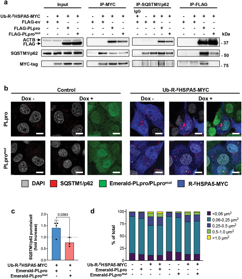
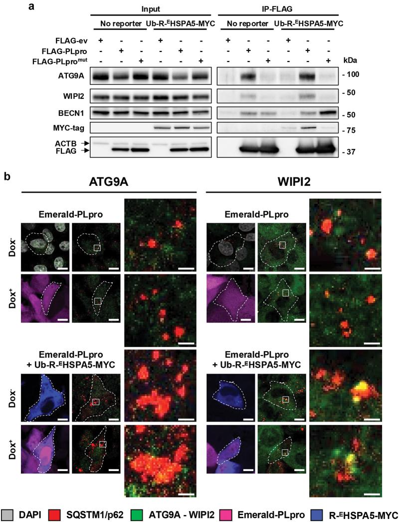
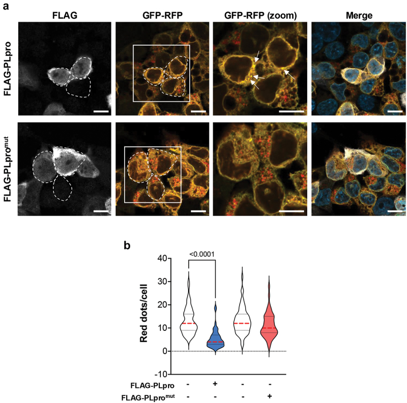
Viral proteases play critical roles in the host cell and immune remodeling that allows virus production. The severe acute respiratory syndrome coronavirus-2 (SARS-CoV-2) papain-like protease (PLpro) encoded in the large nonstructural protein 3 (Nsp3) also possesses isopeptidase activity with specificity for ubiquitin and ISG15 conjugates. Here, we interrogated the cellular interactome of the SARS-CoV-2 PLpro catalytic domain to gain insight into the putative substrates and cellular functions affected by the viral deubiquitinase. PLpro was detected in protein complexes that control multiple ubiquitin and ubiquitin-like (UbL) regulated signaling and effector pathways. By restricting the analysis to cytosolic and membrane-associated ubiquitin ligases, we found that PLpro interacts with N-recognin ubiquitin ligases and preferentially rescues type I N-degron substrates from proteasomal degradation. PLpro stabilized N-degron carrying HSPA5/BiP/GRP78, which is arginylated in the cytosol upon release from the endoplasmic reticulum (ER) during ER stress, and enhanced the Arg-HSPA5-driven oligomerization of the N-recognin SQSTM1/p62 that serves as a platform for phagophore assembly. However, while in addition to Arg-HSPA5 and SQSTM1/p62, ATG9A, WIPI2, and BECN1/Beclin 1 were detected in PLpro immunoprecipitates, other components of the autophagosome biogenesis machinery, such as the ATG12-ATG5-ATG16L1 complex and MAP1LC3/LC3 were absent, which correlated with proteolytic inactivation of ULK1, impaired production of lipidated LC3-II, and inhibition of reticulophagy. The findings highlight a novel mechanism by which, through the reprogramming of autophagy, the PLpro deubiquitinase may contribute to the remodeling of intracellular membranes in coronavirus-infected cells.

Keywords: HSPA5/BiP/GRP78; N-degron; PLpro; SARS-CoV-2; SQSTM1/p62; reticulophagy.


Conflict of interest statement

No potential conflict of interest was reported by the author(s).

Figures

Figure 1.
Figure 1.
PLpro interacts with N-recognin ubiquitin ligases. (a) STRING network diagram of the PLpro-interacting cytosolic and membrane-associated ubiquitin ligases and the shared interacting partners identified by co-immunoprecipitation and mass spectrometry. Mass spectrometry analysis was performed on FLAG-tag immunoprecipitates of U2OS cells transiently transfected with FLAG-ev/PLpro/PLpromut, and bona fide PLpro interactors were identified as described in methods. The PLpro-interacting ligases and shared interacting partners found in the interactome are color-coded. (b) Representative western blots that illustrate the interaction of PLpro with N-recognin ligases. Lysates of HEK293T cells transfected with plasmids expressing FLAG-ev/PLpro/PLpromut were immunoprecipitated with anti-FLAG coated beads, and western blots were probed with the indicated antibodies. β-actin served as a loading control. Each interaction was validated in at least two independent co-immunoprecipitation experiments.
Figure 2.
Figure 2.
PLpro preferentially stabilizes N-degron substrates. (a) schematic illustration of the Ub-fusion reporters. The Ub-X-GFP reporters are cotranslationally cleaved by cellular ubiquitin deconjugases at the Ub-X junction, producing Ub and X-GFP. The Nt-Arg residue generated by the cleavage acts as a degron for N-recognin ubiquitin ligase, leading to ubiquitination and proteasome-dependent degradation. Mutation of the ubiquitin C-terminal gly to val (G76V) prevents cleavage, generating a substrate where a ubiquitin chain is attached to the ubiquitin moiety (UFD reporter). (b) PLpro selectively stabilizes the N-degron reporter Ub-R-GFP. HEK293T cells transiently cotransfected with the Ub-R-GFP or UbG76V-GFP reporters and FLAG-ev/PLpro/PLpromut. FLAG-ev transfected cells were incubated overnight with 100 nM epoxomicin as a control for ubiquitin-dependent proteasomal degradation. Representative western blots from one out of three independent experiments are shown. (c) Densitometric quantification of the GFP-specific bands. The data are displayed as mean ± SD GFP intensity in PLpro/PLpromut transfected and epoxomicin-treated cells relative to FLAG-ev after normalization to the GAPDH loading control. Significance was calculated by unpaired two-tailed Student’s t-tests.
Figure 3.
Figure 3.
PLpro preferentially stabilizes type I N-degron substrates. (a) schematic illustration of the Ub-X-GFP reporters carrying type I (primary, secondary, and tertiary) or type II N-degrons and the control, Ub-M-GFP chimera that, upon ubiquitin cleavage, does not contain a degradation signal. (b) PLpro preferentially stabilizes type I N-degron substrates. HEK293T cells were cotransfected with the indicated Ub-X-GFP and FLAG-ev/PLpro/PLpromut, and expression of the GFP chimeras was analyzed after 24 h by western blot. As a reference for ubiquitin-dependent proteolysis, one aliquot of FLAG-ev transfected cells was treated with 100 nM epoxomicin. Representative western blots from one out of four independent experiments are shown. (c) The intensity of the GFP band was quantified by densitometry in four independent experiments. The mean ± SD relative intensities of the GFP bands in FLAG-PLpro/PLpromut versus FLAG-ev transfected cells after normalization to the GAPDH loading control are shown. Significance was calculated by unpaired two-tailed Student’s t-tests.
Figure 4.
Figure 4.
PLpro stabilizes N-degron-carrying ER chaperones. (a) Cartoon illustrating the generation of N-degron carrying ER chaperones upon induction of ER stress and involvement of N-degron carrying EHSPA5 in the regulation of autophagy via activation of the N-recognin SQSTM1/p62. (b) Schematic illustration of the Ub-X-HSPA5-MYC and Ub-ECALR-GFP reporters. (c) PLpro promotes the stabilization of the EHSPA5 and R-EHSPA5 reporters. HEK293T cells were transiently cotransfected with the FLAG-ev/PLpro/PLpromut and Ub-REHSPA5-MYC or Ub-EHSPA5-MYC plasmids. As a control for proteasome- and lysosome-dependent degradation, the FLAG-ev transfected cells were treated overnight with 100 nM epoxomicin or 100 nM Baf A1, respectively. The expression of the R-EHSPA5 was analyzed 24 h post-transfection by probing western blots with the indicated antibodies. Representative blots from one out of three independent experiments are shown. (d) The intensities of the R-EHSPA5 bands were quantified by densitometry in three independent experiments. The mean ± SD relative intensity of the R-EHSPA5 bands in FLAG-PLpro/PLpromut transfected or epoxomicin/Baf A1 treated cells versus FLAG-ev transfected cells is shown. Significance was calculated by unpaired two-tailed Student t-test. (e) PLpro deubiquitinates R-EHSPA5. HEK293T cells were transiently cotransfected with Ub-R-EHSPA5-MYC and FLAG-ev/PLpro/PLpromut. Cell lysates were immunoprecipitated with anti-MYC agarose beads, and western blots were probed with the indicated antibodies. Blots from one representative experiment out of two are shown in the figure. (f) PLpro stabilizes N-degron carrying CALR. HEK293T cells were cotransfected with FLAG-ev/PLpro/PLpromut and the Ub-ECALR-GFP. Cell lysates were immunoprecipitated with a GFP antibody, followed by capture with protein G-coated sepharose beads. An isotype-matched IgG control was included in the immunoprecipitation to verify specificity. Western blots were probed with the indicated antibodies. Blots from one representative experiment out of three are shown in the figure. (g) The intensities of the GFP bands were quantified by densitometry in three independent experiments. The mean ± SD, the relative intensity of the GFP bands in FLAG-PLpro/PLpromut versus FLAG-ev transfected cells is shown. Significance was calculated by unpaired two-tailed Student’s t-tests.
Figure 5.
Figure 5.
PLpro enhances the R-EHSPA5-driven formation of SQSTM1/p62 aggregates. (a) Reciprocal immunoprecipitation illustrating the interaction of PLpro with R-EHSPA5 and SQSTM1/p62. Equal aliquots of HEK293T lysates cotransfected with FLAG-ev/PLpro/PLpromut and Ub-R-EHSPA5-MYC were immunoprecipitated with anti-FLAG, anti-MYC or anti-SQSTM1/p62 antibody-coated beads. An isotype-matched antibody was used as a control. Western blots were probed with the indicated antibodies. Blots from one representative experiment out of three are shown in the figure. (b) Catalytically active PLpro enhances the formation of large SQSTM1/p62 aggregates in cells expressing R-EHSPA5. Endogenous SQSTM1/p62 (red) was detected by immunofluorescence in control and Dox-treated U2OS Emerald PLpro/PLpromut cells and transfected with the R-EHSPA5-MYC reporter (blue). Images from one representative experiment out of four are shown. (c, d) PLpro enhances the number and size of SQSTM1/p62 aggregates in R-EHSPA5-expressing cells. The number and size of SQSTM1/p62 puncta were determined using the Fiji software and its analysis particle function from 75 confocal images containing approximately 1000 cells per condition. For each image, the brightness and contrast were adjusted to reduce the background noise, and particles of size >0.049 μm2 were scored as puncta. (c) The data are presented as fold change in cells expressing R-EHSPA5 and PLpro/PLpromut relative to cells expressing R-EHSPA5 alone in three (PLpromut) or four (PLpro) independent experiments. Significance was calculated by unpaired two-tailed Student’s t-tests. (d) Microsoft Excell was used to categorize the particles based on size. The data are represented as a percentage (%) of the total number of puncta. Data from the experiment in Figure 5B are shown. Similar results were obtained in three independent experiments. Scale bar: 10 µm.
Figure 6.
Figure 6.
PLpro recruits components of the autophagosome biogenesis machinery to large SQSTM1/p62 aggregates. (a) FLAG immunoprecipitates of HEK293T cells transfected with FLAG-ev/PLpro/PLpromut in the presence or absence of co-transfected Ub-R-EHSPA5-MYC were probed with the indicated antibodies. Western blots from one representative experiment out of three are shown in the figure. (b) Representative confocal images illustrating the colocalization of ATG9A and WIPI2 with large SQSTM1/p62 aggregates. Scale bar: 10 µm.
Figure 7.
Figure 7.
Catalytically active PLpro inhibits the formation of LC3-II. (a) Control and doxycycline-treated U2OS cells stably transfected with plasmids expressing inducible Emerald-ev/PLpro/PLpromut were kept untreated or treated overnight with 100 nM Baf A1 before analysis of protein expression by probing western blots with the indicated antibodies. Blots from one representative experiment out of four are shown. (b) Quantification of the LC3-specific bands. The data are presented as LC3-II:LC3-I ratios in four independent experiments. Significance was calculated by unpaired two-tailed Student’s t-tests.
Figure 8.
Figure 8.
PLpro inhibits reticulophagy. (a) Representative confocal images illustrating the failure to accumulate ER-loaded autophagolysosomes in cells expressing active PLpro. The EATR reporter expresses in-frame the coding sequence of the SERP1/RAMP4 subunit of the ER translocon complex followed by the coding sequences of eGFP and ChFP. Upon ER insertion of the SERP1 domain, eGFP and ChFP face the cytosol and emit equal fluorescence, whereas, due to the selective loss of eGFP fluorescence at low pH, er-loaded autophagosomes appear as distinct red fluorescent dots. Stable HCT116-EATR cells were transfected with plasmids expressing FLAG-PLpro/PLpromut and then starved overnight in EBSS medium before visualizing the formation of er-loaded autophagosomes by confocal microscopy. Yellow dots corresponding to ER membrane aggregates in cells expressing catalytically active PLpro are indicated by arrows. Scale bar: 10 µm. (b) Quantification of the number of red fluorescent dots in FLAG-PLpro/PLpromut positive and negative cells from the same transfection experiments. The cumulative data from two independent experiments where ≥50 PLpro positive and ≥50 negative cells were scored are shown. Significance was calculated by unpaired two-tailed Student t-tests.

References

    1. Jamison DA Jr., Anand Narayanan S, Trovao NS, et al. A comprehensive SARS-CoV-2 and COVID-19 review, part 1: intracellular overdrive for SARS-CoV-2 infection. Eur J Hum Genet. 2022. Aug;30(8):889–898. doi: 10.1038/s41431-022-01108-8 - DOI - PMC - PubMed
    1. Narayanan SA, Jamison DA Jr., Guarnieri JW, et al. A comprehensive SARS-CoV-2 and COVID-19 review, part 2: host extracellular to systemic effects of SARS-CoV-2 infection. Eur J Hum Genet. 2024. Jan;32(1):10–20. doi: 10.1038/s41431-023-01462-1 - DOI - PMC - PubMed
    1. Wu A, Peng Y, Huang B, et al. Genome composition and divergence of the novel coronavirus (2019-nCoV) originating in China. Cell Host Microbe. [2020 Mar 11];27(3):325–328. doi: 10.1016/j.chom.2020.02.001 - DOI - PMC - PubMed
    1. Angelini MM, Akhlaghpour M, Neuman BW, et al. Severe acute respiratory syndrome coronavirus nonstructural proteins 3, 4, and 6 induce double-membrane vesicles. MBio. 2013. Aug 13;4(4). doi: 10.1128/mBio.00524-13 - DOI - PMC - PubMed
    1. Wolff G, Limpens R, Zevenhoven-Dobbe JC, et al. A molecular pore spans the double membrane of the coronavirus replication organelle. Science. 2020. Sep 11;369(6509):1395–1398. doi: 10.1126/science.abd3629 - DOI - PMC - PubMed

Publication types

LinkOut - more resources