. 2025 Jan 15;16(2):241-261.
doi: 10.1021/acschemneuro.4c00796. Epub 2024 Dec 26.

A Copper-Binding Peptide with Therapeutic Potential against Alzheimer's Disease: From the Blood-Brain Barrier to Metal Competition

Affiliations

A Copper-Binding Peptide with Therapeutic Potential against Alzheimer's Disease: From the Blood-Brain Barrier to Metal Competition

Victor E López-Guerrero et al. ACS Chem Neurosci. .

Abstract

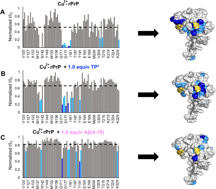
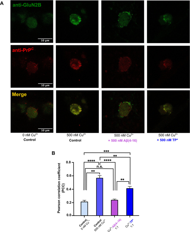
Alzheimer's disease (AD) is the most common form of dementia worldwide. AD brains are characterized by the accumulation of amyloid-β peptides (Aβ) that bind Cu2+ and have been associated with several neurotoxic mechanisms. Although the use of copper chelators to prevent the formation of Cu2+-Aβ complexes has been proposed as a therapeutic strategy, recent studies show that copper is an important neuromodulator that is essential for a neuroprotective mechanism mediated by Cu2+ binding to the cellular prion protein (PrPC). Therefore, in addition to metal selectivity and blood-brain barrier (BBB) permeability, an emerging challenge for copper chelators is to prevent the formation of neurotoxic Cu2+-Aβ species without perturbing the neuroprotective Cu2+-PrPC interaction. Previously, we reported the design of a tetrapeptide (TP) that withdraws Cu2+ from Aβ(1-16) and impacts the Cu2+-induced aggregation of Aβ(1-40). In this study, we improved the drug-like properties of TP in a BBB model, evaluated the metal selectivity of the optimized peptide (TP*), and tested its effect on Cu2+ coordination to PrPC and proteins involved in copper trafficking, such as copper transporter 1 and albumin. Our results show that changing the stereochemistry of the first residue prevents TP degradation in the BBB model and coadministration of TP with a peptide that increases BBB permeability allows its passage through the BBB model. TP* is highly selective toward Cu2+ in the presence of Zn2+ ions, transfers Cu2+ to copper-trafficking proteins, and forms a ternary TP*-Cu2+-PrP species that does not perturb the physiological conformation of PrP and displays only a minor impact in the neuroprotective Cu2+-dependent interaction of PrPC with the N-methyl-d-aspartate receptor. Overall, these results show that TP* displays desirable features for a copper chelator with therapeutic potential against AD. Moreover, this is the first study that explores the effect of a Cu2+ chelator with therapeutic potential for AD on Cu2+ coordination to PrPC (an emerging key player in AD pathology), integrating recent knowledge about metalloproteins involved in AD with the design of copper chelators against AD.

Keywords: Alzheimer′s disease; NMDA receptor; chelation therapy; copper; prion protein.


Conflict of interest statement

The authors declare no competing financial interest.

Figures

Scheme 1
Scheme 1. TP Holds Therapeutic Potential against AD
TP is a tetrapeptide with the sequence MDdWAib. The MD sequence binds Cu2+ with high affinity (A). The hydrophobic dWAib fragment displays β-breaker properties, and it interacts with early intermediate assemblies of Aβ(1–40/42). The presence of both moieties in the same peptide sequence provide bifunctional properties to TP: It removes Cu2+ from Aβ(1–16), as demonstrated by EPR (A), and it impacts the Cu2+-induced aggregation of Aβ(1–40), inhibiting the formation of large Cu2+-bound Aβ(1–40) oligomers (B). These findings support a therapeutic potential for TP against AD. (Figure adapted from (16) ).
Scheme 2
Scheme 2. Strategy to Evaluate and Optimize the Drug-like Properties of TP and Evaluate Its Impact on Cu2+ Binding to Proteins Involved in Copper Homeostasis and Neuroprotection
TP with the sequence MDdWAib was optimized and evaluated using spectroscopic and cell culture assays. This approach includes (1) improving TP permeability in an in vitro BBB cellular model and its optimization to prevent degradation; (2) selectivity towards Cu2+ in the presence of Zn2+ ions; (3) competitions for Cu2+ with peptide models of metal-binding sites of proteins associated to the copper transport and homeostasis, such as HSA, hCtr1, and Aβ(4–40); (4) competitions for Cu2+ with each of the Cu2+-binding sites of the PrPC; and (5) its impact on the Cu2+-dependent cis-interdomain interaction of the PrP; and in (G) the Cu2+- and PrPC-dependent modulation of NMDARs.
Figure 1
Figure 1
Evaluation of TP permeability in a BBB in vitro model. (A) BBB in vitro model consists of a primary culture of rat brain microvascular endothelial cells (RBMECs) plated in the upper chamber of an insert with a semipermeable filter set in a multiwell plate, where conditioned media derived from a primary culture of astrocytes bathe the lower compartment. The electrical circuit that describes the movement of electrical charges through a monolayer of endothelial cells is defined by the transendothelial electrical resistance (TEER), which depends on the paracellular resistance (Rp) and the transcellular resistance (Rt). Rt is due to the resistance to the apical (Ram) and basal (Rbm) membranes, while Rp depends on the resistance of tight junctions (RTJ). The BBB is formed by the endothelial cells TJs; thus, an increase in TEER is directly associated with the sealing of the BBB. (B) TP recovery from apical and basal compartments of the BBB in vitro model, in the absence and presence of P10 after 24 h. (C) TP recovery from both compartments of the BBB in vitro model, after 3.5 and 8 h. (D) TP and DdWAib recoveries from apical and basal compartments of the BBB in vitro model, at 3.5 and 8 h. (E) Proposed degradation mechanism of MDdWAib to DdWAib in the BBB in vitro model. (F) Recovery of TP and its variants TP* and TP** from the apical compartment of the BBB in vitro model, at 8 and 25 h.
Figure 2
Figure 2
TP* binds preferentially Cu2+ ions even in the presence of excess Zn2+ ions. EPR (A) and CD (B) spectra of TP* solutions containing 1 equiv of Cu2+ (blue spectra) and TP* with 1 equiv of stock solutions containing a mixture of Cu2+:Zn2+ ions in either 1:1 (light-gray spectra) or 1:10 ratio (dark-gray spectra). In the three conditions, the EPR signals and the nitrogen superhyperfine coupling pattern (inset in part A) are very similar. EPR (C) and CD (D) spectra of the titration of Cu2+-TP* with 1 or 10 equiv of Zn2+ (light and dark gray, respectively). A values are given in 1 × 10⁻⁴ cm–1. The inset in (C) shows the second derivative of the EPR spectra, showing very similar nitrogen superhyperfine splittings in the three conditions. A coordination model for the 2N2O or 2N2O1S (participation of the axial S ligand is represented as gray dotted lines) for the Cu2+-TP* complex is included in the inset in (B).
Figure 3
Figure 3
Competition for Cu2+ between TP* and proteins involved in copper homeostasis. Coordination modes for Cu2+-hCtr1(1–14) (orange) and Cu2+-HSA (green) complexes (A). EPR (B) and CD (C) spectra of the Cu2+-ATCUN complexes. By EPR (A), Cu2+-ATCUN complexes were titrated with 1 equiv of TP* (overlapped spectra in light shades of colors) and Cu2+-TP* is included for comparison (blue spectrum); the inset shows the second derivative of the perpendicular region of the spectra. A values are given in 1 × 10⁻⁴ cm–1. Representative CD spectra of the titration of the Cu2+-TP* complex (blue spectrum) with increasing amounts of hCtr1(1–14) (dotted light orange spectra) up to 1 equiv (solid light-orange spectrum) (C), and corresponding EPR spectra of the final point of the titration of Cu2+-TP*with hCtr1(1–14) or HSA are shown in (D).
Figure 4
Figure 4
TP* can take Cu2+ away from Cu2+-Aβ(1–16) but not from Cu2+-Aβ(4–16). Full-length Aβ(1–16) is a 40–42 fragment peptide (A, top) that can be proteolytically cleaved to the N-truncated Aβ(4–16) variant. Cu2+ coordination to Aβ(1–16) and Aβ(4–16) can be assessed using the first 16 residues of their peptide sequence (A, blue box). Cu2+-Aβ(1–16) and Cu2+-Aβ(4–16) complexes bind Cu2+ very differently, as illustrated in (B). EPR spectra (C) of Cu2+- Aβ(1–16) (black spectrum) and Cu2+-Aβ(4–16) (dark pink spectrum). As previously reported, TP removes the metal from Cu2+- Aβ(1–16) (gray spectrum). On the other hand, after addition of 1 equiv of TP* to Cu2+-Aβ(4–16), the EPR spectrum remains unchanged (light pink spectrum). A values are given in 1 × 10⁻⁴ cm–1.
Figure 5
Figure 5
Competition for Cu2+ ions between TP* and the OR sites of PrP. EPR (A) spectra that correspond to the addition of 1 equiv of TP* (with respect to the metal ion) to the low- and high-occupancy modes of the Cu2+-PrP(60–91) (olive- and dark-green spectra, respectively) (the A values are given in 1 × 10⁻⁴ cm–1). The corresponding Cu2+-coordination modes are shown in (B). Additions of TP* to both occupancy modes (red spectra) yielded a new set of signals associated with a ternary TP*-Cu2+-PrP(60–91) complex. (C) Addition of TP* to the high-occupancy modes (dark-green spectrum) was also followed by CD spectroscopy. Addition of 1.0 equiv with respect to the metal ion yielded signals similar but not identical bands (red spectrum) to that of the Cu2+-TP* complex (blue spectrum).
Figure 6
Figure 6
Spectroscopic analysis of the ternary TP*-Cu2+-OP complex. EPR (A), CD (B), ESEEM (C), and XANES (D) spectra of the Cu2+-OP (green), TP*-Cu2+-OP (maroon), and Cu2+-TP* (blue) complexes. EPR (A) shows the formation of the ternary TP*-Cu2+-OP complex after the addition of 1 equiv of TP* to the Cu2+-OP complex, and the second derivative of the perpendicular region is shown in the inset. CD-monitored titration (B) of the Cu2+-OP complex with increasing amounts of TP* (dotted green lines) yields the formation of the TP*-Cu2+-OP species. Three-pulse ESEEM spectra (C) of Cu2+-OP, TP*-Cu2+-OP, and Cu2+-TP* complexes: Cu2+-OP and ternary TP*-Cu2+-OP complexes display characteristic features assigned to the participation of a His ligand in their coordination environment. Black arrows indicate participation of backbone coordination to Cu2+ in the Cu2+-OP complex but not in the ternary complex. Normalized copper K-edge XANES spectra (D) of the three Cu2+-complexes. The black arrow represents the pre-edge features of XANES, and the black triangle points to the edge of the spectra. EXAFS and FT of EXAFS (in k2χ(k)) of the ternary TP*-Cu2+-OP complex are shown in (E) and (F), respectively, and the fit of experimental spectra is indicated as dotted dashed lines, using simulation parameters given in Table S10.
Scheme 3
Scheme 3. Coordination modes of (A) Cu2+-TP and (B) ternary TP-Cu2+-OP complexes at pH = 7.4, with possible 3N1O or 3N1O1S coordination modes
Figure 7
Figure 7
Competition for Cu2+ between TP and the rPrP. EPR (A), CD (B), and ESEEM spectra (C) of the ternary TP*-Cu2+-rPrP complex. (A) Cu2+-rPrP complexes (green lines) were prepared using a Cu2+-rPrP ratio of 1:1 and mixtures of component 3 and Cu2+-His96/His111 are evident. Addition of 1 equiv of TP* results in the formation of ternary TP*-Cu2+-rPrP species (red lines). The spectra were deconvoluted as a mixture of species represented in black dotted lines. The second derivative of the perpendicular region is shown in the inset. (B) CD spectra are consistent with the formation of ternary TP*-Cu2+-rPrP species. (C) Three-pulse ESEEM spectra of Cu2+-rPrP, TP*-Cu2+-rPrP, and Cu2+-TP* complexes: Cu2+-rPrP and ternary TP*-Cu2+-rPrP complexes display characteristic features assigned to the participation of an imidazole (His) ligand in their coordination sphere. A values are given in 1 × 10⁻⁴ cm–1.
Figure 8
Figure 8
Observed PREs of the cis-interdomain interaction in Cu2+-rPrP complexes. Bar plots showing the magnitude of the 1H–15N HSQC NMR peak intensity reduction derived from the PREs on specific residues in the rPrP. Normalized I/IO values for rPrP-Cu2+ (A), rPrP-Cu2+ + 1.0 equiv TP* (B), and rPrP-Cu2+ + Aβ(4–16) are plotted against rPrP residues. Gray bars represent peaks unaffected by PREs, light blue represents relatively weak PREs, and dark blue represents relatively strong PREs. These PREs are mapped onto the surface of PrP (PDB: 1XYX), where His139 and His176 are in yellow.
Figure 9
Figure 9
Effect of TP* in the Cu2+-dependent colocalization of PrPC and NMDAR in SK-N-SH cells. Colocalization analysis was performed by immunocytochemistry using fluorescently labeled secondary antibodies: green for the GluN2B and red for PrPC. Representative images of colocalization in conditions without copper (control); 500 nM Cu2+ (control); 500 nM Cu2+/Aβ(4–16); and 500 nM Cu2+/TP* in a ratio of 1:1 of Cu2+:peptide. The Pearson correlation coefficient (PCC) was employed to quantify the colocalization between both fluorescent probes. Comparison of the PCC values of the different experimental conditions (B). All comparisons show statistical difference, except comparison between no copper control and Cu2+:Aβ(4–16) condition (data are the means ± SEM, one-way ANOVA, post hoc Tukey; *P < 0.05, ***P < 0.001, ****P < 0.0001).
Scheme 4
Scheme 4. Schematic Representation of the Principal Results of This Work
In Summary, Our Findings Indicate That (A, B) TP Does Not Cross the BBB In Vitro Model and Is Degraded. However, Its Coadministration with P10 Allow Its Passage Using the Paracellular Pathway and the Substitution of L-to-D Met in the Sequence of TP Prevents Its Degradation. (C) Metal Binding of Optimized TP* is Highly Selective for Cu2+ in the Presence of High Amounts of Zn2+. (D) TP* Does Not Compete for Cu2+ with Proteins/Peptides Associated to Copper Trafficking, Such as hCtr1, HSA, and Aβ(4–40); Instead, TP* Can Transfer the Metal Ion to These Species. (E) TP* Forms Ternary TP*-Cu2+-PrP Complexes with Peptide Models of Each of the Cu2+-Binding Sites of PrP and with the Full-Length rPrP; however, (F) Formation of Ternary Species Does Not Perturb Importantly the cis-Interdomain Interaction of the Protein. Finally, (G) TP* Exerts Only Mild Effects in the Cu2+-Dependent Colocalization of PrPC and NMDARs.

Similar articles

Cited by

References

    1. Organization, W. H. Dementia. 2019. https://www.who.int/news-room/fact-sheets/detail/dementia. (accessed July 4, 2024).
    1. Organization, W. H. Causes of Death. 2019. https://www.who.int/news-room/fact-sheets/detail/the-top-10-causes-of-death. (accessed July 4, 2024).
    1. DeTure M. A.; Dickson D. W. The neuropathological diagnosis of Alzheimer’s disease. Mol. Neurodegener. 2019, 14 (1), 3210.1186/s13024-019-0333-5. - DOI - PMC - PubMed
    1. Lopez J. A. S.; Gonzalez H. M.; Leger G. C. Alzheimer’s disease. Handb. Clin. Neurol. 2019, 167, 231–255. 10.1016/B978-0-12-804766-8.00013-3. - DOI - PubMed
    1. Wang H.; Wang M.; Wang B.; Li M.; Chen H.; Yu X.; Yang K.; Chai Z.; Zhao Y.; Feng W. Immunogold labeling and X-ray fluorescence microscopy reveal enrichment ratios of Cu and Zn, metabolism of APP and amyloid-beta plaque formation in a mouse model of Alzheimer’s disease. Metallomics 2012, 4 (10), 1113–1118. 10.1039/c2mt20056b. - DOI - PubMed
    2. From NLM Medline

MeSH terms