. 2025 Jan 31;91(1):e0205024.
doi: 10.1128/aem.02050-24. Epub 2024 Dec 26.

Regulation mechanism of the long-chain n-alkane monooxygenase gene almA in Acinetobacter venetianus RAG-1

Affiliations

Regulation mechanism of the long-chain n-alkane monooxygenase gene almA in Acinetobacter venetianus RAG-1

Shuai Chen et al. Appl Environ Microbiol. .

Abstract

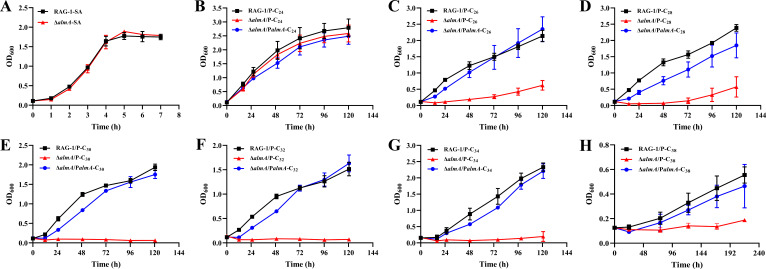
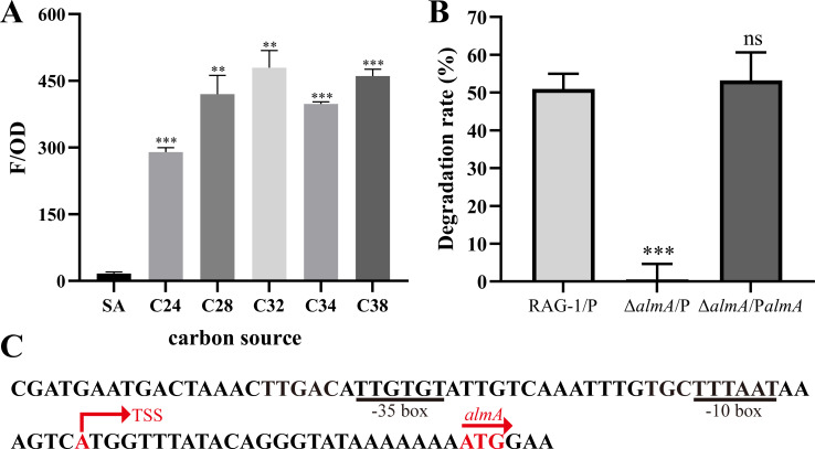
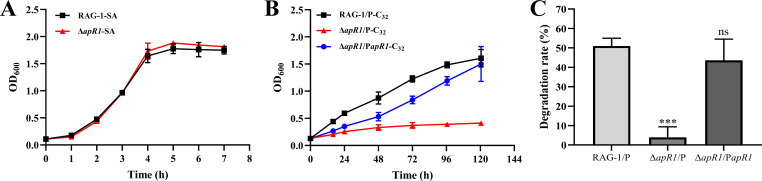
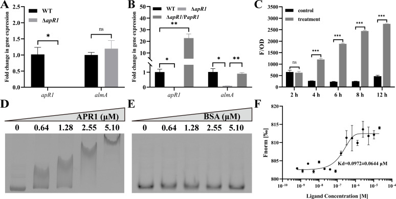
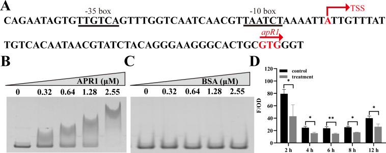
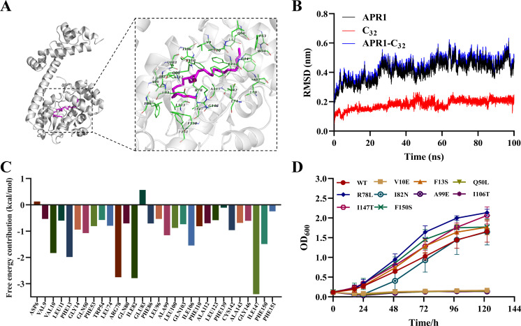
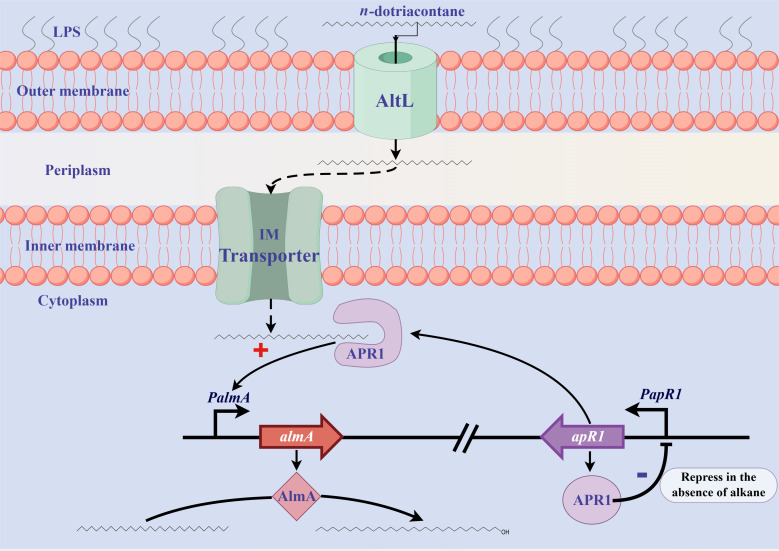
As toxic pollutants, n-alkanes are pervasively distributed in most environmental matrices. Although the alkane monooxygenase AlmA plays a critical role in the metabolic pathway of solid long-chain n-alkanes (≥C20) that are extremely difficult to degrade, the mechanism regulating this process remains unclear. Here, we characterized the function of AlmA in Acinetobacter venetianus RAG-1, which was mainly involved in the degradation of long-chain n-alkanes (C26-C38), among which, n-C32 induced the almA promoter activity most. APR1 (AlmA Positive Regulator) that it is an AraC/XylS-type transcription regulator, a potential transcriptional regulator of almA, was screened by DNA-pull down, which was determined by conserved domain analysis. The deletion of apR1 severely reduced the capacity of strain RAG-1 to utilize long-chain n-alkanes (C22-C38), indicating the involvement of APR1 in n-alkanes degradation. The results of the APR1-dependent reporter system, electrophoretic mobility shift assay, and microscale thermophoresis further proved that APR1 was able to directly bind to the almA promoter region, thus activating the almA transcription. Furthermore, APR1 could inhibit self-expression through autoregulation in the absence of long-chain n-alkanes. n-C32 acted as a ligand of APR1, and the amino acid residues Val10, Gln50, Ala99, and Ile106 at the N-terminus of APR1 were necessary for binding n-C32. In addition, the key amino acid residues of APR1 within the C-terminal helix-turn-helix motif that bound to the downstream promoter region were confirmed by multiple sequence alignment and site-directed mutagenesis. The homologs of AlmA and APR1 shared a similar evolutionary course in the Proteobacteria; thus, this mode of regulation might be relatively conserved.

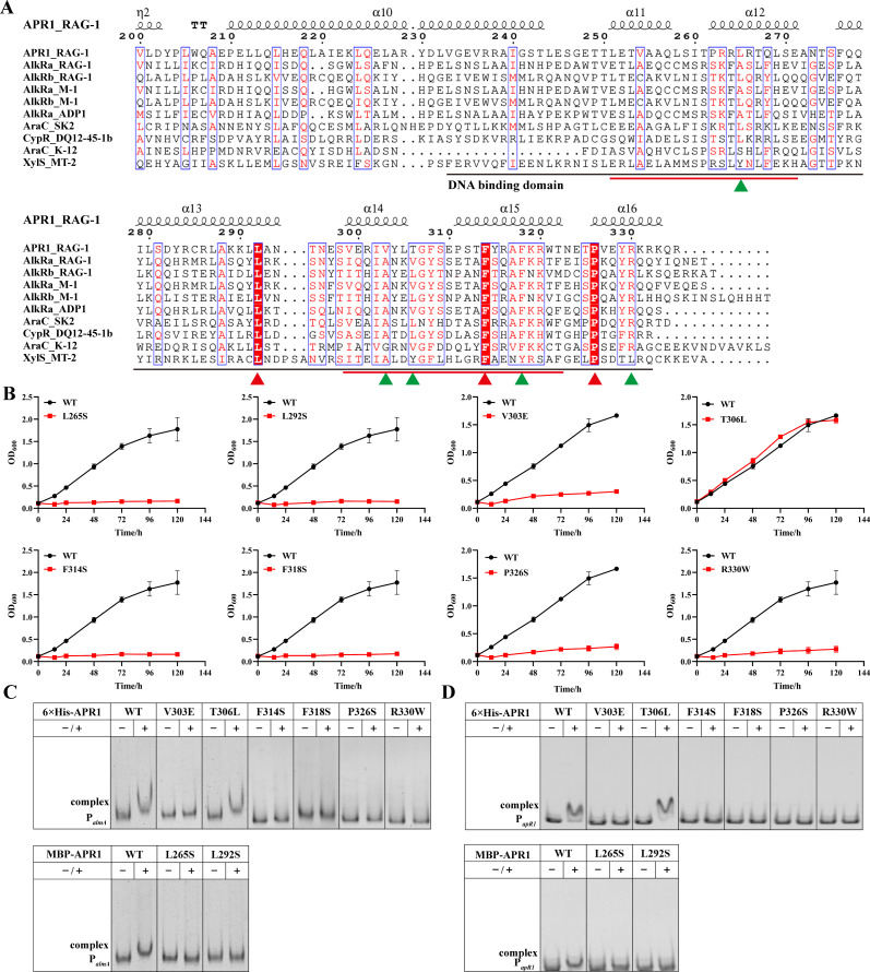
Importance: The extreme hydrophobicity of long-chain n-alkanes ({greater than or equal to}C20) presents a significant challenge to their degradation in natural environments. It is, therefore, imperative to elucidate the regulatory mechanisms of the metabolic pathways of long-chain n-alkanes, which will be of great significance for the future application of hydrocarbon-degrading bacteria to treat oil spills. However, the majority of current studies have focused on the regulatory mechanisms of short- and medium-chain n-alkanes, with long-chain n-alkanes receiving comparatively little attention. In this study, we identified APR1, a transcriptional regulator of the alkane monooxygenase AlmA in Acinetobacter venetianus RAG-1, and characterized its function and regulatory mechanism. In the presence of ligand n-C32, APR1 could directly activate the transcription of almA, which was involved in the n-C32 metabolism. The amino acid residue unique to the C-terminal DNA-binding domain of AraC/XylS type n-alkanes transcription regulators was also identified. These findings further improved our understanding of the long-chain n-alkanes degradation mechanism, which is important for the management of petroleum pollution.

Keywords: APR1; Acinetobacter venetianus RAG-1; AlmA; AraC/XylS family transcriptional regulator; long-chain n-alkanes.


Conflict of interest statement

The authors declare no conflict of interest.

Figures

Fig 1
Fig 1
Effects of AlmA on various long-chain n-alkanes utilization. Growth curves of RAG-1/P (RAG-1 containing pMK vector), ΔalmA/P (ΔalmA containing pMK vector), and ΔalmA/PalmAalmA containing almA complemented plasmid) strains cultured in basal salt medium (BSM) with sodium acetate (SA) (A), n-C24 (B), n-C26 (C), n-C28 (D), n-C30 (E), n-C32 (F), n-C34 (G), n-C38 (H) as the sole carbon source. Values are shown as means ± SD (n = 3).
Fig 2
Fig 2
Analysis of almA promoter activity. (A) The almA gene promoter activity induced by SA and various long-chain n-alkanes was by red fluorescence expression during the logarithmic growth phase. The red fluorescence was calculated as relative fluorescence units (F/OD), which represent fluorescence values per OD600. (B) Degradation rates of RAG-1/P, △almA/P, and △almA/PalmA strains cultured in BSM medium with n-C32 as the sole carbon source for 5 days. Values are shown as mean ± SD (n = 3), and the asterisk indicates a significant difference (**P < 0.01; ***P < 0.001; ns, not significant) by Student’s t-test. (C) DNA elements of the almA gene promoter. The putative −35 and the −10 regions of the almA gene promoter are shown underlined. The transcription start site (TSS) and start codon are shown in red.
Fig 3
Fig 3
Effect of APR1 on the ability of RAG-1 to utilize n-C32. (A) Growth curves of RAG-1 and △apR1 strains in BSM medium with SA as the sole carbon source; Growth curves (B) and degradation rates (C) of RAG-1/P, △apR1/P, and △apR1/PapR1 strains cultured in BSM medium with n-C32 as the sole carbon source for 5 days. Values are shown as mean ± SD (n = 3), and the asterisk indicates a significant difference (***P < 0.001; ns, not significant) by Student’s t-test.
Fig 4
Fig 4
Binding of APR1 to almA gene promoter. (A and B) qRT-PCR analysis of apR1 and almA gene transcription levels in WT, △apR1, and △apR1/PapR1 strains grown in BSM medium with SA (A) and n-C32 (B) as the sole carbon source. The 16S rRNA gene was used as an internal standard gene. WT, RAG-1. (C) Detection of red fluorescence intensity from the co-transformed cells at different times. Reporter vectors with the almA gene promoter and apR1 expression vectors were co-transformed into E. coli DH5α to determine red fluorescence intensity. Control, pUC18 + PalmA-mcherry; treatment, pUC18-apR1 + PalmA-mcherry. The red fluorescence was calculated as relative fluorescence units (F/OD), which represent fluorescence values per OD600. (D and E) (EMSAs) on the binding of APR1 (D) and BSA (E) to almA gene promoter region. EMSAs with the DNA probe (110 nM) and purified APR1 and BSA (0–5.10 µM). (F) Binding affinities between APR1 and almA gene promoter region were measured by MST. NT-647-NHS labeled APR1 (10 µM) was incubated with 0.64–41,400 nM almA gene promoter. Values are shown as mean ± SD (n = 3), and the asterisk indicates a significant difference (*P < 0.05; **P < 0.01; ***P < 0.001; ns, not significant) by Student’s t-test.
Fig 5
Fig 5
Binding of APR1 to its own gene promoter. (A) DNA elements of the apR1 gene promoter. The putative −35 and the −10 regions of the apR1 gene promoter are shown underlined. The TSS and start codon are shown in red. (B and C) EMSAs on the binding of APR1 (B) and BSA (C) to apR1 gene promoter region. EMSAs with the DNA probe (140 nM) and purified APR1 and BSA (0–2.55 µM). (D) Detection of red fluorescence intensity from the co-transformed cells at different times. Reporter vectors with the apR1 gene promoter and apR1 expression vectors were co-transformed into E. coli DH5α to determine red fluorescence intensity. Control, pUC18 + PapR1-mcherry; treatment, pUC18-apR1 + PapR1-mcherry. The red fluorescence was calculated as relative fluorescence units (F/OD), which represent fluorescence values per OD600. Values are shown as mean ± SD (n = 3), and an asterisk indicates a significant difference (*P < 0.05; **P < 0.01) by Student’s t-test.
Fig 6
Fig 6
Molecular docking and dynamic simulation results of APR1-C32. (A) Docking analysis of APR1 with substrate n-C32. The n-C32 is shown in purple. (B) Root-mean-square deviation (RMSD) values of APR1-C32 complex systems over 100 ns. (C) Energy contribution of hot residue during simulation of APR1 and n-C32 dynamics. (D) Growth curves of WT and its point mutants (V10E, F13S, Q50L, R78L, I82N, A99E, I106T, I147T, F150S) cultured in BSM medium with n-C32 as the sole carbon source for 5 days. Values are shown as means ± SD (n = 3).
Fig 7
Fig 7
Key sites for APR1 binding to downstream promoters. (A) MSAs of APR1 and other Arac/XylS family transcriptional regulators of hydrocarbon-degrading bacteria. The secondary structural elements of APR1_RAG-1 are listed above the sequence. The red-filled boxes represent completely conserved sequences in these strains, and the unfilled boxes represent relatively conserved sequences. The DNA-binding domain and motifs are underlined in black and red, respectively. Red and green triangles indicate sites where point mutations were performed. (B) Growth curves of WT and its point mutants (L265S, L292S, V303E, T306L, F314S, F318S, F326S, R330W) cultured in BSM medium with n-C32 as the sole carbon source for 5 days. Values are shown as means ± SD (n = 3). The binding of the WT 6 × His-APR1 and its point mutants or MBP-APR1 and its point mutants to (C) the almA promoter region (110 nM) or (D) the apR1 promoter region (140 nM) was measured by EMSA. −, no APR1 was added; +, 2.55 µM APR1 was added.
Fig 8
Fig 8
Phylogenetic analysis based on the complete amino acid sequences of AlmA and APR1. (A) AlmA and its homologs or (B) APR1 and its homologs from different strains in Proteobacteria were used to construct a phylogenetic tree by the neighbor-joining algorithm (1,000 replicates). The AlmA and APR1 from A. venetianus RAG-1 are marked with red. Bootstrap values (%) are indicated at the branch nodes, and the scale bar represents 0.05 substitutions per site.
Fig 9
Fig 9
The proposed regulatory mechanism of AlmA in RAG-1 cells. Please refer to the initial paragraph of the Discussion section for further details.

Similar articles

Cited by

References

    1. Varjani SJ. 2017. Microbial degradation of petroleum hydrocarbons. Bioresour Technol 223:277–286. doi:10.1016/j.biortech.2016.10.037 - DOI - PubMed
    1. Liu J, Chen S, Zhao B, Li G, Ma T. 2022. A novel FadL homolog, AltL, mediates transport of long-chain alkanes and fatty acids in Acinetobacter venetianus RAG-1. Appl Environ Microbiol 88:e0129422. doi:10.1128/aem.01294-22 - DOI - PMC - PubMed
    1. Xu X, Liu W, Tian S, Wang W, Qi Q, Jiang P, Gao X, Li F, Li H, Yu H. 2018. Petroleum hydrocarbon-degrading bacteria for the remediation of oil pollution under aerobic conditions: a perspective analysis. Front Microbiol 9:2885. doi:10.3389/fmicb.2018.02885 - DOI - PMC - PubMed
    1. Fuentes S, Méndez V, Aguila P, Seeger M. 2014. Bioremediation of petroleum hydrocarbons: catabolic genes, microbial communities, and applications. Appl Microbiol Biotechnol 98:4781–4794. doi:10.1007/s00253-014-5684-9 - DOI - PubMed
    1. Rojo F. 2009. Degradation of alkanes by bacteria. Environ Microbiol 11:2477–2490. doi:10.1111/j.1462-2920.2009.01948.x - DOI - PubMed

MeSH terms

LinkOut - more resources