. 2025 Jan;34(1):e70022.
doi: 10.1002/pro.70022.

The structural influence of the oncogenic driver mutation N642H in the STAT5B SH2 domain

Affiliations

The structural influence of the oncogenic driver mutation N642H in the STAT5B SH2 domain

Liam Haas-Neill et al. Protein Sci. 2025 Jan.

Abstract

The point mutation N642H of the signal transducer and activator of transcription 5B (STAT5B) protein is associated with aggressive and drug-resistant forms of leukemia. This mutation is thought to promote cancer due to hyperactivation of STAT5B caused by increased stability of the active, parallel dimer state. However, the molecular mechanism leading to this stabilization is not well understood as there is currently no structure of the parallel dimer. To investigate the mutation's mechanism of action, we conducted extensive all-atom molecular dynamics simulations of multiple oligomeric forms of both STAT5B and STAT5BN642H, including a model for the parallel dimer. The N642H mutation directly affects the hydrogen bonding network within the phosphotyrosine (pY)-binding pocket of the parallel dimer, enhancing the pY-binding interaction. The simulations indicate that apo STAT5B is highly flexible, exploring a diverse conformational space. In contrast, apo STAT5BN642H accesses two distinct conformational states, one of which resembles the conformation of the parallel dimer. The simulation predictions of the effects of the mutation on structure and dynamics are supported by the results of hydrogen-deuterium exchange (HDX) mass spectrometry measurements carried out on STAT5B and STAT5BN642H in which a phosphopeptide was used to mimic the effects of parallel dimerization on the SH2 domain. The molecular-level information uncovered in this work contributes to our understanding of STAT5B hyperactivation by the N642H mutation and could help pave the way for novel therapeutic strategies targeting this mutation.

Keywords: SH2 domains; STAT proteins; intrinsically disordered regions; molecular dynamics simulations; oncogenic mutation; protein dynamics; signal transducer and activator of transcription.


Figures

FIGURE 1
FIGURE 1
3D structure of STAT5B and its SH2 domain. (a) Structure of the STAT5B monomer core fragment shown in cartoon representation with each domain labeled (coiled‐coil domain: 138–331, DNA binding domain: 332–471, linker domain: 498–591, SH2 domain 592–685). The structure shown is chain B from the crystal structure of the STAT5B antiparallel dimer, PDB 6MBZ (de Araujo, Erdogan, et al., 2019). (b) 3D structures of the SH2 domain from STAT5B (pY and pY + 3 pockets in surface representation; pY and pY + 3 residues in licorice representation). (c) Structure of the STAT5B antiparallel dimer core fragment (PDB 6MBZ [de Araujo, Erdogan, et al., 2019]) shown in cartoon representation with each domain labeled. (d) SH2 domain with the secondary structure elements labeled. Key residues are shown in licorice representation. (e) Structure of the STAT6 parallel dimer core fragment (PDB 4Y5U [Li et al., 2016]). (f) Structure of the STAT5B parallel dimer SH2 domain and phosphotyrosine motif region (including residues 589–709). Residue pY699 from chain A (green, licorice representation) interacts with the pY‐binding pocket of chain B (blue, surface representation). The structure of the SH2 domain shown in panels (b, d and f) corresponds to the SH2 domain in the homology model of the parallel dimer. The simulations of the homology model also include the linker domain, which is not shown here for clarity. The crystal structures of STAT5B and STAT6 in (a, c, and e) include only the core fragment and are missing the N‐terminal oligomerization and transactivation domains.
FIGURE 2
FIGURE 2
A model for the STAT5B parallel dimer and effects of the N642H mutation on the parallel dimer interface. (a) The conformational landscape of mammalian STAT SH2 domains is shown. The simulation trajectory of the parallel dimer equilibration simulation is projected onto the space, with each 20 ns of the equilibration simulation indicated as a different color (see legend). Regions of the conformational space encapsulating each STAT protein are indicated as shaded regions. Phosphate‐bound crystal structures (including parallel dimers and phosphopeptides) are shown in orange, while apo crystal structures are shown in gray. Key structures from the conformational space are shown along the side, and are indicated on the conformational space by stars with colors matching the structures. The same conformational space with all STAT crystal structures labeled is provided in Figure S3. (b) Representative configuration of residue R618, the pY residue and residue 642 (N642, left; N642H, right) to illustrate the main difference in the hydrogen bonding network in the pY pocket due to the mutation. (c) Average number of hydrogen bonds between the mutation site residue (642) and other SH2 domain residues in simulations of STAT5B (dark green) and STAT5BN642H (light green) parallel dimers. (d) Probability distribution of the denticity of the pY‐R618 hydrogen bond in simulations of STAT5B (dark green) and STAT5BN642H (light green) parallel dimers. (e) Conditional probability distribution of denticity of the pY‐R618 hydrogen bond in the STAT5BN642H simulations, conditioned on the N642H‐R618 hydrogen bond being formed (blue) or not formed (gray). Error bars in panels (c–e) indicate standard error of the mean obtained by treating each SH2 domain in the simulations as an independent measurement.
FIGURE 3
FIGURE 3
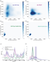
Differences in the structural flexibility of the SH2 domain due to the mutation and parallel dimerization. (a–d) Two‐dimensional distribution of pY pocket volume and pY + 3 pocket volume for apo STAT5B (a), apo STAT5BN642H (b), STAT5B parallel dimer (c), and STAT5BN642H parallel dimer (d). The color of each pixel indicates the frequency of sampling each region. Darker colors indicate more frequently sampled regions. Different color scales are used in panels (a, b) and (c, d). (e, f) Root‐mean‐square fluctuation (RMSF) profiles (e) and Radius of gyration (R g) distributions (f) for STAT5B parallel dimer (dark green), STAT5BN642H parallel dimer (teal), apo STAT5B (blue), and apo STAT5BN642H (plum) ensembles. The vertical shaded areas indicate the average value of R g ± the standard error of the mean. In (e, f), shaded areas indicate standard error of the mean obtained by treating each SH2 domain in the simulations as an independent measurement.
FIGURE 4
FIGURE 4
Comparison of N642H mutant antiparallel dimer A‐state and B‐state trajectories with pY‐bound parallel dimer ensemble. (a) Average number of contacts between the mutation site residue (642) and other SH2 domain residues. (b) β propensity of each residue in the SH2 domain. (c) Root mean squared fluctuation of each residue in the SH2 domain. The secondary structure is shown below the x‐axis. In (a–c), shaded regions indicate standard error of the mean obtained by treating each SH2 domain in the simulations as an independent measurement. (d) Principal component analysis of all N642H mutant antiparallel dimer and both WT and N64H mutant pY‐bound parallel dimer trajectories. The B‐state ensemble partly overlaps with the parallel dimer ensemble, which is disjoint with the A‐state ensemble. (e–g) Close‐up view of representative structures from the parallel dimer (e), B‐state (f), and A‐state (g) ensembles. The mutation site residue (642) is shown in licorice representation. In all figure panels, results for the A‐state, B‐state and parallel dimer are indicated in yellow, orange and green, respectively. Trajectories are separated into A‐state and B‐state trajectories based on the occurrence of any β structure in the βD‐strand.
FIGURE 5
FIGURE 5
The STAT SH2 conformational landscape. (a–f) Every structure from all simulation trajectories was projected onto the conformational space of STAT SH2 domains (the same principal components as in Figure 2a). Each point represents one structure. Structural ensembles consisting of 24 aligned structures selected at random from simulation trajectories are also shown for each system: STAT5B parallel dimer (a), antiparallel dimer (b), and monomer (c); STAT5BN642H parallel dimer (d), antiparallel dimer (e), and monomer (f).
FIGURE 6
FIGURE 6
HDX experiments show a difference in deuterium uptake upon pY binding and with the N642H mutation. (a) The difference in deuterium uptake between apo STAT5B and phosphopeptide‐bound STAT5B. Green bars indicate peptides having significantly greater deuterium uptake in the apo case. Uncertainty is indicated by dashed lines in panels (a, c). (b) A structure of the SH2 domain taken from the first frame of the wild‐type parallel dimer simulation. Residues belonging to peptide sequences that show greater deuterium incorporation in the apo state relative to pY bound are shown in green. In both panels (b, d), residues belonging to peptide sequences that show no difference in deuterium incorporation are shown in gray, regions for which there is no deuterium incorporation data are shown as transparent, Cα atoms of pocket residues are shown as yellow spheres, and the N642 residue is shown in a ball‐and‐stick representation. (c) The difference in deuterium uptake between apo STAT5B and apo STAT5BN642H. Plum bars indicate peptides having significantly greater deuterium uptake in wild type. (d) A structure of the SH2 domain taken from the first frame of the N642H parallel dimer simulation. Peptide sequences that show decreased deuterium incorporation in the N642H mutant are shown in plum.
FIGURE 7
FIGURE 7
Common cancer mutations in the STAT5B SH2 domain. (a)The STAT5B SH2 domain and is shown in cartoon representation. The pY and pY + 3 pockets are shown in surface representation, in yellow and pink, respectively. Sites of mutations that commonly occur in cancer (Bhattacharya et al., 2022) are shown in ball‐and‐stick representation colored in red. Residue N642 is also a mutation site, but it is excluded for clarity. Mutations identified at these sites include: T628S, Y665F, P685R, P702A, and Q706L (Bhattacharya et al., 2022). (b) The parallel dimer of the STAT5B SH2 domain is shown in cartoon representation, colored by residue type. Negative residues are shown in red, positive residues are shown in blue, polar residues are shown in green, and hydrophobic residues are shown in white. Sites of mutations that commonly occur in cancer are colored red and shown in ball‐and‐stick representation on one of the monomers. The pY and pY + 3 are shown in surface representation and colored in yellow, and pink, respectively.

Similar articles

References

    1. Anselmi M, Calligari P, Hub JS, Tartaglia M, Bocchinfuso G, Stella L. Structural determinants of phosphopeptide binding to the N‐terminal Src homology 2 domain of the SHP2 phosphatase. J Chem Inf Model. 2020;60(6):3157–3171. - PMC - PubMed
    1. Ariyoshi K, Nosaka T, Yamada K, Onishi M, Oka Y, Miyajima A, et al. Constitutive activation of STAT5 by a point mutation in the SH2 domain. J Biol Chem. 2000;275(32):24407–24413. - PubMed
    1. Awasthi N, Liongue C, Ward AC. STAT proteins: a kaleidoscope of canonical and non‐canonical functions in immunity and cancer. J Hematol Oncol. 2021;14:198. - PMC - PubMed
    1. Ball DP, Lewis AM, Williams D, Resetca D, Wilson DJ, Gunning PT. Signal transducer and activator of transcription 3 (STAT3) inhibitor, S3I‐201, acts as a potent and non‐selective alkylating agent. Oncotarget. 2016;7(15):20669–20679. - PMC - PubMed
    1. Bandapalli OR, Schuessele S, Kunz JB, Rausch T, Stutz AM, Tal N, et al. The activating STAT5B N642H mutation is a common abnormality in pediatric T‐cell acute lymphoblastic leukemia and confers a higher risk of relapse. Haematologica. 2014;99(10):e188–e192. - PMC - PubMed

Substances

LinkOut - more resources