. 2025 Jan 9;68(1):448-464.
doi: 10.1021/acs.jmedchem.4c02043. Epub 2024 Dec 26.

Probing the Histamine H1 Receptor Binding Site to Explore Ligand Binding Kinetics

Affiliations

Probing the Histamine H1 Receptor Binding Site to Explore Ligand Binding Kinetics

Sebastiaan Kuhne et al. J Med Chem. .

Abstract

Analysis of structure-kinetic relationships (SKR) can contribute to an improved understanding of receptor-ligand interactions. Here, fragment 1 (4-(2-benzylphenoxy)-1-methylpiperidine) was used in different fragment growing approaches to mimic the putative binding mode of the long residence time (RT) ligands olopatadine, acrivastine, and levocetirizine at the histamine H1 receptor (H1R). SKR analyses reveal that introduction of a carboxylic acid moiety can increase RT at H1R up to 11-fold. Ligand efficiency (LE) decreases upon the introduction of the negatively charged group, whereas kinetic efficiency (KE) increases up to 8.5-fold. The olopatadine/acrivastine mimics give up to 15-fold differences in the RT, while the levocetirizine mimics afford similar RTs with only a 3-fold difference. Therefore, the levocetirizine mimics are less sensitive to structural changes. This study illustrates that for H1R, there are several ways to increase RT but the different strategies differ significantly in SKR.


Conflict of interest statement

The authors declare no competing financial interest.

Figures

Figure 1
Figure 1
(A) Antihistamines targeting H1R with corresponding binding affinities (pKi) and RT, as determined in this study. Ligand efficiency (LE) = −R·T·ln(Ki)/N, where N is the number of heavy atoms (HA, i.e., non-hydrogen atoms), and R = (8.31447215 J K–1 mol–1 and 4184 J = 1 kcal), and T = 298.15 K. Kinetic efficiency (KE) = RT/N, where RT is the residence time, and N is the number of HAs. (B) H1R X-ray structure (PDB-code: 3RZE) with the cocrystallized ligand doxepin (green carbon atoms) and the proposed binding mode of fragment 1 (magenta carbon atoms). Important binding site residues are represented as ball-and-sticks with light gray carbon atoms. Nitrogen, oxygen, and hydrogen atoms are colored blue, red, and cyan, respectively. Polar hydrogen atoms of the ligands are shown, but are absent for the binding site residues. H1R binding site surface is shown and colored to designate the four different regions of the binding site, i.e., the amine binding region, the lower aromatic binding region, the upper aromatic binding region, and the phosphate binding region. UniProt numbers (first) and the Ballesteros–Weinstein numbering (second in superscript) are reported for each residue throughout this manuscript. (C) Schematic 2D representation of fragment 1, binding affinity and kinetic parameters (kon, koff, RT) as determined in this study, and the three different design strategies to probe the H1R SKR.
Figure 2
Figure 2
(A) Fragment growing of hit fragment 1, mimicking the H1R reference compounds olopatadine, acrivastine, and levocetirizine, to study the H1R SKR. Proposed binding mode of the H1R reference compounds (B) olopatadine (gray carbon atoms), (C) acrivatine (gray carbon atoms), and (D) levocetirizine (gray carbon atoms) in H1R. Proposed binding mode of the (E) olopatadine mimics 2b (blue carbon atoms) and 3b (orange carbon atoms), (F) acrivastine mimics 2c (blue carbon atoms) and 3c (orange carbon atoms), and (G) levocetirizine mimics binding of 4c (green carbon atoms) to H1R. Binding modes were obtained by docking the compounds in the H1R crystal structure (pdb: 3RZE). Rendering and color-coding are the same as in Figure 1.
Scheme 1
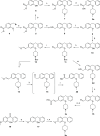
Scheme 1. Synthesis of Ligands to Probe the Olopatadine and Acrivastine Binding Region: Growing from the 5-Position of the Aromatic Ring
Reagents and conditions: (a) AlCl3, benzene, rt, 165 min, 73%. (b) BBr3, DCM, 0 °C to rt, 3 h, 70%. (c) 1-methylpiperidin-4-ol, PPh3, DEAD (40 wt % in toluene), THF, 0 °C to rt, 24 h, 31%. (d) 1.0 M NaOH (aq), MeOH, reflux, 5 h, 33%. (e) 1.0 M LAH in THF, THF, −78 °C to rt, 2.5 h, 96%. (f) MnO2, DCM, rt, 42 h, 93%. (g) EtSNa, DMF, 120 °C, 3 h, 61%. (h) tert-butyl 4-((methylsulfonyl)oxy)piperidine-1-carboxylate, Cs2CO3, DMF, 60 °C, 16 h, 74%. (i) t-BuOK, (methoxymethyl)triphenylphosphonium chloride, THF, rt, 6 h, 66%. (j) 1.0 M LAH in THF, THF, 0–45 °C, 8.5 h, 92%. (k) 1.0 M HCl in dioxane, H2O, rt, 1 h. (l) NaH2PO4·H2O, 2-methylbut-2-ene, NaClO2, t-BuOH, H2O, rt, 1 h, 6% over two steps. (m) 1.0 M LAH in THF, THF, 0 °C to reflux, 1.5 h, 38%. (n) MnO2, DCM, rt, 16 h. (o) Methyl (triphenylphosphoranylidene)acetate, toluene, 0 °C to rt, 48 h. (p) 2.0 M NaOH (aq), MeOH, reflux, 6 h, 7% over three steps. (q) Et3SiH, TFA, DCM, rt, 17 h, 52%. (r) tert-Butyl 4-((methylsulfonyl)oxy)piperidine-1-carboxylate, Cs2CO3, DMF, 65 °C, 22 h. (s) 1.0 M LAH in THF, THF, 0–50 °C, 4 h, 54% over two steps. Compound 2d was converted to a hemifumaric acid salt and compound 2e to a fumaric acid salt.
Scheme 2
Scheme 2. Synthesis of Ligands to Probe the Olopatadine and Acrivastine Binding Region: Growing from the 6-Position of the Aromatic Ring
Reagents and conditions: (a) MgCl2, TEA, paraformaldehyde, THF, reflux, 4 h, 66%. (b) tert-butyl 4-((methylsulfonyl)oxy)piperidine-1-carboxylate, Cs2CO3, DMF, 60 °C, 32 h, 70%. (c) 1.0 M LAH in THF, THF, 0–60 °C, 4 h, 17%. (d) MnO2, DCM, rt, 42 h, 80%. (e) NaH2PO4·H2O, 2-methylbut-2-ene, NaClO2, t-BuOH, H2O, rt, 2 h, 2%. (f) t-BuOK, (methoxymethyl)triphenylphosphonium chloride, THF, rt, 16 h, 73%. (g) 1.0 M HCl in dioxane, H2O, rt, 1 h. (h) NaH2PO4.H2O, 2-methylbut-2-ene, NaClO2, t-BuOH, H2O, rt, 1 h, 9% over two steps. (i) Methyl (triphenylphosphoranylidene)acetate, THF, 0 °C to rt, 48 h. (j) 2.0 M NaOH (aq), MeOH, reflux, 6 h, 24% over two steps.
Scheme 3
Scheme 3. Synthesis of Ligands to Probe the Levocetirizine Binding Region: Growing from the Basic Nitrogen Atom
Reagents and conditions: (a) corresponding bromo-ester, DMF, K2CO3, 80 °C, 3–9 h, 32–71%. (b) [1] 2.0 M NaOH (aq), MeOH, reflux, 3 h. [2] 1.0 M HCl (aq), rt, 30 min, 30–50%. (c) 5-Bromopentanenitrile, K2CO3, DMF, 85 °C, 2 h, 28% as fumarate salt. (d) NaN3, NH4Cl, DMF, 100 °C, 72 h, 28%.
Figure 3
Figure 3
Characterization of ligand binding to a homogenate of HEK293T cells expressing H1R using [3H]-mepyramine. (A) HEK293T cell homogenate expressing H1R was incubated with increasing concentrations of [3H]-mepyramine for 4 h in the absence (red) or presence (black) of 10–5 M mianserin. (B) Cell homogenate was incubated with 2.9 nM (blue), 1.3 nM (red), 0.5 nM (green), and 0.3 nM (brown) of [3H]-mepyramine for various incubation times. (C) Cell homogenate was incubated with a single concentration [3H]-mepyramine with increasing concentrations of unlabeled ligand for 4 h. Ligands are depicted by the red curve (1), green curve (2c), brown curve (4c) and blue curve (levocetirizine). (D) Single concentration [3H]-mepyramine was coincubated for various times with cell homogenate in the absence (black) or presence of an approximate concentration of 10 times the respective Ki of these unlabeled ligands (red curve (1), green curve (2c), brown curve (4c) and blue curve (levocetirizine)). Representative graphs are shown of N ≥ 3 experiments with mean and SD of triplicate values (A, C) or duplicate values (B, D).
Figure 4
Figure 4
(A) Correlation between the kon (y-axis) and the koff (x-axis) for 1, the ligands probing the olopatadine and acrivastine binding region 2b–e and 3a–d, and olopatadine (olo) and acrivastine (acri). Ligands substituted at the 5-position are colored blue (2b–e), and ligands substituted at the 6-position are colored orange (3a–d). Ligand 2a is omitted for clarity purposes (kon: 0.06 × 10⁶ M–1·min–1; koff: 1 min–1). Parallel dashed lines represent constant KD values (KD = koff/kon) as indicated for the respective lines in the graph. Color of the molecule numbers corresponds to the color-coding in Figure 2A. (B) Correlation between the kon (y-axis) and the koff (x-axis) for 1, the ligands probing the levocetirizine binding region (4a–f, green), and levocetirizine (levo). Corresponding pharmacological data can be found in Table 1 and 2.
Figure 5
Figure 5
Correlation plot between the log kon (y-axis) and the pKa (x-axis) for all analogs of 1 with a pKa below 5.5 (2a–c, 3a–c, 4a–f, 27). Ligands probing the acrivastine/olopatadine binding region are depicted in blue and orange for analogs of 1 substituted at the 5-position (2b–e) or substituted at the 6-position (3a–d), respectively. Ligands probing the levocetirizine binding region (4a–f) are depicted in green, while 27 is depicted in gray. pKa values were calculated using MarvinSketch. Corresponding pharmacological data can be found in Tables 1 and 2.

Similar articles

Cited by

References

    1. Yang D.; Zhou Q.; Labroska V.; Qin S.; Darbalaei S.; Wu Y.; Yuliantie E.; Xie L.; Tao H.; Cheng J.; et al. G protein-coupled receptors: structure- and function-based drug discovery. Signal Transduct Target Ther 2021, 6 (1), 7.10.1038/s41392-020-00435-w. - DOI - PMC - PubMed
    1. Addis P.; Bali U.; Baron F.; Campbell A.; Harborne S.; Jagger L.; Milne G.; Pearce M.; Rosethorne E. M.; Satchell R.; et al. Key aspects of modern GPCR drug discovery. SLAS Discov 2024, 29 (1), 1–22. 10.1016/j.slasd.2023.08.007. - DOI - PubMed
    1. Cheng L.; Xia F.; Li Z.; Shen C.; Yang Z.; Hou H.; Sun S.; Feng Y.; Yong X.; Tian X.; et al. Structure, function and drug discovery of GPCR signaling. Mol. Biomed 2023, 4 (1), 46.10.1186/s43556-023-00156-w. - DOI - PMC - PubMed
    1. van der Velden W. J. C.; Heitman L. H.; Rosenkilde M. M. Perspective: Implications of Ligand-Receptor Binding Kinetics for Therapeutic Targeting of G Protein-Coupled Receptors. ACS Pharmacol Transl Sci. 2020, 3 (2), 179–189. 10.1021/acsptsci.0c00012. - DOI - PMC - PubMed
    1. Egyed A.; Kiss D. J.; Keseru G. M. The Impact of the Secondary Binding Pocket on the Pharmacology of Class A GPCRs. Front Pharmacol 2022, 13, 84778810.3389/fphar.2022.847788. - DOI - PMC - PubMed

Publication types

MeSH terms

LinkOut - more resources