. 2024 Dec 26;18(12):e0012704.
doi: 10.1371/journal.pntd.0012704. eCollection 2024 Dec.

Dermatopathological findings of Bothrops atrox snakebites: A case series in the Brazilian Amazon

Affiliations

Dermatopathological findings of Bothrops atrox snakebites: A case series in the Brazilian Amazon

Fabiane Bianca Albuquerque Barbosa et al. PLoS Negl Trop Dis. .

Abstract

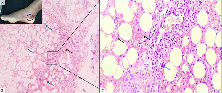
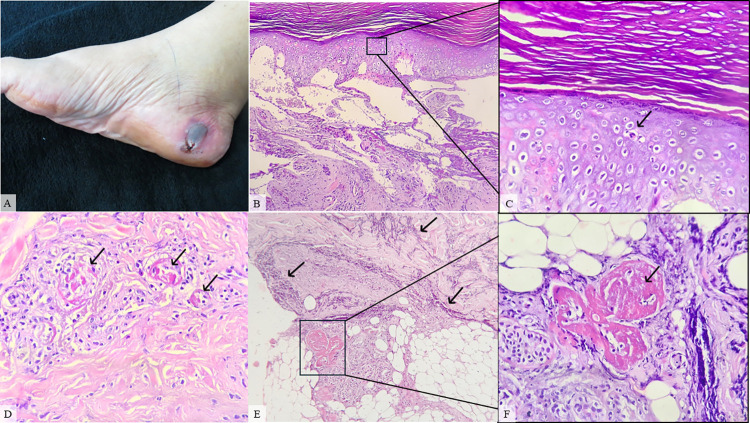
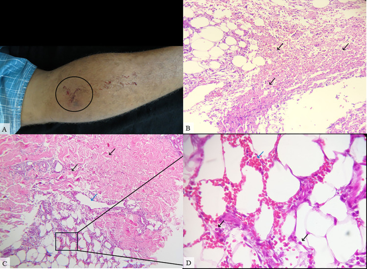
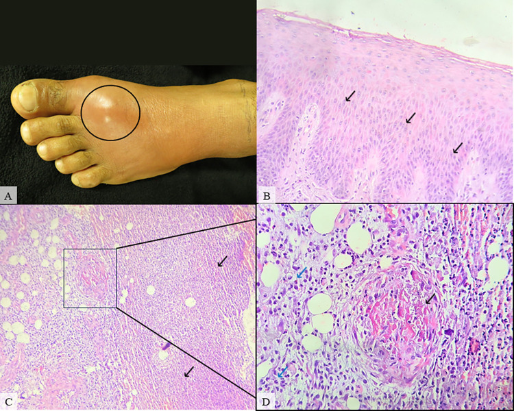
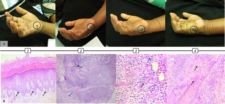
Background: Bothrops venom consists primarily of metalloproteinase and phospholipase A2 toxins, which are responsible for the acute inflammatory, coagulant and hemorrhagic action following snakebite. The local effects of snakebite envenomation by Bothrops species are particularly prevalent yet poorly studied, but include pain, edema, erythema, blistering, bleeding, and ecchymosis.

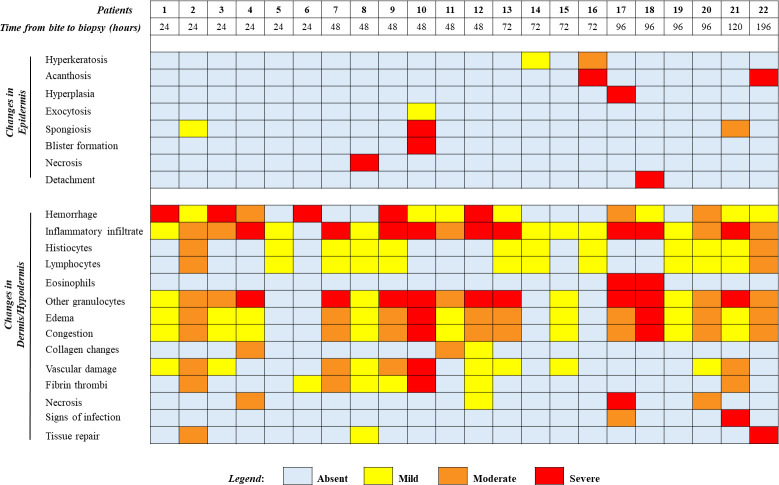
Methods and findings: In this study, we describe the dermatopathological findings observed in a series of 22 patients diagnosed with Bothrops envenomation treated in a tertiary hospital of Manaus, in the Brazilian Amazon. Clinically, pain and edema were observed in all patients, followed by fang marks (63.6%), secondary infection (36.3%), ecchymosis (31.8%), erythema (22.7%), blister (13.6%), and necrosis (4.5%). Regarding histopathological findings, epidermal alterations such as spongiosis, acanthosis and hyperkeratosis were the most observed characteristics in our cases series, with isolated cases of hyperplasia, hemorrhagic intraepidermal blister and severe necrosis. Changes in dermis and hypodermis consisted mainly of hemorrhage, inflammatory infiltrate, edema, congestion, and vascular damage, whereas cases of collagen damage, necrosis, abscess, and signs of tissue repair, indicated by the presence of granulation tissue, were also observed, with a persistence of inflammatory and hemostatic alterations even days after antivenom administration. Therefore, the tissue damage resulting from Bothrops envenomation could be related to both direct venom activity as well as inflammatory response or presence of infectious process. The histopathological analysis of human skin injury can enlighten the pathological and endogenous effects of local envenomation and could underpin new strategies, including novel treatments, adjuvants or changes in clinical management, that lead to better outcomes in snakebite patients.


Conflict of interest statement

The authors have declared that no competing interests exist.

Figures

Fig 1
Fig 1. Patient #9, male, 23, classified as a moderate case.
The time elapsed between snakebite and antivenom treatment was 8 hours. A. Lower right limb presenting moderate edema, ecchymosis, and biopsy suture (black circle) on day 4 after envenomation. B. Skin biopsy showing severe inflammatory infiltrate (black arrow), hemorrhage (blue arrow) and moderate vascular damage (inset) on hypodermis (HE, 100X). C. A magnified view of the erythrocytes (black arrow) and vascular damage (blue arrow) is shown in the inset (HE, 400X).
Fig 2
Fig 2. Patient #10, female, 60, classified as a moderate case.
Alcohol was used as a self-care procedure at the bite site and the time elapsed between snakebite and antivenom treatment was 17h30min. A. Lower right limb presenting hemorrhagic blister (black circle) on day 4 after envenomation. B. Skin biopsy showing severe spongiosis that evolved to hemorrhagic blister and neutrophil exocytosis (HE, 100X). C. A magnified view of the neutrophil exocytosis (black arrow) is shown in the inset (HE, 400X). D. Vascular damage (black arrow) in the dermis (HE, 400x). E. Dermis and hypodermis presenting severe inflammatory infiltrate (black arrow) (HE, 100x). F. Inset showing a magnified view of fibrin thrombi (black arrow) in the deep dermis (HE, 400x).
Fig 3
Fig 3. Patient #20, male, 20, classified as a mild case.
Pirarucu’s fat (Arapaima gigas) was used as a self-care procedure at the bite site and the time elapsed between snakebite and antivenom treatment was 5 hours. A. Lower left limb presenting mild edema and bleeding (black circle) on hospital admission. B. Hypodermis presenting severe necrosis (black arrow) (HE, 200X). C. Skin biopsy showing moderate dermal edema (black arrow), vascular damage (blue arrow), moderate hemorrhage and inflammatory infiltrate on hypodermis (HE, 100X). D. A magnified view of the inflammatory cells (black arrow) and erythrocytes (blue arrow) is shown in the inset (HE, 400X).
Fig 4
Fig 4. Patient #21, male, 28, classified as a severe case.
A tourniquet was applied as a self-care procedure near the bite site and the time elapsed between snakebite and antivenom treatment was 4 hours. A. Lower left limb presenting cellulitis (black circle) on day 6 after envenomation. B. Skin biopsy showing moderate spongiosis (black arrow) (HE, 200X). C. Deep dermis presenting severe abscess (black circle), vascular damage and fibrin thrombi (HE, 100X). D. Vascular damage (black arrow) and severe inflammatory infiltrate (blue arrow) in the deep dermis (HE, 400x).
Fig 5
Fig 5. Patient #22, female, 29, classified as a severe case.
An infusion of different plants in vegetal oil, known as “balsam”, was made as a self-care procedure at the bite site and the time elapsed between snakebite and antivenom treatment was 6 hours. Tissue damage evolution and microscopical alterations observed after envenomation. A1. 2 days after snakebite presenting severe edema (from hand to upper arm) snakebite marks (black circle), serum secretion and blister formation. A2. Day 3 with severe edema, blisters on dorsal region spreading to hand palm and necrotic area (black circle). A3. Day 4 with edema, blisters, and necrosis on snakebite side (black circle). A4. Day 6 with edema, blisters with pus, necrosis on the snakebite site (black circle). A debridement was made to remove necrotic area on day 8. Tissue samples for histological analysis were obtained at that moment. B1. Biopsy presenting severe irregular acanthosis (black arrow) (HE, 100x). B2. Mixed inflammatory infiltrate (black arrow) and formation of new capillaries (blue arrow) responsible for collagen production in tissue repair process (HE, 100x). B3. Mixed inflammatory infiltrate (black arrow) and formation of new capillaries (blue arrow) (HE, 200x) B4. Activated fibroblasts (black arrow) (HE, 200x).
Fig 6
Fig 6. Histopathological findings of Bothrops envenomation patients according to severity grade: absent, mild, moderate, and severe.
Patients were distributed in ascending order of time (in hours) from bite to the day of biopsy procedure.

Similar articles

Cited by

References

    1. Warrell DA. Snake bite. Lancet [Internet]. 2010;375(9708):77–88. Available from: doi: 10.1016/S0140-6736(09)61754-2 - DOI - PubMed
    1. Gutiérrez JM, Calvete JJ, Habib AG, Harrison RA, Williams DJ, Warrell DA. Snakebite envenoming. Nat Rev Dis Prim. 2017;3(1). - PubMed
    1. Longbottom J, Shearer FM, Devine M, Alcoba G, Chappuis F, Weiss DJ, et al.. Vulnerability to snakebite envenoming: a global mapping of hotspots. Lancet. 2018;392(10148):673–84. doi: 10.1016/S0140-6736(18)31224-8 - DOI - PMC - PubMed
    1. de Oliveira SS, de Souza Sampaio V, de Almeida Gonçalves Sachett J, Campos Alves E, da Silva VC, Alcântara de Lima JA, et al.. Snakebites in the Brazilian Amazon: Current Knowledge and Perspectives. 2018;73–99.
    1. Magalhães SFV, Peixoto HM, Moura N, Monteiro WM, De Oliveira MRF. Snakebite envenomation in the Brazilian Amazon: A descriptive study. Trans R Soc Trop Med Hyg. 2019;113(3):143–51. doi: 10.1093/trstmh/try121 - DOI - PubMed

LinkOut - more resources