. 2024 Dec 26;20(12):e1011408.
doi: 10.1371/journal.pgen.1011408. eCollection 2024 Dec.

RcsF-independent mechanisms of signaling within the Rcs phosphorelay

Affiliations

RcsF-independent mechanisms of signaling within the Rcs phosphorelay

Anushya Petchiappan et al. PLoS Genet. .

Abstract

The Rcs (regulator of capsule synthesis) phosphorelay is a conserved cell envelope stress response mechanism in enterobacteria. It responds to perturbations at the cell surface and the peptidoglycan layer from a variety of sources, including antimicrobial peptides, beta-lactams, and changes in osmolarity. RcsF, an outer membrane lipoprotein, is the sensor for this pathway and activates the phosphorelay by interacting with an inner membrane protein IgaA. IgaA is essential; it negatively regulates the signaling by interacting with the phosphotransferase RcsD. We previously showed that RcsF-dependent signaling does not require the periplasmic domain of the histidine kinase RcsC and identified a dominant negative mutant of RcsD that can block signaling via increased interactions with IgaA. However, how the inducing signals are sensed and how signal is transduced to activate the transcription of the Rcs regulon remains unclear. In this study, we investigated how the Rcs cascade functions without its only known sensor, RcsF, and characterized the underlying mechanisms for three distinct RcsF-independent inducers. Previous reports showed that Rcs activity can be induced in the absence of RcsF by a loss of function mutation in the periplasmic oxidoreductase DsbA or by overexpression of the DnaK cochaperone DjlA. We identified an inner membrane protein, DrpB, as a multicopy RcsF-independent Rcs activator in E. coli. The loss of the periplasmic oxidoreductase DsbA and the overexpression of the DnaK cochaperone DjlA each trigger the Rcs cascade in the absence of RcsF by weakening IgaA-RcsD interactions in different ways. In contrast, the cell-division associated protein DrpB uniquely requires the RcsC periplasmic domain for activation; this domain is not needed for RcsF-dependent signaling. This suggests the possibility that the RcsC periplasmic domain acts as a sensor for some Rcs signals. Overall, the results add new understanding to how this complex phosphorelay can be activated by diverse mechanisms.


Conflict of interest statement

The authors have declared that no competing interests exist.

Figures

Fig 1
Fig 1. RcsF-independent activators of Rcs signaling.
A. Components of the Rcs phosphorelay: The Rcs pathway is comprised of the hybrid histidine kinase RcsC, the phosphotransferase RcsD, and the response regulator RcsB as well as the upstream regulatory components RcsF and IgaA. Under non-inducing growth conditions, IgaA represses signaling by interacting with RcsD. RcsF senses damage to the envelope and triggers the RcsC-RcsD-RcsB phosphorelay by interacting with IgaA, altering its interaction with RcsD. B. Rcs activation by overexpression of DjlA and DrpB: All strains carry a rprA promoter fusion to mCherry (PrprA::mCherry); mCherry fluorescence acts as an indicator for Rcs activation. For the PrprA::mCherry assay, the strains overexpressing DjlA (pBAD-DjlA/pPSG961) or DrpB (pBAD-DrpB/pDSW1977) were grown in MOPS minimal glycerol medium containing chloramphenicol (25 μg/ml) and either 0.2% glucose or 0.02% arabinose at 37°C. The RFU at OD 0.4 compared to the uninduced vector control (set to 1) is plotted. The strains used are: WT (EAW8), rcsF::kan (AP51), and rcsB::kan (EAW31). C. Rcs signaling in dsbA mutants: For the PrprA::mCherry assay, the cells were grown in MOPS minimal glucose medium at 37°C. The RFU at OD 0.4 as compared to WT uninduced, set to 1, is depicted here. The cells were treated with either 1mM DTT (blue bars) or 20μg/ml PMBN (brown bars) from the beginning of growth. The strains used were: WT (EAW8), rcsF::cat (EAW32), rcsB::kan (EAW31), dsbA::kan (EAW62), dsbA::kan rcsF::cat (EAW67), and ΔdsbA rcsB::kan (AP12). Details of the assay are described in Materials and Methods. Data from three independent experiments is plotted as mean with error bars indicating the standard deviation. Values were statistically analyzed using multiple unpaired t-tests. Statistical significance is indicated as follows: ns (P > 0.05; non- significant), * (P < 0.05), ** (P ≤ 0.01), *** (P ≤ 0.001), and **** (P ≤ 0.0001).
Fig 2
Fig 2. Requirement of RcsC periplasmic domain for RcsF-independent signaling by DrpB.
A. dsbA mutants do not need the RcsC periplasmic domain for signaling: For the PrprA::mCherry assay, the cells were grown in MOPS minimal glucose medium at 37°C. The RFU at OD 0.4 as compared to WT uninduced, set to 1, is depicted here. The cells were treated with either 1mM DTT or 20μg/ml PMBN. B. DrpB, but not DjlA, requires the RcsC periplasmic domain for Rcs activation: For the PrprA::mCherry assay, the strains overexpressing DjlA (pBAD-DjlA/pPSG961) or DrpB (pBAD-DrpB/pDSW1977) were grown in MOPS minimal glycerol medium containing chloramphenicol (25 μg/ml) and either 0.2% glucose (-Ara) or 0.02% arabinose at 37°C. The RFU at OD 0.4 compared to the uninduced vector control is plotted. The strains used were: WT (EAW8), dsbA::kan (EAW62), rcsC::tet (EAW18), rcsC::tet dsbA::kan (EAW63), rcsCΔperi (EAW70), and dsbA::kan rcsCΔperi (EAW74). Values (mean ± SD) were statistically analyzed using multiple unpaired t-tests. Statistical significance is shown as: ns (P > 0.05; non- significant), ** (P ≤ 0.01), and **** (P ≤ 0.0001).
Fig 3
Fig 3. Effect of RcsD T411A mutation on RcsF-independent signaling.
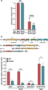
A. RcsD T411A mutation blocks induction by dsbA: For the PrprA::mCherry assay, the cells were grown in MOPS minimal glucose medium at 37°C. The RFU at OD 0.4 as compared to the uninduced WT control, set to 1, is depicted here. The cells were treated with either 1mM DTT or 20μg/ml PMBN. Details of the assay are described in Materials and Methods. B. DjlA, but not DrpB, can overcome the RcsD T411A mutation for Rcs activation: For the PrprA::mCherry assay, the strains overexpressing DjlA (pDjlA/pPSG961) or DrpB (pDrpB/pDSW1977) were grown in MOPS minimal glycerol medium containing chloramphenicol (25 μg/ml) and either 0.2% glucose or 0.02% arabinose at 37°C. The RFU at OD 0.4 compared to the vector uninduced control, set to 1, is plotted. The strains used were: WT (EAW8), dsbA::kan (EAW62), ΔrcsD (EAW19), ΔdsbA rcsD541::kan (AP13), rcsD T411A (EAW121), and rcsD T411A dsbA::kan (AP14). Statistical significance was calculated using multiple unpaired t-tests and is shown as follows: ns (P > 0.05; non- significant), * (P < 0.05), ** (P ≤ 0.01), and **** (P ≤ 0.0001).
Fig 4
Fig 4. DsbA is needed for efficient IgaA-RcsD periplasmic interactions.
A. IgaA-RcsD interactions are weakened in ΔdsbA. Beta-galactosidase activity was measured in a cyaA mutant strain (BTH101 or AP 58 (BTH101 ΔdsbA)) expressing two plasmids encoding the T18 and T25 domains of adenylate cyclase fused to the proteins of interest and the expression measured compared to vector background. The IgaA/RcsD protein fusion plasmids paired with their cognate vector had very low activity; the three controls were averaged and used as “background” for normalization. The IgaA-RcsD interaction was normalized to 1 and other interactions were plotted relative to this interaction in WT. In both Fig 4A and 4B, the interaction of IgaA-RcsD was 577 units, while the vector control was 29; these units are 1000x the slope of OD420 (see Materials and Methods). This data is compiled from separate sets of assays, each normalized relative to the IgaA/RcsD signal in that experiment. The P values are relative to the dsbA+ values. B. Periplasmic interactions are affected in ΔdsbA and a T411A mutation strengthens this IgaA interaction. IgaA and RcsD/T411A were fused to the T18 and T25 domains, respectively. The IgaA-RcsD interaction in WT was normalized to 1 and all other interactions are plotted relative to this interaction. The RcsD T411A mutation tightens the IgaA-RcsD interaction in WT as well as ΔdsbA. The plasmids used were pEAW1 (IgaA-T18), pEAW8 (RcsD-T25), pEAW2 (IgaA-T25), pEAW7 (RcsD-T18), pAP101 (IgaA Δcyt), pEAW1peri (IgaA Δperi), and pEAW8T (RcsD T411A-T25). Values (mean ± SD) from independent experiments were statistically analyzed using multiple unpaired t-tests. Statistical significance is shown as follows: ns (P > 0.05; non- significant), *** (P ≤ 0.001), and **** (P ≤ 0.0001).
Fig 5
Fig 5. DjlA weakens IgaA-RcsD interactions.
A. DjlA loosens IgaA-RcsD interactions: Beta-galactosidase activity was measured in a cyaA mutant strain (BTH101) in the presence of the indicated plasmids. IgaA and RcsD/RcsD T411A were fused to the T18 and T25 domains, respectively. DjlA or the H233Q mutant of DjlA, inactive as a co-chaperone, was cloned downstream of IgaA under the same promoter control. The IgaA-RcsD interaction was normalized to 1 and all other interactions are plotted relative to this interaction. In both Fig 5A and B, the interaction of IgaA-RcsD was 545 units, while the vector control was 18; these units are 1000x the slope of OD420 (see Materials and Methods). B. DjlA can disrupt cytoplasmic interactions of IgaA and RcsD: The IgaA-RcsD interaction was normalized to 1 and all other interactions are plotted relative to this interaction. C. RcsB, RcsC, RcsF are not needed for DjlA to weaken interactions. The IgaA-RcsD interaction was tested in BTH101 (WT), EAW 1 (BTH 101 rcsB::tet), EAW2 (BTH 101 rcsC::tet) and EAW4 (BTH 101 rcsF::cat). Plasmids used in this set of experiments were pEAW1 (IgaA-T18), pEAW8 (RcsD-T25), pEAW8T (RcsD T411A-T25), pAP804 (RcsD T411A Δperi -T25), pAP1401 (IgaA-T18 + DjlA), and pAP1402 (IgaA-T18 + DjlA H233Q). The WT interaction here was 533 units, with the vector control as 22 units. Statistical significance was calculated using multiple unpaired t-tests and is shown as follows: ** (P ≤ 0.01), *** (P ≤ 0.001), and **** (P ≤ 0.0001).
Fig 6
Fig 6. DrpB signaling is independent of ftsEX.
A. Role of DrpB as a ftsEX suppressor is independent of its role as an Rcs activator: Strains transformed with the pBAD33 vector or pDrpB (pDSW1977) were grown overnight in LB Miller media with chloramphenicol at 37°C. The cultures were diluted to an OD600 of 1 and 4 ul dilutions were spotted on LB Miller (permissive), LB without NaCl, and LB with 0.2% arabinose with no NaCl. All plates contained chloramphenicol (25 μg/ml). Plates were imaged after 16h incubation at 37°C. The strains used are: WT (EC251), rcsB::kan (AP158, ΔrcsB), ΔftsEX (EC1215), and ΔftsEX rcsB::kan (AP159, ΔftsEX ΔrcsB). B. DrpB can activate Rcs signaling in ΔftsEX: For the PrprA::mCherry assay, the WT (EAW8) and ftsE::kan (AP154, ΔftsEX) strains transformed with the pBAD33 vector or pDrpB (pDSW1977) were grown in MOPS minimal glycerol medium (with 150 mM NaCl) containing chloramphenicol and either 0.2% glucose or 0.02% arabinose at 37°C. The RFU at OD 0.4 is plotted relative to uninduced WT with the vector, set to 1. Statistical significance was calculated using multiple unpaired t-tests and is shown as follows: ns (P > 0.05; non- significant),** (P ≤ 0.01), *** (P ≤ 0.001).
Fig 7
Fig 7. Diverse modes of RcsF-independent activation by DsbA, DjlA, and DrpB.
First, dsbA mutants activate the Rcs phosphorelay in response to DTT and PMBN. The absence of DsbA likely leads to defective disulfide bond formation and misfolding of the IgaA periplasmic domain. Thus, DsbA is needed to maintain proper IgaA-RcsD interactions in the periplasm and in its absence, IgaA cannot effectively regulate Rcs signaling. Second, DjlA acts as a cochaperone for IgaA-RcsD interactions. Overexpression of DjlA weakens the IgaA-RcsD interactions in the cytoplasm leading to Rcs activation. Third, DrpB overexpression induces the Rcs cascade but it does not act directly on IgaA-RcsD interactions like DsbA and DjlA. Uniquely, DrpB requires the RcsC periplasmic domain to activate Rcs signaling and either activates it by direct interaction with RcsC or via an indirect mechanism involving RcsC as the sensor instead of RcsF.

Update of

Similar articles

Cited by

References

    1. Silhavy TJ, Kahne D, Walker S. The bacterial cell envelope. Cold Spring Harb Perspect Biol. 2010;2(5):a000414. Epub 20100414. doi: 10.1101/cshperspect.a000414 ; PubMed Central PMCID: PMC2857177. - DOI - PMC - PubMed
    1. Wall E, Majdalani N, Gottesman S. The Complex Rcs Regulatory Cascade. Annu Rev Microbiol. 2018;72:111–39. Epub 20180613. doi: 10.1146/annurev-micro-090817-062640 . - DOI - PubMed
    1. Guo XP, Sun YC. New Insights into the Non-orthodox Two Component Rcs Phosphorelay System. Front Microbiol. 2017;8:2014. Epub 20171017. doi: 10.3389/fmicb.2017.02014 ; PubMed Central PMCID: PMC5651002. - DOI - PMC - PubMed
    1. Watanabe N, Savchenko A. Molecular insights into the initiation step of the Rcs signaling pathway. Structure. 2024. Epub 20240625. doi: 10.1016/j.str.2024.06.003 . - DOI - PubMed
    1. Rogov VV, Rogova NY, Bernhard F, Löhr F, Dötsch V. A disulfide bridge network within the soluble periplasmic domain determines structure and function of the outer membrane protein RCSF. J Biol Chem. 2011;286(21):18775–83. Epub 20110406. doi: 10.1074/jbc.M111.230185 ; PubMed Central PMCID: PMC3099694. - DOI - PMC - PubMed

MeSH terms

LinkOut - more resources