. 2024 Dec 26;19(12):e0311976.
doi: 10.1371/journal.pone.0311976. eCollection 2024.

Sub-toxic cisplatin concentrations induce extensive chromosomal, nuclear and nucleolar abnormalities associated with high malignancy before acquired resistance develops: Implications for clinical caution

Affiliations

Sub-toxic cisplatin concentrations induce extensive chromosomal, nuclear and nucleolar abnormalities associated with high malignancy before acquired resistance develops: Implications for clinical caution

John G Delinassios et al. PLoS One. .

Abstract

Aim: This study investigates the impact of sub-toxic cisplatin levels on nuclear and nucleolar abnormalities and chromosome instability in HeLa cells since our current knowledge of cisplatin effects on these parameters is based on studies with high concentrations of cisplatin.

Materials and methods: HeLa cells were exposed to gradually increasing sub-toxic doses of cisplatin (0.01 to 0.2 μg/ml). Cells treated with 0.1 and 0.2 μg/ml, termed HeLaC0.1 and HeLaC0.2, were not cisplatin-resistant, only exhibiting a slightly reduced viability, and were termed "cisplatin-sensitized cells." Giemsa and silver staining were used to detect nuclear and nucleolar abnormalities and chromosomal alterations.

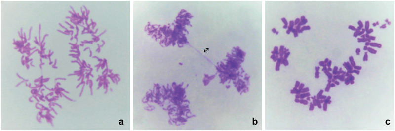
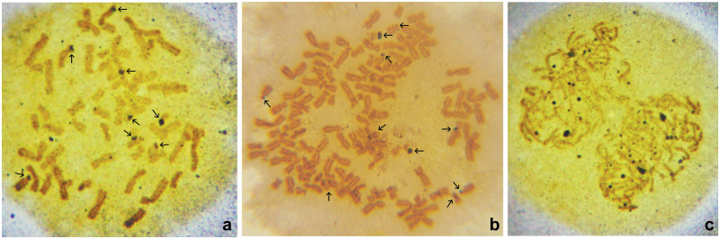
Results: Notable abnormalities were observed in HeLaC0.1 and HeLaC0.2 cells after treatment with sub-toxic concentrations of cisplatin: nuclei showed abnormal shapes, blebs, micronuclei, fragmentation, pulverization, and multinucleation; nucleoli exhibited irregular shapes and increased numbers; anaphase cells showed more nucleolar organizing regions. Abnormal chromosome segregation, heightened aneuploidy (81-140 chromosomes), polyploidy, double minutes, dicentrics, chromatid exchanges, chromatid separations, pulverization, and chromosome markers were prominently noted. These abnormalities were intensified in cells pre-sensitized to 0.02 or 0.08 μg/ml cisplatin for seven days, then exposed to 0.03 or 0.1 μg/ml cisplatin for 24 hours, and finally cultured in cisplatin-free medium for 24 hours before chromosome analysis.

Conclusion: HeLa cells subjected to increasing concentrations of sub-toxic cisplatin exhibited large-scale, multiple-type abnormalities in nuclei, nucleoli, chromosomes, and chromosomal numbers, indicating genetic/chromosomal instability associated with high malignancy, before the development of cisplatin resistance. These results suggest that low doses of cisplatin administration in the clinical setting may promote malignancy and caution should be used with this type of treatment.


Conflict of interest statement

The authors have declared that no competing interests exist.

Figures

Fig 1
Fig 1. Morphological features of nuclei from untreated and low-concentration cisplatin-treated HeLa cells.
a: Untreated HeLa parental cells. b: HeLaCO.1. c: HeLaCO.2. d: HeLa cells sensitized to 0.02 μg/ml and transferred to 0.03 μg/ml cisplatin. e: Multinucleated HeLa cells with abnormal and fragmented nuclei after transfer from 0.02 to 0.03 μg/ml cisplatin. f: Abnormal HeLa nuclei with blebs, chromatin bridges and micronuclei after transfer from 0.02 to 0.03 μg/ml cisplatin. g: Abnormal HeLa nuclei with blebs, chromatin bridges, and micronuclei after transfer from 0.08 to 0.1 μg/ml cisplatin. h: Multinucleated HeLaCO.1 cell with chromatin bridges, and micronuclei. i: Multinucleated HeLaCO.2 cells with abnormal nuclei and four large-size micronuclei. Giemsa; a, b, c, d, g, ×220; e, h ×420; f, i, ×1,000. Marks in the figures: ➝: blebs; ⇒: abnormal nuclear shape; ↔: chromatin bridge; *: multinucleated cell; +: micronucleus.
Fig 2
Fig 2. Abnormal chromosomal segregation.
a: HeLaC0.1, chromosomal segregation following tetrapolar mitosis. b: HeLaC0.1, abnormal tripolar mitosis with anaphase bridge (↔). c: HeLaC0.2, segregation of metaphase chromosomes.
Fig 3
Fig 3. Morphological features of nucleoli.
a: HeLa cells. b: HeLaC0.1 cells. c: HeLaC0.2 cells. d: Nucleoli in HeLa cells sensitized to 0.02 and transferred to 0.03 μg/ml cisplatin. Over 20 dots of nucleolar organizing regions (NORs) can be seen in the nucleus at the lower left corner. e: HeLaC0.2 cells. Left, a nucleus with over 20 NOR dots. Right, late anaphase with over 20 NOR dots in the newly formed nuclei. f: Nucleus of a HeLa cell sensitized to 0.08 and transferred to 0.1 μg/ml cisplatin. Over 25 nucleoli are visible with variable sizes, appearing as single dots or assemblies of 2–3 dots. Silver staining; a, ×100; b, ×110; c, ×110; d, ×500; e, ×700; f, ×1,200.
Fig 4
Fig 4. Nucleolar organizing regions (NORs) on chromosomes.
a: Metaphase of a HeLa cell sensitized to 0.02 and transferred to 0.03 μg/ml cisplatin. Distinctly activated NORs are present on eight acrocentric chromosomes (arrows), with one of them being the most intensely stained on a small acrocentric chromosome. b: Metaphase of a HeLaC0.2 cell showing 10 NORs (arrows), with two of them intensely stained on two small acrocentric chromosomes. c: Early anaphase of a HeLa cell sensitized to 0.02 and transferred to 0.03 μg/ml cisplatin. Over 24 NORs of various sizes are visible, with two of them being more intensely stained.
Fig 5
Fig 5. Chromosomal abnormalities.
a: Metaphase of a cell sensitized to 0.02 and transferred to 0.03 μg/ml cisplatin, containing two dicentric chromosomes (single arrows), one marker (double arrow), and numerous double minutes (DMs) (asterisk). b: Metaphase of a cell sensitized to 0.08 and transferred to 0.1 μg/ml cisplatin, containing numerous DMs, two marker chromosomes (arrows) and chromosome segregations. c: Metaphase of a HeLa cell sensitized to 0.02 μg/ml cisplatin, containing two marker chromosomes (short arrows) and one DM with two far-separated dots (arrows). d: Metaphase of a HeLa cell sensitized to 0.02 μg/ml cisplatin, containing three large DMs (asterisks) and one marker chromosome (arrow). e: Metaphase of a HeLa cell sensitized to 0.08 μg/ml cisplatin, containing sister chromatid separations. f: Metaphase of a HeLa cell sensitized to 0.08 μg/ml cisplatin, with two DMs (long arrows), one marker chromosome (short arrow), and several chromosomes with a centromere separation. g: Metaphase of a HeLa cell sensitized to 0.07 μg/ml cisplatin, showing a ring chromosome (arrow), sister-chromatid separations, and unaffected chromosomes. h: Metaphase of a HeLa cell sensitized to 0.02 μg/ml cisplatin, showing sister-chromatid separations, chromosome pulverization, and chromosome segregations. i: Metaphase of a HeLa cell sensitized to 0.02 μg/ml cisplatin, showing over 600 chromosomes and chromosomal segregations.
Fig 6
Fig 6. Metaphases with marker chromosomes.
a: HeLa cell (control) metaphase, with two associated small acrocentric chromosomes (arrow). b: HeLaC0.2 cell metaphase containing two marker chromosomes (arrows). c: HeLaC0.2 cell metaphase containing one dicentric (asterisk) and one marker chromosome (arrow). d: Metaphase of a HeLa cell sensitized to 0.02 μg/ml cisplatin, containing one marker chromosome (arrow). e: Metaphase of a HeLa cell sensitized to 0.02 and transferred to 0.03 μg/ml cisplatin, containing two marker chromosomes (arrows). f: Metaphase of a HeLa cell sensitized to 0.08 μg/ml and transferred to 0.1 μg/ml cisplatin, containing numerous double minutes (DMs) and two marker chromosomes (arrows). g: Metaphase of a HeLa cell sensitized to 0.08 μg/ml and transferred to 0.1 μg/ml cisplatin, containing one marker chromosome with extended long arm (arrow). h: Metaphase of a HeLa cell sensitized to 0.02 μg/ml cisplatin, with over 140 chromosomes including DMs and more than four marker chromosomes (arrows). i: G-banding of metaphase of a HeLa cell sensitized to 0.08 μg/ml cisplatin, showing two marker chromosomes (arrows).

Similar articles

References

    1. Stewart DJ. Mechanisms of resistance to cisplatin and carboplatin. Crit Rev Oncol Hematol. 2007;63(1):12–31. doi: 10.1016/j.critrevonc.2007.02.001 - DOI - PubMed
    1. Shen DW, Pouliot LM, Hall MD, Gottesman MM. Cisplatin resistance: A cellular self-defense mechanism resulting from multiple epigenetic and genetic changes. Pharmacol Rev. 2012;64(3):706–721. doi: 10.1124/pr.111.005637 - DOI - PMC - PubMed
    1. Wang D, Lippard SJ. Cellular processing of platinum anticancer drugs. Nat Rev Drug Discov. 2005;4(4):307–320. doi: 10.1038/nrd1691 - DOI - PubMed
    1. Skowron MA, Melnikova M, van Roermund JGH, Romano A, Albers P, Thomale J, et al.. Multifaceted mechanisms of cisplatin resistance in long-term treated urothelial carcinoma cell lines. Int J Mol Sci. 2018;19(2). doi: 10.3390/ijms19020590 - DOI - PMC - PubMed
    1. Zdraveski ZZ, Mello JA, Marinus MG, Essigmann JM. Multiple pathways of recombination define cellular responses to cisplatin. Chem Biol. 2000;7(1):39–50. doi: 10.1016/s1074-5521(00)00064-8 - DOI - PubMed

MeSH terms

LinkOut - more resources