. 2024 Dec 26;135(4):e183592.
doi: 10.1172/JCI183592.

Tagless LysoIP for immunoaffinity enrichment of native lysosomes from clinical samples

Affiliations

Tagless LysoIP for immunoaffinity enrichment of native lysosomes from clinical samples

Daniel Saarela et al. J Clin Invest. .

Abstract

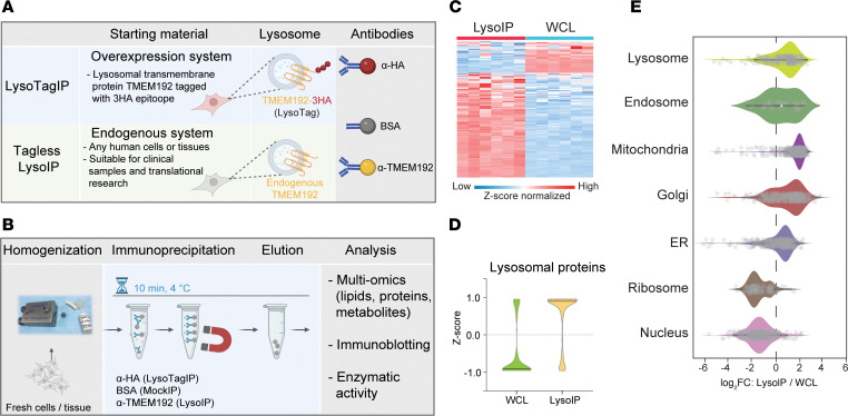
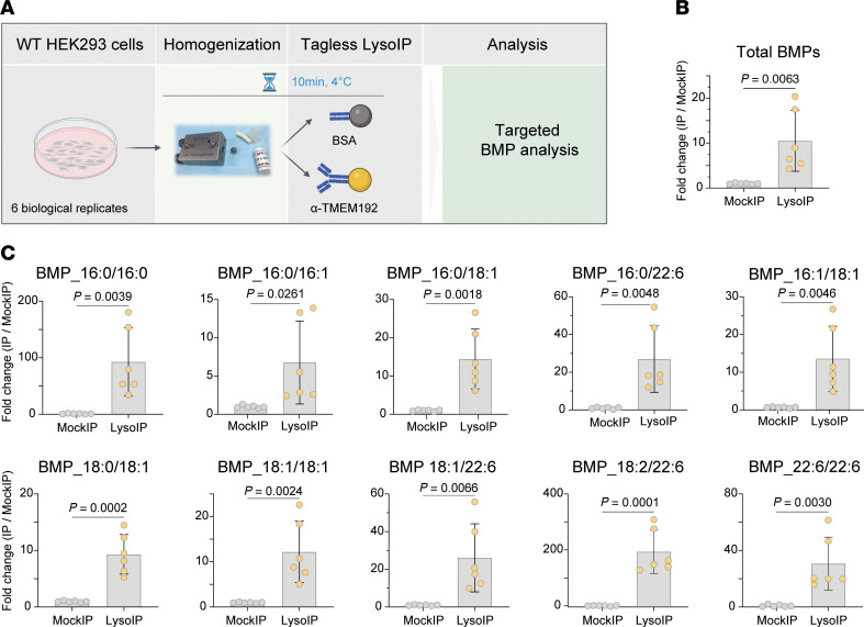
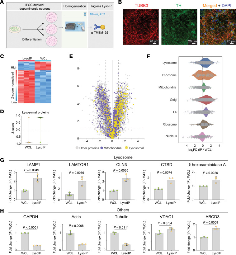
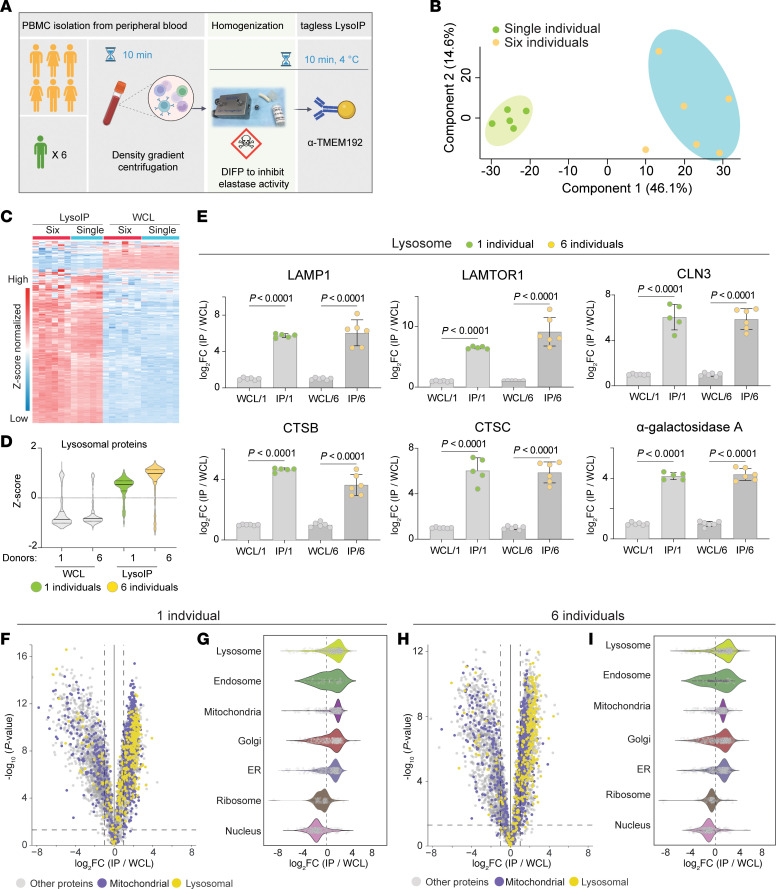
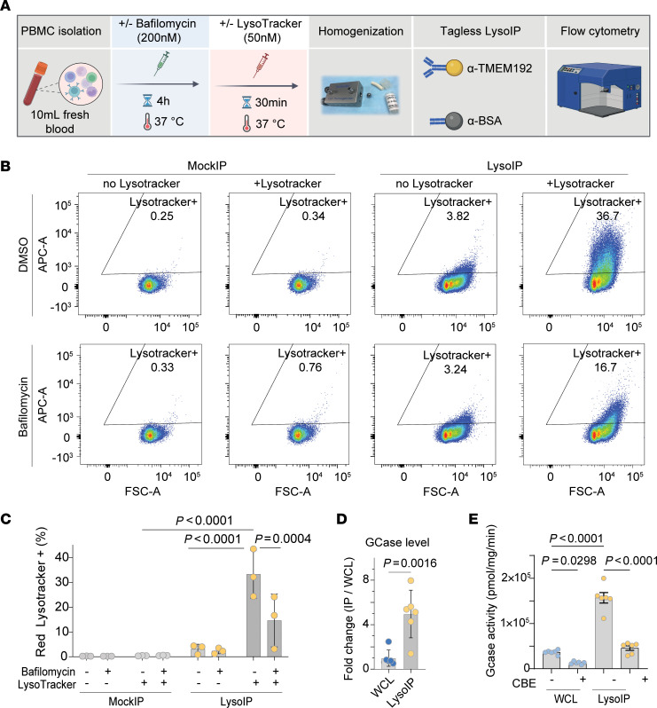
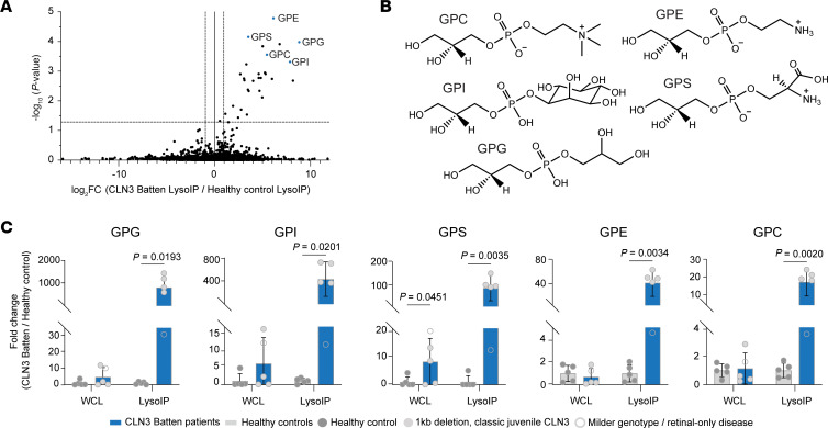
Lysosomes are implicated in a wide spectrum of human diseases, including monogenic lysosomal storage disorders (LSDs), age-associated neurodegeneration, and cancer. Profiling lysosomal content using tag-based lysosomal immunoprecipitation (LysoTagIP) in cell and animal models has substantially moved the field forward, but studying lysosomal dysfunction in patients remains challenging. Here, we report the development of the 'tagless LysoIP' method, designed to enable the rapid enrichment of lysosomes, via immunoprecipitation, using the endogenous integral lysosomal membrane protein TMEM192, directly from clinical samples and human cell lines (e.g., induced pluripotent stem cell-derived neurons). Isolated lysosomes were intact and suitable for subsequent multimodal omics analyses. To validate our approach, we applied the tagless LysoIP to enrich lysosomes from peripheral blood mononuclear cells derived from fresh blood of healthy donors and patients with CLN3 disease, an autosomal recessive neurodegenerative LSD. Metabolic profiling of isolated lysosomes revealed massive accumulation of glycerophosphodiesters (GPDs) in patients' lysosomes. Interestingly, a patient with a milder phenotype and genotype displayed lower accumulation of lysosomal GPDs, consistent with their potential role as disease biomarkers. Altogether, the tagless LysoIP provides a framework to study native lysosomes from patient samples, identify disease biomarkers, and discover human-relevant disease mechanisms.

Keywords: Cell biology; Genetic diseases; Lysosomes; Neurodegeneration; Neuroscience.


Conflict of interest statement

Conflict of interest: The authors have declared that no conflict of interest exists.

Figures

Figure 1
Figure 1. Tagless LysoIP for enriching lysosomes from clinical samples.
(A) Concept of the tagless LysoIP method via immunoprecipitation of the lysosomal transmembrane TMEM192 protein compared with the LysoTag system, which relies on overexpression of 3HA epitopes at the TMEM192 C-terminus. (B) Tagless LysoIP workflow. (C) Protein profile heatmap and (D) Violin plots of lysosomal proteins enriched via the tagless LysoIP in the immunoprecipitates and whole cell lysates from WT HEK293 cells. (E) Organelle profiling demonstrates marked enrichment of lysosomal but also nonlysosomal proteins.
Figure 2
Figure 2. Bis(monoacylglycero)phosphate lipids are enriched in LysoIP samples from WT HEK293 cells.
(A) Experimental design to study lysosomal lipids using tagless LysoIP followed by targeted Bis(monoacylglycero)phosphate (BMP) analysis. (B) Targeted analysis of accumulated BMPs in lysosomes derived from WT HEK293 cells using tagless LysoIP (n = 6). Relative enrichment of total BMPs quantified in the LysoIP samples and the MockIP samples. Data presented as mean ± SD (n = 6). Statistical analysis was performed using 2-tailed unpaired t test. (C) Enrichment of specific BMP species in the LysoIP samples.
Figure 3
Figure 3. Tagless LysoIP from human iPSC-derived dopaminergic neurons.
(A) Workflow. (B) 78-day–old iPSC-derived dopaminergic neurons were stained for tyrosine hydroxylase ([TH] green), a marker of dopaminergic neurons, TUBB3 (red), a neuronal-specific βIII tubulin marker, and DAPI (blue), indicating nuclear DNA. (C) Protein profile heatmap. (D) Violin plots of lysosomal proteins enriched via the tagless LysoIP in the immunoprecipitates from iPSC-derived dopaminergic neurons. (E) Volcano plot of proteins enriched/depleted in the LysoIPs compared with whole cell extracts. Data were analyzed via Curtain: https://curtain.proteo.info/#/cefde280-9d48-4157-b959-cf2c3b0cc9e7 (F) Organelle profiling demonstrates enrichment of lysosomal proteins and modest enrichment of other organelles. Bar plots of representative (G) lysosomal proteins and (H) cytosolic, mitochondrial, and Golgi proteins in LysoIPs and their respective whole cell extracts. Data presented as mean ± SD (n = 3). Statistical analysis was performed using 2-tailed unpaired t test.
Figure 4
Figure 4. Tagless LysoIP in human peripheral blood mononuclear cells.
(A) Workflow (B) Principal component analysis of DIA mass spectrometry data of tagless LysoIPs from PBMCs from a single donor (6 replicates) and 6 different donors. (C) Protein profile heatmap. (D) Lysosomal protein enrichment. (E) Bar plots of representative lysosomal transmembrane (top panel) and intraluminal (lower panel) proteins in LysoIPs from 1 and 6 donors compared with the respective whole cell extracts. Data presented as mean ± SD (n = 6). Multiple unpaired t tests with 2-stage step-up method of Benjamini, Krieger, and Yekutieli (1% FDR) used to correct for multiple comparisons between the groups. Volcano and violin plots of LysoIPs compared with whole cell extracts and organelle profiling from the single (F and G) and multiple (H and I) donor experiments. Data were analyzed via Curtain: https://curtain.proteo.info/#/b573afb8-df5d-4a39-b5ba-88eb9488820d (F), https://curtain.proteo.info/#/83006f89-901d-44db-b078-5b8504844ee6 (H).
Figure 5
Figure 5. Tagless LysoIP enriches intact and functional lysosomes from healthy donor PBMCs.
(A) Workflow of flow cytometry analysis of magnetic beads bound to lysosomes enriched via tagless LysoIPs or MockIPs from PBMC homogenates pretreated with and without the V-ATPase inhibitor bafilomycin A1 (200 nM), prior to staining with Red Lysotracker (50 nM). (B) Representative scatter plot from 1 of the 3 donors, (Y-axis) and bead size (Forward scatter, FSC, X-axis), (C) Quantification of the percentage of beads positive for Lysotracker fluorescence. Data presented as mean ± SD (n = 3 donors) and analysed by ordinary 2-way ANOVA with Dunnet’s multiple comparison test. (D) GCase (glucocerebrosidase, GBA1) protein enrichment quantified by DIA mass-spectrometry in LysoIPs from 6 donors. Data presented as mean ± SD (n = 6). 2-tailed unpaired t test. (E) GCase activity measured by 4-methylumbelliferone (4-MU) assay in lysosomes enriched from PBMCs from 6 donors. Data presented as mean ± SD and 2-way ANOVA with Dunnet’s test for multiple comparison.
Figure 6
Figure 6. Striking GPD accumulation in lysosomes enriched from patients with CLN3 disease.
(A) Volcano plot comparing untargeted metabolomic LysoIP data derived from PBMCs from patients with CLN3 and healthy controls. (B) The chemical structure of the annotated GPDs in this study. (C) Targeted analyses of GPDs in the LysoIPs and corresponding WCLs. Data presented as mean ± SD (n = 5). Multiple unpaired t tests with 2-stage step-up method of Benjamini, Krieger, and Yekutieli (1% FDR) used to correct for multiple comparisons between the groups.

Comment in

  • Purifying and profiling lysosomes to expand understanding of lysosomal dysfunction–associated diseases

References

    1. Settembre C, et al. Signals from the lysosome: a control centre for cellular clearance and energy metabolism. Nat Rev Mol Cell Biol. 2013;14(5):283–296. doi: 10.1038/nrm3565. - DOI - PMC - PubMed
    1. Xu H, Ren D. Lysosomal physiology. Annu Rev Physiol. 2015;77:57–80. doi: 10.1146/annurev-physiol-021014-071649. - DOI - PMC - PubMed
    1. Ballabio A, Bonifacino JS. Lysosomes as dynamic regulators of cell and organismal homeostasis. Nat Rev Mol Cell Biol. 2020;21(2):101–118. doi: 10.1038/s41580-019-0185-4. - DOI - PubMed
    1. Platt FM, et al. Lysosomal storage diseases. Nat Rev Dis Primers. 2018;4(1):27. doi: 10.1038/s41572-018-0025-4. - DOI - PubMed
    1. Perera RM, Zoncu R. The lysosome as a regulatory hub. Annu Rev Cell Dev Biol. 2016;32:223–253. doi: 10.1146/annurev-cellbio-111315-125125. - DOI - PMC - PubMed

MeSH terms