. 2024 Dec 26;20(12):e1011518.
doi: 10.1371/journal.pgen.1011518. eCollection 2024 Dec.

Decreased Hsp90 activity protects against TDP-43 neurotoxicity in a C. elegans model of amyotrophic lateral sclerosis

Affiliations

Decreased Hsp90 activity protects against TDP-43 neurotoxicity in a C. elegans model of amyotrophic lateral sclerosis

Laura Garcia-Toscano et al. PLoS Genet. .

Abstract

Neuronal inclusions of hyperphosphorylated TDP-43 are hallmarks of disease for most patients with amyotrophic lateral sclerosis (ALS). Mutations in TARDBP, the gene coding for TDP-43, can cause some cases of familial inherited ALS (fALS), indicating dysfunction of TDP-43 drives disease. Aggregated, phosphorylated TDP-43 may contribute to disease phenotypes; alternatively, TDP-43 aggregation may be a protective cellular response sequestering toxic protein away from the rest of the cell. The heat shock responsive chaperone Hsp90 has been shown to interact with TDP-43 and stabilize its normal conformation; however, it is not known whether this interaction contributes to neurotoxicity in vivo. Using a C. elegans model of fALS mutant TDP-43 proteinopathy, we find that loss of function of HSP-90 protects against TDP-43 neurotoxicity and subsequent neurodegeneration in adult animals. This protection is accompanied by a decrease in both total and phosphorylated TDP-43 protein. We also find that hsp-90 mutation or inhibition upregulates key stress responsive heat shock pathway gene expression, including hsp-70 and hsp-16.1, and we demonstrate that normal levels of hsp-16.1 are required for hsp-90 mutation effects on TDP-43. We also observe that the neuroprotective effect due to HSP-90 dysfunction does not involve direct regulation of proteasome activity in C. elegans. Our data demonstrate for the first time that Hsp90 chaperone activity contributes to adverse outcomes in TDP-43 proteinopathies in vivo using a whole animal model of ALS.


Conflict of interest statement

The authors have declared that no competing interests exist.

Figures

Fig 1
Fig 1. HSP-90 mutation or inhibition protects against hTDP-43 driven motor dysfunction in C. elegans.
A. Radial locomotion assays were used to measure motor function following temperature-based inactivation of HSP-90. Animals were shifted to the restrictive temperature at L4 stage and assessed after 24 hours of HSP-90 inactivation. Animals expressing fALS TDP-43(M337V) combined with the hsp-90(p673) mutation show an improvement in their motility. Error bars represent Mean with 95% confidence interval (CI): N = 3 independent experimental replicates; total n>100. Statistical significance as determined using Student’s t-test. (**** p<0.0001). B. hsp-90(p673) animals did not move significantly differently from wildtype (N2) animals, which were used as non-transgenic (non-Tg) controls. N = 3 independent experimental replicates; total n>100. Statistical significance as determined using Student’s t-test. (ns = not significant). C-D. Radial locomotion assays were used to measure motor function following pharmacological inhibition of HSP-90. Treatment with the Hsp90 inhibitor 17-AAG improves motor dysfunction caused by the expression of mutant hTDP-43 in C. elegans. Two independent transgenic strains, expressing either TDP-43(M337V) (C) or TDP-43(A315T) (D) exhibit improved motility after 48h of 17-AAG treatment (7.5 μM) when scored at 30 minutes, but this effect diminishes or is lost by 60 minutes or 24 hours post-removal of drug. Error bars represent Mean with 95% CI. N = 4; n>100 for the TDP-43(M337V) treated worms, and n>100 when using the TDP-43(A315T) strain. Statistical significance as determined using a Mixed-effects analysis (Bonferroni’s multiple comparisons test post hoc test). (*** p<0.001; *p<0.05). E-F. Control worms showed no difference in their motility assessment after 48h of treatment with 17-AAG (7.5 μM). Results shown combined data from multiple experiments. Error bars represent Mean with 95% CI. N = 3; n>100. Statistical significance was determined using a Mixed-effects analysis (Bonferroni’s multiple comparisons test post hoc test). G. Radial locomotion assays were used to measure motor function in older TDP-43(M337V) at day 3, 5, or 7 of adulthood following a 24 hour temperature-based inactivation of HSP-90. TDP-43(M337V);hsp-90(p673) animals show an improvement in their motility at day 3 of adulthood, but this is lost at days 5 and 7. Error bars represent Mean with 95% CI: N = 3 independent experimental replicates; total n>65. Statistical significance as determined using Student’s t-test. (**** p<0.0001).
Fig 2
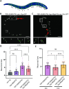
Fig 2. hsp-90 mutation rescues neurodegeneration observed in TDP-43(M337V) transgenic animals.
GABAergic neurons marked by GFP (unc-25p::GFP) were counted in adult transgenic animals after a 24h incubation at 25°C. A. Cartoon model shows the positions of 19 GABAergic neurons scored using the GFP reporter. Fig 2A was created in BioRender (2024) https://BioRender.com/g76d973. B-C. Representative images from B. TDP-43(M337V) and C. TDP-43(M337V);hsp-90(p673) animals. White arrows indicate degenerating neurons; white stars indicate dead neurons. Scale bar = 75μm. D. Quantification of neuronal loss following a shift to the restrictive temperature of 25°C. Neurons were counted and the total number was subtracted from 19, the average number in a developmentally normal wild-type animal, to obtain the number of neurons lost. Some animals were scored with greater than 19 neurons, which may represent developmental variants or GFP artifacts, and resulted in a negative number of neurons lost. Error bars represent SEM: N = 3; n = 58–61. E. Quantification of neuronal loss without a shift to the restrictive temperature. Error bars represent SEM: N = 2; n = 45–49. Statistical significance was determined using a non-parametric Kruskal-Wallis test (Dunn’s multiple comparisons post hoc test). (**** p<0.0001; **p<0.01).
Fig 3
Fig 3
Hsp90 mutation or inhibition decreases accumulation of phosphorylated TDP-43 A. Representative immunoblots for total and phosphorylated TDP-43 in C. elegans samples. B-C. Mutant hsp-90 significantly decreases the levels of total and phosphorylated species of TDP-43 (pTDP-43). Error bars represent SEM: N = 4. Statistical significance as determined using unpaired t-test. (*p<0.05, **p< 0.01) D. The ratio of phosphorylated TDP-43 to total TDP-43 (pTDP-43/ total TDP-43) is unchanged in TDP43(M337V) versus TDP43(M337V);hsp-90(p673). E. Representative immunoblots for endogenous total and phosphorylated TDP-43 in HEK293T cells pre-treated with increasing amounts of the Hsp90 inhibitor 17-AAG and exposed to Ethacrynic acid 150μM. F-G. Quantification of replicate immunoblots shows no significant differences in total TDP-43 protein levels but a trend towards a reduction in the levels of pTDP-43 in a dose-dependent manner, reaching statistical significance at the highest dose tested, 10μM. Results combined data from multiple experiments. Error bars represent SEM. N = 3. Statistical significance was determined using the One-way ANOVA test (Dunnett’s multiple comparisons post hoc test) (* p<0.05).
Fig 4
Fig 4. hsp-90 mutation does not increase proteasome activation.
A. Representative photomicrographs of proteasome reporter rpt-3p::GFP near the tail of non-Tg, hsp-90(p673), TDP43(M337V), and TDP43(M337V);hsp-90(p673) animals. Scale bars = 10μm. B. TDP-43(M337V) expressing C. elegans increase rpt-3p::GFP reporter expression, but hsp-90 mutation does not affect reporter expression either independently or in combination with TDP-43(M337V). Error bars represent SEM: N = 3, n = 25–31. Statistical significance as determined using the non-parametric Kruskal-Wallis test (Dunn’s multiple comparisons post hoc test).
Fig 5
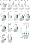
Fig 5
hsp-90 mutation selectively increases heat shock protein expression A-O. qRT-PCR was used to detect mRNA expression changes in heat shock proteins of interest. No significant expression differences were observed for hsf-1 (A), dnj-24 (C), dnj-14 (D), dnj-27 (E), hsp-4 (F), dnj-12 (G), hsp-25 (H), hsp-90 (I), enpl-1 (J), hsp-75 (K). Both hsp-70 (B) and hsp-16.1 (O) have increased expression in hsp-90(p673) and TDP43(M337V); daf-21(p673) animals. hsp-16.2 (L), hsp-16.48 (M), and hsp-16.41 (N) were increased in hsp-90(673) animals, but this expression increase was attenuated in TDP43(M337V); daf-21(p673). Error bars represent SEM. N = 3–4; Statistical significance as determined using the One-way ANOVA test (Tukey’s multiple comparisons post-hoc test) or a non-parametric Kruskal-Wallis test (Dunn’s multiple comparisons post hoc test) when required. (*p<0.05; **p<0.01; *** p<0.001). P. TDP43(M337V) and TDP43(M337V);hsp-90(p673) were treated with control (empty vector), hsp-70, or hsp-16.1 targeting RNAi via feeding exposure starting at hatching (L1 stage). At L4 stage, animals were shifted to the restrictive temperature (25°C) for 24 hours. Radial locomotion assays were used to measure motor function. Error bars represent Mean with 95% CI: N = 3 independent experimental replicates; total n>65. Statistical significance as determined using One-way ANOVA test (Tukey’s multiple comparisons post-hoc test). (** p<-.-1, **** p<0.0001, ns = not significant).

Similar articles

References

    1. Masrori P, Van Damme P. Amyotrophic lateral sclerosis: a clinical review. Eur J Neurol. 2020;27(10):1918–29. doi: 10.1111/ene.14393 - DOI - PMC - PubMed
    1. Smukowski SN, Maioli H, Latimer CS, Bird TD, Jayadev S, Valdmanis PN. Progress in Amyotrophic Lateral Sclerosis Gene Discovery: Reflecting on Classic Approaches and Leveraging Emerging Technologies. Neurol Genet. 2022;8(3):e669. doi: 10.1212/NXG.0000000000000669 - DOI - PMC - PubMed
    1. Marsh AP. Molecular mechanisms of proteinopathies across neurodegenerative disease: a review. Neurol Res Pract. 2019;1:35. doi: 10.1186/s42466-019-0039-8 - DOI - PMC - PubMed
    1. Prasad A, Bharathi V, Sivalingam V, Girdhar A, Patel BK. Molecular Mechanisms of TDP-43 Misfolding and Pathology in Amyotrophic Lateral Sclerosis. Front Mol Neurosci. 2019;12:25. doi: 10.3389/fnmol.2019.00025 - DOI - PMC - PubMed
    1. Ayala YM, De Conti L, Avendano-Vazquez SE, Dhir A, Romano M, D’Ambrogio A, et al.. TDP-43 regulates its mRNA levels through a negative feedback loop. EMBO J. 2011;30(2):277–88. doi: 10.1038/emboj.2010.310 - DOI - PMC - PubMed

MeSH terms

Substances