Review
. 2025 Mar 1;11(3):317-328.
doi: 10.1001/jamaoncol.2024.5506.

Molecular Testing for the World Health Organization Classification of Central Nervous System Tumors: A Review

Affiliations
Review

Molecular Testing for the World Health Organization Classification of Central Nervous System Tumors: A Review

Craig Horbinski et al. JAMA Oncol. .

Abstract

Importance: Molecular techniques, including next-generation sequencing, genomic copy number profiling, fusion transcript detection, and genomic DNA methylation arrays, are now indispensable tools for the workup of central nervous system (CNS) tumors. Yet there remains a great deal of heterogeneity in using such biomarker testing across institutions and hospital systems. This is in large part because there is a persistent reluctance among third-party payers to cover molecular testing. The objective of this Review is to describe why comprehensive molecular biomarker testing is now required for the accurate diagnosis and grading and prognostication of CNS tumors and, in so doing, to justify more widespread use by clinicians and coverage by third-party payers.

Observations: The 5th edition of the World Health Organization (WHO) classification system for CNS tumors incorporates specific molecular signatures into the essential diagnostic criteria for most tumor entities. Many CNS tumor types cannot be reliably diagnosed according to current WHO guidelines without molecular testing. The National Comprehensive Cancer Network also incorporates molecular testing into their guidelines for CNS tumors. Both sets of guidelines are maximally effective if they are implemented routinely for all patients with CNS tumors. Moreover, the cost of these tests is less than 5% of the overall average cost of caring for patients with CNS tumors and consistently improves management. This includes more accurate diagnosis and prognostication, clinical trial eligibility, and prediction of response to specific treatments. Each major group of CNS tumors in the WHO classification is evaluated and how molecular diagnostics enhances patient care is described.

Conclusions and relevance: Routine advanced multidimensional molecular profiling is now required to provide optimal standard of care for patients with CNS tumors.


Figures

Figure 1.
Figure 1.. Requirement of Advanced Molecular Testing to Accurately Classify and Treat Diagnostically Ambiguous Gliomas, Part 1
A woman in her late 40s presented with new onset of aphasia and left arm paresthesia. Preoperative T2-weighted fluid-attenuated inversion recovery (FLAIR) magnetic resonance imaging (MRI) showed a hyperintense lesion with minimal enhancement on postcontrast imaging centered in the left mesial temporal lobe and insula (arrowhead). Surgical resection of the lesion was performed that revealed a histologically low-grade glial or glioneuronal neoplasm (original magnification ×100), for a which a consensus diagnosis of ganglioglioma, World Health Organization grade 1 central nervous system tumor was favored after careful review of the clinical, imaging, and microscopic features, including an extensive immunohistochemical staining panel. The chromosomal copy number profile revealed gain/trisomy of chromosome 7, loss/monosomy of chromosome 10, focal homozygous/biallelic deletion of the CDKN2A and CDKN2B tumor suppressor genes on chromosome 9p21.3, and focal homozygous/biallelic deletion of the NF1 tumor suppressor gene on chromosome 17q11.2. See Figure 2 for the remaining elements to this case.
Figure 2.
Figure 2.. Requirement of Advanced Molecular Testing to Accurately Classify and Treat Diagnostically Ambiguous Gliomas, Part 2
A woman in her late 40s presented with new onset of aphasia and left arm paresthesia. A, Targeted next-generation DNA sequencing revealed a hotspot substitution variant (c.-124C>T) in the promoter region of the TERT gene and a deleterious missense variant (p.Y68H) in the PTEN tumor suppressor gene with loss of the remaining wild-type allele, along with the above-mentioned focal deletions involving CDKN2A/B and NF1. B, Genome-wide DNA methylation interrogation revealed an epigenomic profile that aligned with isocitrate dehydrogenase (IDH) wild-type glioblastoma (mesenchymal subclass). While the histologic features suggested a low-grade glial or glioneuronal neoplasm, such as ganglioglioma, the molecular features indicated an IDH wild-type glioblastoma and led to an accurate final integrated diagnosis of glioblastoma, IDH wild-type, central nervous system World Health Organization grade 4. Despite aggressive external beam radiation and chemotherapy, the patient experienced local disease recurrence at 16 months after initial diagnosis which then demonstrated conventional histologic features of glioblastoma (not shown). ID indicates identifier; ITD, internal tandem duplication; NA, not applicable; PXA, pleomorphic xanthoastrocytoma; RTK, receptor tyrosine kinase; t-SNE, t-distributed stochastic neighbor embedding; UMAP, uniform manifold approximation and projection.
Figure 3.
Figure 3.. Histone H3 K27M Glioma With Concomitant BRAF Alteration
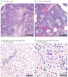
A man in his early 30s developed a mass in the pineal region that was resected. Initial hematoxylin-eosin staining was of a low-grade glioma (A; original magnification ×100) with low Ki67 proliferation index (B; original magnification ×100). Next-generation sequencing showed that the tumor had an H3-3A K27M alteration and an activating K601N alteration in BRAF. Despite the H3-3A variant normally associated with short patient survival, the tumor did not recur until 6 years later, with obvious progression in grade by hematoxylin-eosin staining (C; original magnification ×100) and Ki67 proliferation index (D; original magnification ×100).
Figure 4.
Figure 4.. Examples of Advanced Molecular Testing Enhancing Patient Care
A man in his mid-50s had a right-sided frontal parietal mass that, by histopathology using hematoxylin-eosin staining, had abundant geographic necrosis (A; original magnification ×40) and apparent perivascular pseudorosettes (B; original magnification ×40). Isocitrate dehydrogenase wild-type glioblastoma and grade 3 ependymoma were in the original differential diagnosis, but upon revisiting the case years later, next-generation sequencing detected a BRAF V600E variant and matched to pleomorphic xanthoastrocytoma by methylation profiling. Seven years after initial surgery, the tumor still had not recurred or spread elsewhere in the central nervous system. In another case, a woman in her mid-20s had a history of seizures since childhood that recently became uncontrollable with antiepileptic drugs. An ill-defined right-sided parietal mass was biopsied, and on hematoxylin-eosin staining, there were some gliomalike cells, perivascular inflammation, and reactive gliosis (C [original magnification ×100] and D [original magnification ×200]), but no mitoses, necrosis, or microvascular proliferation. The only abnormality detected by next-generation sequencing, copy number variant array, methylation profiling, and fusion screening was copy-neutral loss of heterozygosity involving 5q21.1-5q21.3. Thus, the final diagnosis was low-grade glioma vs glial-glioneuronal malformation, with the recommendation of watchful waiting and withholding adjuvant therapy until the lesion recurred.

References

    1. Price M, Ryan K, Shoaf ML, et al. Childhood, adolescent, and adult primary brain and central nervous system tumor statistics for practicing healthcare providers in neuro-oncology, CBTRUS 2015-2019. Neurooncol Pract. 2023;11(1):5–25. doi: 10.1093/nop/npad061 - DOI - PMC - PubMed
    1. Ostrom QT, Price M, Neff C, et al. CBTRUS statistical report: primary brain and other central nervous system tumors diagnosed in the United States in 2016-2020. Neuro Oncol. 2023;25(12)(suppl 2):iv1–iv99. doi: 10.1093/neuonc/noad149 - DOI - PMC - PubMed
    1. Burnet NG, Jefferies SJ, Benson RJ, Hunt DP, Treasure FP. Years of life lost (YLL) from cancer is an important measure of population burden--and should be considered when allocating research funds. Br J Cancer. 2005;92(2):241–245. doi: 10.1038/sj.bjc.6602321 - DOI - PMC - PubMed
    1. Yabroff KR, Lamont EB, Mariotto A, et al. Cost of care for elderly cancer patients in the United States. J Natl Cancer Inst. 2008;100(9):630–641. doi: 10.1093/jnci/djn103 - DOI - PubMed
    1. Yabroff KR, Lund J, Kepka D, Mariotto A. Economic burden of cancer in the United States: estimates, projections, and future research. Cancer Epidemiol Biomarkers Prev. 2011;20(10):2006–2014. doi: 10.1158/1055-9965.EPI-11-0650 - DOI - PMC - PubMed

MeSH terms

Substances