Review
. 2025 Apr 1;98(1168):485-495.
doi: 10.1093/bjr/tqae252.

The role and potential of digital breast tomosynthesis in neoadjuvant systemic therapy evaluation for optimising breast cancer management: a pictorial essay

Affiliations
Review

The role and potential of digital breast tomosynthesis in neoadjuvant systemic therapy evaluation for optimising breast cancer management: a pictorial essay

Luciano Mariano et al. Br J Radiol. .

Abstract

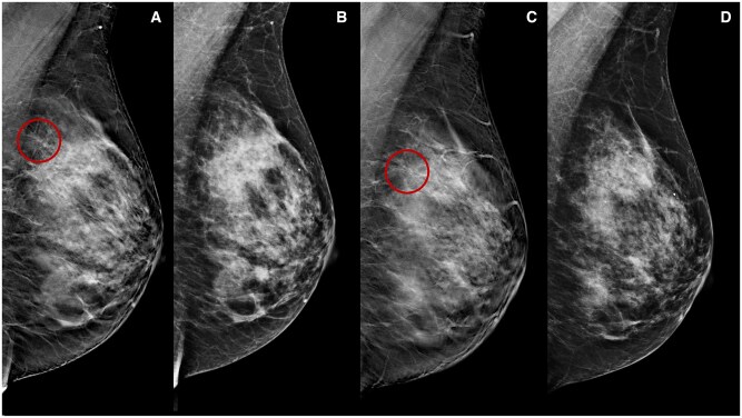
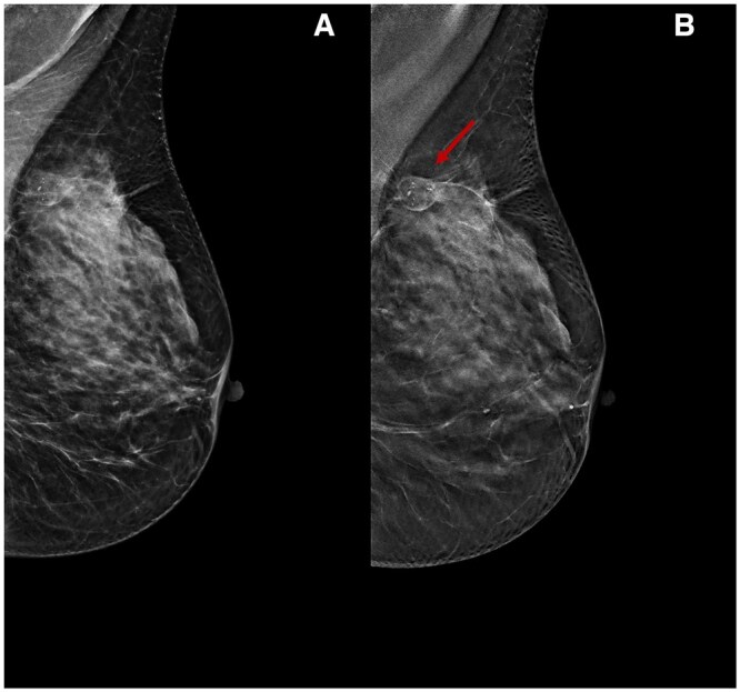
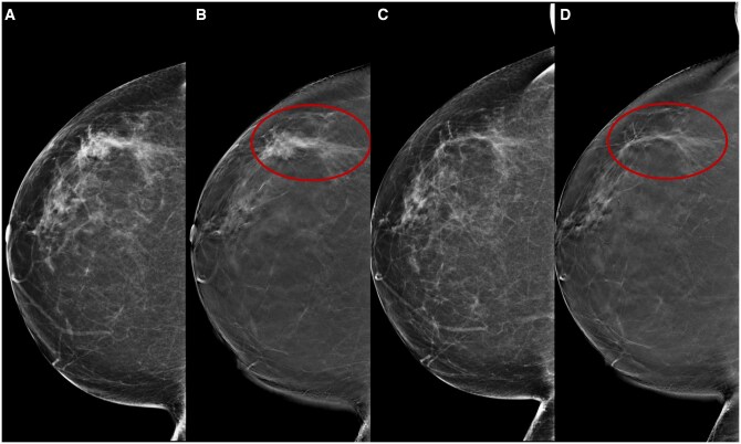
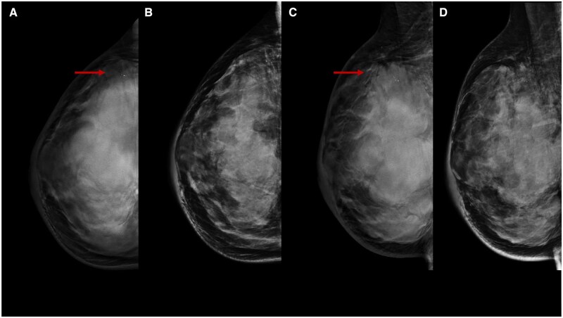
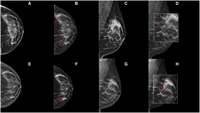
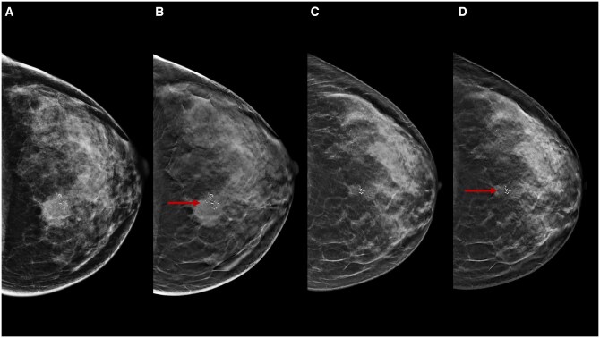
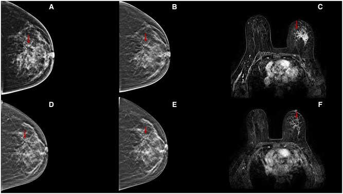
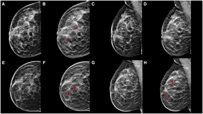
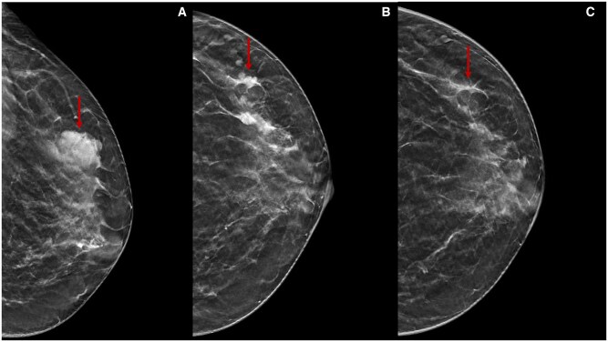
Neoadjuvant therapy (NT) has become the gold standard for treating locally advanced breast cancer (BC). The assessment of pathological response (pR) post-NT plays a crucial role in predicting long-term survival, with contrast-enhanced MRI currently recognised as the preferred imaging modality for its evaluation. Traditional imaging techniques, such as digital mammography (DM) and ultrasonography (US), encounter difficulties in post-NT assessments due to breast density, lesion changes, fibrosis, and molecular patterns. Digital breast tomosynthesis (DBT) offers solutions to prevalent challenges in DM, such as tissue overlap, and facilitates a comprehensive assessment of lesion morphology, dimensions, and margins. Studies suggest that DBT correlates more accurately with pathology than DM and US, showcasing its potential advantages. This pictorial essay demonstrates the potential of DBT as a complementary tool to DM for assessing pR after NT, including instances of true- and false-positive assessments correlated with histopathological findings. In conclusion, DBT emerges as a valuable adjunct to DM, effectively addressing its limitations in post-NT assessment. The technology's potential to diminish tissue overlap, improve discrimination, and provide multi-dimensional perspectives demonstrates promising results, indicating its utility in scenarios where MRI is contraindicated or inaccessible.

Keywords: breast cancer; neoadjuvant therapy; tomosynthesis.


Conflict of interest statement

The authors declare no conflict of interest.

Figures

Figure 1.
Figure 1.
A 46-year-old woman underwent NT (four cycles of epirubicin-cyclophosphamide followed by weekly paclitaxel for 12 weeks) for an invasive ductal carcinoma cT1cN0, G3 (ER 0%, PgR 0%, Ki-67 65%, HER2 1+). Medio-lateral-oblique DBT before (A) and following (C) NT show a parenchymal distortion in the upper half of the left breast (rims) obscured by high fibroglandular tissue density on DM (B, D).
Figure 2.
Figure 2.
A 55-year-old woman underwent NT (four cycles of epirubicin-cyclophosphamide followed by weekly paclitaxel for 12 weeks) for an invasive ductal carcinoma cT2cN0, G2 (ER 10%, PgR 0%, Ki-67 40%, HER2 0). Medio-lateral-oblique DM (A) and DBT (B) following NT. There is a lucent oval area with a radiopaque rim and some microcalcifications inside in the upper quadrants of the left breast (arrow), in keeping with liponecrotic phenomena which is seen on DBT but not clearly seen on DM.
Figure 3.
Figure 3.
A 49-year-old woman underwent NT (four cycles of epirubicin-cyclophosphamide followed by weekly paclitaxel for 12 weeks) for an invasive ductal carcinoma cT2cN0, G2 (ER 5%, PgR 0%, Ki-67 50%, HER2 0). Cranio-caudal DBT before (A) and following (C) NT show a dimensional and density reduction of a lobulated lesion in the lower half of the left breast (arrows), an indication of treatment response, which is partially obscured by fibroglandular tissue density on DM (B, D).
Figure 4.
Figure 4.
A 66-year-old woman underwent NT (four cycles of epirubicin-cyclophosphamide followed by weekly trastuzumab + paclitaxel for 12 weeks) for an invasive ductal carcinoma cT1cN0, G3 (ER 95%, PgR 0%, Ki-67 36%, HER2 3+). Cranio-caudal DBT before (B) and following (D) NT better show a residual fibrotic streak (rims) in the outer quadrants of the right breast which is not clearly seen on DM (A, B). A significant advantage of DBT is the better detection of planar lesions, such as breast structural distortions.
Figure 5.
Figure 5.
A 40-year-old woman underwent NT (four cycles of epirubicin-cyclophosphamide followed by weekly paclitaxel for 12 weeks) for an invasive ductal carcinoma cT1cN0, G3 (ER 0%, PgR 0%, Ki-67 85%, HER2 0). Cranio-caudal (A) and medio-lateral-oblique (C) DBT of the right breast before NT show an irregular opacity lesion with punctate microcalcifications in the upper outer quadrant of the right breast (arrows), which is not clearly detectable on DM (B, D). DBT allows a better assessment of lesions with irregular margins.
Figure 6.
Figure 6.
A 54-year-old woman underwent NT (four cycles of epirubicin-cyclophosphamide followed by weekly trastuzumab + paclitaxel for 12 weeks) for an invasive ductal carcinoma cT1cN0, G2 (ER 20%, PgR 1%, Ki-67 35%, HER2 3+). Cranio-caudal and medio-lateral-oblique DBT before (B, D) and following (F, H) NT better highlight many parenchymal distortion areas with spot-like microcalcifications inside in the upper quadrants of the left breast (arrows), which is not clearly seen on DM (A, C, E, G). DBT can reduce tissue overlap and the masking effect of tissue density by better evaluating residual microcalcifications.
Figure 7.
Figure 7.
A 47-year-old woman underwent NT (four cycles of epirubicin-cyclophosphamide followed by weekly paclitaxel for 12 weeks) for an invasive ductal carcinoma cT3cN0, G3 (ER 0%, PgR 0%, Ki-67 70%, HER2 2+, FISH−). Cranio-caudal DBT before (B) and following (D) NT show an irregular lesion with pleomorphic microcalcifications associated in the upper inner quadrant of the left breast (arrows), which is better seen on DBT compared to DM (A, C). DBT facilitates discrimination between lesion opacity and calcification, emphasising their changes.
Figure 8.
Figure 8.
A 44-year-old woman underwent NT (four cycles of epirubicin-cyclophosphamide followed by weekly trastuzumab + paclitaxel for 12 weeks) for an invasive ductal carcinoma cT2cN0, G3 (ER 40%, PgR 0%, Ki-67 25%, HER2 3+). Cranio-caudal DBT before (B) and following (E) NT better highlight an extensive area of a pleomorphic microcalcification with increased parenchymal density in the upper half of the left breast (arrows), compared to DM (A, D). At the end of therapy, DBT allows a better assessment of the areas of radiolucency for concentric shrinkage, confirmed on MRI subtraction image (F).
Figure 9.
Figure 9.
A 46-year-old woman underwent NT (four cycles of epirubicin-cyclophosphamide followed by weekly trastuzumab + paclitaxel for 12 weeks) for an invasive ductal carcinoma cT3cN0, G3 (ER 0%, PgR 0%, Ki-67 24%, HER2 3+). Cranio-caudal and medio-lateral-oblique DBT before NT (B, D) better highlight many parenchymal distortions with pleomorphic microcalcifications associated (arrows) in the upper outer breast quadrant of the right breast compared to DM (A, C). After NT, mild structural distortion with residual microcalcifications is observed on cranio-caudal and medio-lateral-oblique DBT (F, H). Histological evaluation after skin mastectomy confirms a ductal intraepithelial neoplasia (DIN 2) in fibrosis area diagnosis (ypTisN0), with a pathological complete response.
Figure 10.
Figure 10.
A 38-year-old woman underwent NT (four cycles of epirubicin-cyclophosphamide followed by weekly paclitaxel for 12 weeks) for an invasive ductal carcinoma cT1cN0, G3 (ER 0%, PgR 0%, Ki-67 70%, HER2 0). Cranio-caudal DBT before (A), during (B), and after (C) NT show an irregular lesion (arrows) in the upper outer quadrant of the left breast. After NT, the lesion is not observed. Histological evaluation after conservative surgery confirms a fibrosis area and residual tumour inside diagnosis (ypT1aN0), with pathological partial response.

Similar articles

Cited by

References

    1. Untch M, Harbeck N, Huober J, et al. Primary therapy of patients with early breast cancer: evidence, controversies, consensus. Geburtshilfe Frauenheilkd. 2015;75(6):556-565. 10.1055/s-0035-1546120 - DOI - PMC - PubMed
    1. Mathew J, Asgeirsson KS, Cheung KL, Chan S, Dahda A, Robertson JFR. Neoadjuvant chemotherapy for locally advanced breast cancer: a review of the literature and future directions. Eur J Surg Oncol (EJSO). 2009;35(2):113-122. 10.1016/j.ejso.2008.03.015 - DOI - PubMed
    1. Beasley GM, Olson JA. What’s new in neoadjuvant therapy for breast cancer? Adv Surg. 2010;44:199-228. 10.1016/j.yasu.2010.05.013 - DOI - PubMed
    1. Ahmed MI, Lennard TWJ. Breast cancer: role of neoadjuvant therapy. Int J Surg. 2009;7(5):416-420. 10.1016/j.ijsu.2009.06.001 - DOI - PubMed
    1. Cortazar P, Zhang L, Untch M, et al. Pathological complete response and long-term clinical benefit in breast cancer: the CTNeoBC pooled analysis. Lancet. 2014;384(9938):164-172. 10.1016/S0140-6736(13)62422-8 - DOI - PubMed