. 2024 Dec 26;151(1):21.
doi: 10.1007/s00432-024-06051-3.

CTSG is a prognostic marker involved in immune infiltration and inhibits tumor progression though the MAPK signaling pathway in non-small cell lung cancer

Affiliations

CTSG is a prognostic marker involved in immune infiltration and inhibits tumor progression though the MAPK signaling pathway in non-small cell lung cancer

Qian Dai et al. J Cancer Res Clin Oncol. .

Abstract

Purpose: This study aims to investigate the biological roles and molecular mechanisms of Cathepsin G (CTSG) in the progression of non-small cell lung cancer (NSCLC).

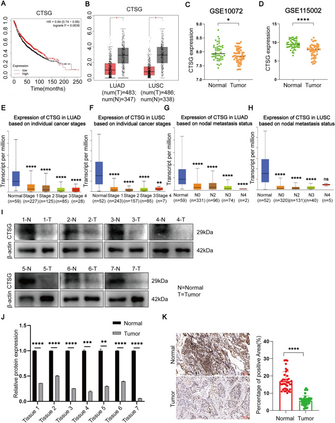
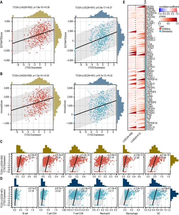
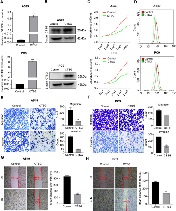
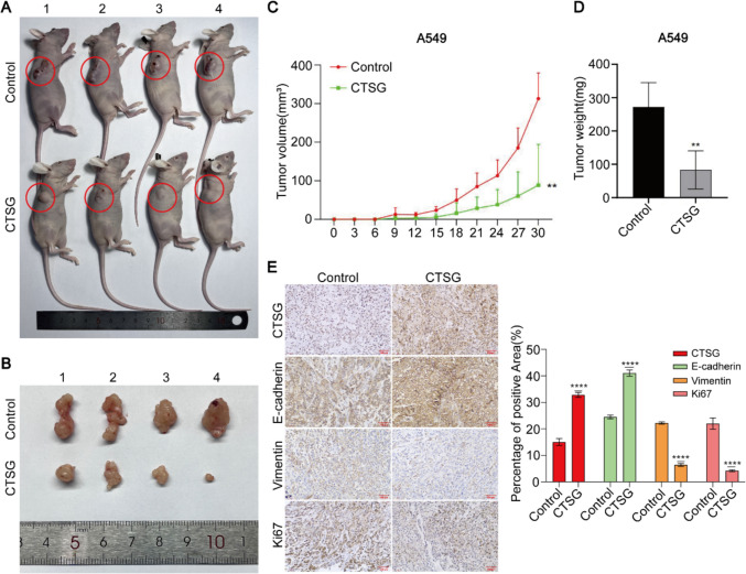
Methods: Western blotting and immunohistochemistry analyses of clinical samples were performed to determine the expression levels of CTSG in patients with NSCLC. Bioinformatic analysis of clinical datasets was conducted to evaluate the correlation between CTSG and lymph node metastasis, tumor stage, and immune cell infiltration. Gain-of-function assays and tumor implantation experiments were employed to determine the effects of CTSG on malignant behaviors of NSCLC cells. Transcriptome sequencing and subsequent bioinformatic analysis were performed to explore the signaling pathways regulated by CTSG. Western blotting and qPCR were utilized to assess the influence of CTSG on the MAPK and EMT signaling pathways.

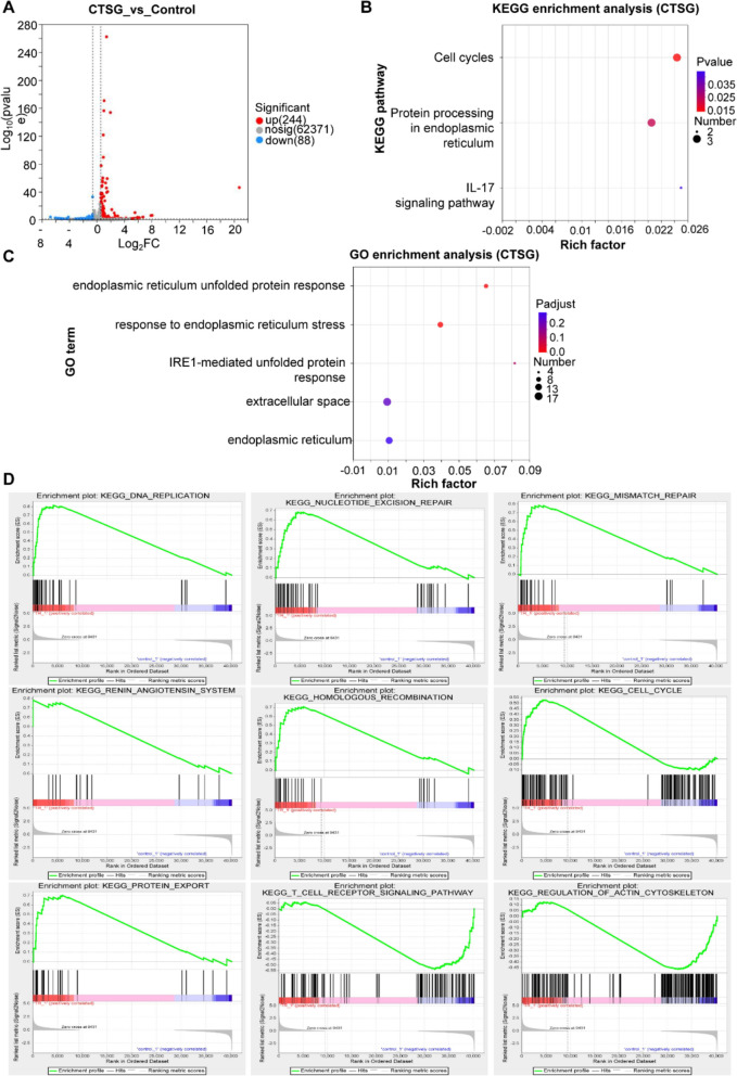
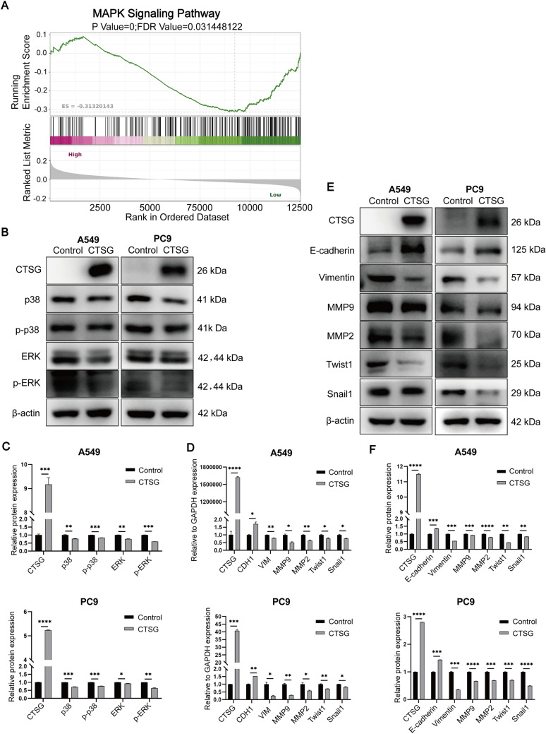
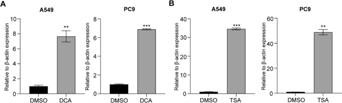
Results: CTSG is expressed at low levels and serves as a prognostic marker in NSCLC. The downregulation of CTSG expression was associated with lymph node metastasis, tumor stage, and immune cell infiltration. CTSG inhibits NSCLC cell proliferation, migration, and invasion as well as tumor growth in nude mice. There exists a significant correlation between CTSG expression and endoplasmic reticulum function, cell cycling, and the IL-17 signaling pathway. CTSG suppresses the MAPK and EMT signaling pathways in NSCLC cells. Moreover, DNA methylation and histone deacetylation have been identified as crucial mechanisms contributing to the decreased expression of CTSG.

Conclusion: CTSG inhibits NSCLC development by suppressing the MAPK signaling pathway and is also associated with tumor immunity.

Keywords: Biomarker; CTSG; MAPK; NSCLC; Prognostic.


Conflict of interest statement

Declarations. Conflict of interest: The authors declare no competing interests. Ethical approval: All experiments involved in clinical samples and animals were reviewed and approved by the Ethics Committee of Animal Experiments of Anhui Medical University.

Figures

Fig. 1
Fig. 1
CTSG is a prognostic marker and is under-expressed in NSCLC tissue. A Kaplan‒Meier survival plots depicting the survival curve of patients with NSCLC based on CTSG expression. B Differences in CTSG expression in NSCLC and normal tissues from the GEPIA database. C-D Relative abundance of CTSG protein in NSCLC tissues based on the GEO database. EF CTSG expression grouped by tumor pathological stage. G-H CTSG expression grouped by tumor nodal metastasis. I Western blotting analysis of CTSG level in human NSCLC and normal tissues. J Quantification of CTSG protein. K IHC staining of CTSG in human NSCLC and normal tissues (n = 9; scale bar = 100 μm). *p < 0.05, **p < 0.01, ***p < 0.001, ****p < 0.0001
Fig. 2
Fig. 2
CTSG expression correlates with NSCLC tumor immune infiltrates. A-B Associations between CTSG expression and stromal and immune scores in NSCLC tumors based on the ESTIMATE method. C-D Association between CTSG expression and various immune cell abundance based on the TIMER database. E Correlation between CTSG expression and immune checkpoint-associated genes. *p < 0.05
Fig. 3
Fig. 3
CTSG overexpression suppresses NSCLC cell proliferation, migration, and invasion. A-B qPCR and western blotting results of CTSG expression in A549 and PC9 cells. C-D CCK-8 and Ki67 staining results showing impaired cell proliferation in CTSG-overexpressing A549 and PC9 cells. EF Transwell assay results displaying decreased migration and invasion in CTSG-overexpressing A549 and PC9 cells. G-H Wound-healing assay showing reduced migration of CTSG-overexpressing A549 and PC9 cells. *p < 0.05, **p < 0.01, ***p < 0.001
Fig. 4
Fig. 4
CTSG overexpression inhibits tumor growth in vivo. A-B Subcutaneous and xenograft tumors in the control and CTSG-overexpressing groups (n = 4). C-D Weight and volume of xenograft tumors in the control and CTSG-overexpressing groups. E Representative IHC staining of CTSG, Ki67, E-cadherin, and Vimentin proteins (n = 4; scale bar = 200 μm). *p < 0.05, **p < 0.01, ***p < 0.001, ****p < 0.0001
Fig. 5
Fig. 5
CTSG-related differentially expressed genes and functional enrichment analysis. A Volcano plot of differentially expressed genes. B GO enrichment analysis. C KEGG enrichment analysis. D GSEA pathway analysis.
Fig. 6
Fig. 6
CTSG regulates the MAPK and EMT signaling pathways. A GSEA results showing enrichment of the MAPK signaling pathway. B Western blotting analysis results showing the altered abundance of proteins in the MAPK signaling pathway. C Quantification of CTSG, p38, p-p38, ERK and p-ERK proteins. D qPCR analysis of EMT-related genes: CDH1 (encode E-cadherin), VIM (encode Vimentin), MMP9, MMP2, Twist1, and Snail1. E Western blotting analysis of the indicated proteins involved in the EMT signaling pathway. F Quantification of CTSG, E-cadherin, Vimentin, MMP9, MMP2, Twist1, and Snail1 proteins
Fig. 7
Fig. 7
DNA methylation and histone deacetylation suppress CTSG expression. A-B qPCR results of CTSG expression in DCA- and TSA-treated A549 and PC9 cells. A549 and PC9 cells were incubated with 1 µM TSA for 24 h; A549 and PC9 cells were incubated with 1 mM DCA for 48 h. *p < 0.05, **p < 0.01, ***p < 0.001

Similar articles

Cited by

References

    1. Bajbouj K, Al-Ali A, Ramakrishnan RK, Saber-Ayad M, Hamid Q et al (2021) Histone modification in NSCLC: molecular mechanisms and therapeutic targets. Int J Mol Sci. 10.3390/ijms222111701 - PMC - PubMed
    1. Bank U, Ansorge S et al (2001) More than destructive: neutrophil-derived serine proteases in cytokine bioactivity control. J Leukoc Biol 69:197–206 - PubMed
    1. Bi KW, Wei XG, Qin XX, Li B et al (2020) BTK has potential to be a prognostic factor for lung adenocarcinoma and an indicator for tumor microenvironment remodeling: a study based on TCGA data mining. Front Oncol 10:424. 10.3389/fonc.2020.00424 - PMC - PubMed
    1. Breznik B, Limbaeck Stokin C, Kos J, Khurshed M, Hira VVV, Bosnjak R, Lah TT, Van Noorden CJF et al (2018) Cysteine cathepsins B, X and K expression in peri-arteriolar glioblastoma stem cell niches. J Mol Histol 49:481–497. 10.1007/s10735-018-9787-y - PMC - PubMed
    1. Brix K, Dunkhorst A, Mayer K, Jordans S et al (2008) Cysteine cathepsins: cellular roadmap to different functions. Biochimie 90:194–207. 10.1016/j.biochi.2007.07.024 - PubMed

MeSH terms

Substances

LinkOut - more resources