. 2025 Jan 22;66(1):97-102.
doi: 10.1093/jrr/rrae091.

Ultra-high dose rate (FLASH) carbon ion irradiation inhibited immune suppressive protein expression on Pan02 cell line

Affiliations

Ultra-high dose rate (FLASH) carbon ion irradiation inhibited immune suppressive protein expression on Pan02 cell line

Shohei Katsuki et al. J Radiat Res. .

Abstract

Recently, ultra-high dose rate (> 40 Gy/s, uHDR; FLASH) radiation therapy (RT) has attracted interest, because the FLASH effect that is, while a cell-killing effect on cancer cells remains, the damage to normal tissue could be spared has been reported. This study aimed to compare the immune-related protein expression on cancer cells after γ-ray, conventionally used dose rate (Conv) carbon ion (C-ion), and uHDR C-ion. B16F10 murine melanoma and Pan02 murine pancreas cancer were irradiated with γ-ray at Osaka University and with C-ion at Osaka HIMAK. The dose rates at 1.16 Gy/s for Conv and 380 Gy/s for uHDR irradiation. The expressed calreticulin (CRT), major histocompatibility complex class (MHC)-I, and programmed cell death 1 ligand (PD-L1) were evaluated by flow cytometry. Western blotting and PCR were utilized to evaluate endoplasmic reticulum (ER) stress, DNA damage, and its repair pathway. CRT, MHC-I on B16F10 was also increased by irradiation, while only C-ion increased MHC-I on Pan02. Notably, PD-L1 on B16F10 was increased after irradiation with both γ-ray and C-ion, while uHDR C-ion suppressed the expression of PD-L1 on Pan02. The present study indicated that uHDR C-ion has a different impact on the repair pathway of DNA damage and ER than the Conv C-ion. This is the first study to show the immune-related protein expressions on cancer cells after uHDR C-ion irradiation.

Keywords: FLASH; PD-L1; calreticulin (CRT); carbon ion beam; ultra-high dose rate.


Conflict of interest statement

There are no conflicts of interest.

Figures

Fig. 1
Fig. 1
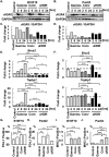
The impact of uHDR C-ion irradiation on the ER. (A, B) SF of B16F10 (A) and Pan02 (B) calculated by colony formation assay were shown. Solid line, dot line, and dashed line indicate γ-ray, Conv, and uHDR C-ion, respectively. For each SF curve, we conducted three independent experiments. Data are indicated mean ± S.E. The curves were fitted with an LQ model. (C-F) Phosphorylated eIF2a and GAPDH in control at the indicated time pot-irradiation in B16F10 and Pan02 were shown, and the ratios of these proteins’ expression were calculated. (G, H) Calreticulin-positive populations on B16F10 (G) and Pan02 (H) at 48 hrs after irradiation were shown. *P < 0.05, **P < 0.01, ***P < 0.001.
Fig. 2
Fig. 2
The impact of uHDR C-ion on DSB repair pathway. (A-D) γH2AX and GAPDH were shown, and GAPDH is the same loading control in Fig. 1. The ratios between γH2AX and GAPDH were shown. (E, F) Fold expression changes of Brca2 and Topbp1 were normalized with the control group in both cells. The dashed lines indicated in the bar graphs mean the value of 1, the same as the control. Data are indicated mean ± S.E. (G-J) The population of PD-L1-positive and MHC-I-positive cells were shown. Data are indicated mean ± S.E. *P < 0.05, **P < 0.01, ***P < 0.0001.

Similar articles

References

    1. Curtin NJ. DNA repair dysregulation from cancer driver to therapeutic target. Nat Rev Cancer 2012;12:801–17. 10.1038/nrc3399. - DOI - PubMed
    1. Kagawa K, Murakami M, Hishikawa Y et al. Preclinical biological assessment of proton and carbon ion beams at Hyogo Ion Beam Medical Center. Int J Radiat Oncol Biol Phys 2002;54:928–38. 10.1016/S0360-3016(02)02949-8. - DOI - PubMed
    1. Komatsu S, Fukumoto T, Demizu Y et al. Clinical results and risk factors of proton and carbon ion therapy for hepatocellular carcinoma. Cancer 2011;117:4890–904. 10.1002/cncr.26134. - DOI - PubMed
    1. Favaudon V, Caplier L, Monceau V et al. Ultrahigh dose-rate FLASH irradiation increases the differential response between normal and tumor tissue in mice. Sci Transl Med 2014;6:245ra93. 10.1126/scitranslmed.3008973. - DOI - PubMed
    1. Diffenderfer ES, Verginadis II, Kim MM et al. Design, implementation, and in vivo validation of a novel proton FLASH radiation therapy system. Int J Radiat Oncol Biol Phys 2020;106:440–8. 10.1016/j.ijrobp.2019.10.049. - DOI - PMC - PubMed

Grants and funding