

[Role of the nuclear factor-kappa B signaling pathway in the repair of white matter injury in neonatal rats through human umbilical cord mesenchymal stem cell transplantation]
- PMID: 39725400
- PMCID: PMC11684829
- DOI: 10.7499/j.issn.1008-8830.2408099
[Role of the nuclear factor-kappa B signaling pathway in the repair of white matter injury in neonatal rats through human umbilical cord mesenchymal stem cell transplantation]
Abstract
Objectives: To observe the reparative effects of human umbilical cord mesenchymal stem cell (hUC-MSC) transplantation on white matter injury (WMI) in neonatal rats and explore its mechanism through the nuclear factor-kappa B (NF-κB) signaling pathway mediated by microglial cells.
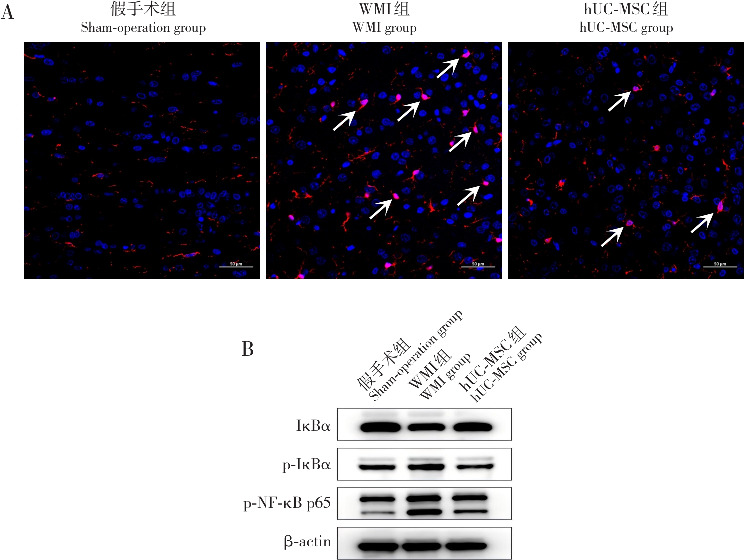
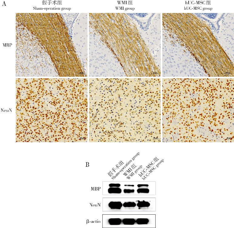
Methods: Sprague-Dawley rats, aged 2 days, were randomly divided into three groups: sham-operation,WMI, and hUC-MSC (n=18 each). Fourteen days after modeling, hematoxylin-eosin staining was used to observe pathological changes in the white matter, and immunofluorescence staining was used to measure the expression level of ionized calcium-binding adapter molecule 1 (Iba1). Western blotting was used to measure the protein expression levels of inhibitory subunit of nuclear factor-kappa B alpha (IκBα), phosphorylated IκBα (p-IκBα), phosphorylated NF-κB p65 (p-NF-κB p65), myelin basic protein (MBP), and neuron-specific nuclear protein (NeuN). Quantitative real-time PCR was used to assess the mRNA expression levels of tumor necrosis factor-α (TNF-α), interleukin-1β (IL-1β), MBP, and NeuN. Immunohistochemistry was used to measure the protein expression levels of MBP and NeuN. On day 28, the Morris water maze test was used to evaluate spatial cognitive ability.
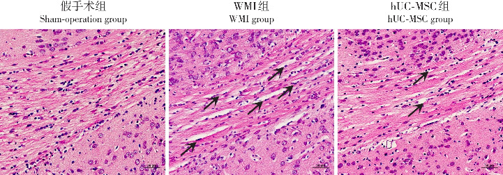
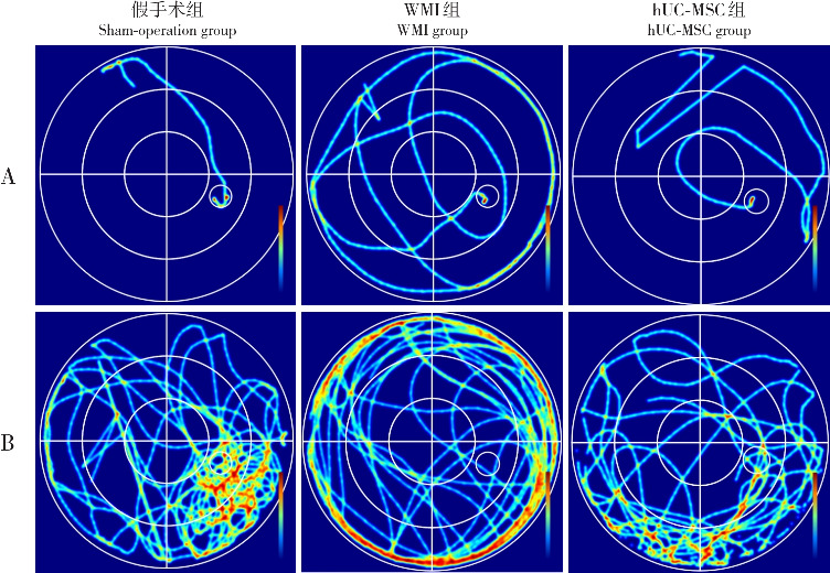
Results: Fourteen days after modeling, the sham-operation group exhibited intact white matter structure with normal cell morphology and orderly nerve fiber arrangement. In the WMI group, large-scale cell degeneration and necrosis were observed, and nerve fiber arrangement was disordered. The hUC-MSC group showed relatively normal cell morphology and more orderly nerve fibers. Compared with the sham-operation group, the WMI group had significantly higher proportions of Iba1-positive cells, increased protein levels of p-IκBα and p-NF-κB p65, and higher mRNA levels of TNF-α and IL-1β. The protein expression of IκBα and the positive expression of MBP and NeuN, as well as their protein and mRNA levels, were significantly reduced in the WMI group (P<0.05). Compared with the WMI group, the hUC-MSC group showed reduced proportions of Iba1-positive cells, decreased protein levels of p-IκBα and p-NF-κB p65, and lower mRNA levels of TNF-α and IL-1β. Furthermore, IκBα protein expression and MBP and NeuN expression (both at the protein and mRNA levels) were significantly increased in the hUC-MSC group (P<0.05). On day 28, the Morris water maze results showed that compared with the sham-operation group, the WMI group had significantly longer escape latency and fewer platform crossings (P<0.05). In contrast, the hUC-MSC group had significantly shorter escape latency and more platform crossings than the WMI group (P<0.05).
Conclusions: hUC-MSC transplantation can repair WMI in neonatal rats, promote the maturation of oligodendrocytes, and support neuronal survival, likely by inhibiting activation of the NF-κB signaling pathway mediated by microglial cells.
目的: 观察人脐带间充质干细胞(human umbilical cord mesenchymal stem cell, hUC-MSC)移植对新生大鼠脑白质损伤(white matter injury, WMI)的修复作用,并通过小胶质细胞介导的核因子κB(nuclear factor kappa B, NF-κB)信号通路探讨其机制。方法: 将2日龄Sprague-Dawley新生大鼠随机分为假手术组、WMI组和hUC-MSC组,每组18只。建模后14 d,通过苏木精-伊红染色法观察脑白质病理变化,免疫荧光染色法检测离子钙结合适配器分子1(ionized calcium-binding adapter molecule 1, Iba1)表达,免疫印迹法检测核因子κB抑制蛋白α(inhibitory subunit of nuclear factor-kappa B alpha, IκBα)、磷酸化IκBα(p-IκBα)、磷酸化NF-κB p65(p-NF-κB p65)、髓鞘碱性蛋白(myelin basic protein, MBP)、神经元核蛋白(neuron-specific nuclear protein, NeuN)蛋白表达,实时荧光定量聚合酶链反应法检测肿瘤坏死因子(tumor necrosis factor-α, TNF-α)、白介素-1β(interleukin-1β, IL-1β)、MBP、NeuN mRNA表达,免疫组化法检测MBP、NeuN蛋白表达水平;建模后28 d,利用Morris水迷宫实验评估大鼠空间认知能力。结果: 建模后14 d,假手术组脑白质区域组织结构完整,细胞形态正常、神经纤维排列规则;WMI组大量细胞变性坏死、神经纤维排列紊乱;hUC-MSC组细胞形态相对正常,神经纤维排列较整齐。WMI组Iba1阳性细胞占比、p-IκBα和p-NF-κB p65蛋白表达,以及TNF-α和IL-1β mRNA表达较假手术组增加,IκBα蛋白表达及MBP和NeuN阳性表达、蛋白和mRNA表达较假手术组减少(P<0.05);hUC-MSC组Iba1阳性细胞占比、p-IκBα和p-NF-κB p65蛋白表达,以及TNF-α、IL-1β mRNA表达较WMI组减少,IκBα蛋白表达及MBP和NeuN阳性表达、蛋白和mRNA表达较WMI组增加(P<0.05)。建模后28 d,水迷宫结果显示与假手术组比较,WMI组逃避潜伏期时间延长、穿越平台次数较减少(P<0.05);与WMI组比较,hUC-MSC组逃避潜伏期时间缩短、穿越平台次数增加(P<0.05)。结论: hUC-MSC可修复新生大鼠WMI,促进少突胶质细胞成熟和神经元存活,其机制可能与抑制小胶质细胞介导的NF-κB信号通路激活相关。.
Keywords: Human umbilical cord mesenchymal stem cell; Neonatal rat; Nuclear factor-kappa B pathway; White matter injury.
Conflict of interest statement
所有作者声明不存在利益冲突。
Figures
Similar articles
-
[Repair effect of different doses of human umbilical cord mesenchymal stem cells on white matter injury in neonatal rats].Zhongguo Dang Dai Er Ke Za Zhi. 2024 Apr 15;26(4):394-402. doi: 10.7499/j.issn.1008-8830.2310081. Zhongguo Dang Dai Er Ke Za Zhi. 2024. PMID: 38660904 Free PMC article. Chinese.
-
[Protective effect of Liujing Toutong Tablets on rats with permanent cerebral ischemia via NF-κB signaling pathway].Zhongguo Zhong Yao Za Zhi. 2023 Nov;48(21):5871-5880. doi: 10.19540/j.cnki.cjcmm.20230710.705. Zhongguo Zhong Yao Za Zhi. 2023. PMID: 38114183 Chinese.
-
[Effect of Chaihu Jia Longgu Muli Decoction on apoptosis in rats with heart failure after myocardial infarction through IκBα/NF-κB pathway].Zhongguo Zhong Yao Za Zhi. 2025 Apr;50(8):2184-2192. doi: 10.19540/j.cnki.cjcmm.20241217.401. Zhongguo Zhong Yao Za Zhi. 2025. PMID: 40461228 Chinese.
-
Downregulation of Nogo-B ameliorates cerebral ischemia/reperfusion injury in mice through regulating microglia polarization via TLR4/NF-kappaB pathway.Neurochem Int. 2023 Jul;167:105553. doi: 10.1016/j.neuint.2023.105553. Epub 2023 May 23. Neurochem Int. 2023. PMID: 37230196 Review.
-
Molecular mechanisms of system control of NF-kappaB signaling by IkappaBalpha.Biochemistry. 2010 Mar 2;49(8):1560-7. doi: 10.1021/bi901948j. Biochemistry. 2010. PMID: 20055496 Free PMC article. Review.
References
Publication types
MeSH terms
Substances
LinkOut - more resources
Full Text Sources
Miscellaneous