. 2025 Apr 7;74(5):804-814.
doi: 10.1136/gutjnl-2024-333368.

Mutational signatures define immune and Wnt-associated subtypes of ampullary carcinoma

Affiliations

Mutational signatures define immune and Wnt-associated subtypes of ampullary carcinoma

Ekaterina Zhuravleva et al. Gut. .

Abstract

Background and objective: Ampullary carcinoma (AMPAC) taxonomy is based on morphology and immunohistochemistry. This classification lacks prognostic reliability and unique genetic associations. We applied an approach of integrative genomics characterising patients with AMPAC exploring molecular subtypes that may guide personalised treatments.

Design: We analysed the mutational landscapes of 170 patients with AMPAC. The discovery included 110 tumour/normal pairs and the validation comprised 60 patients. In a tumour subset, we interrogated the transcriptomes and DNA methylomes. Patients were stratified based on mutational signatures and associated with molecular and clinical features. To evaluate tumour and immune cellularity, 22 tumours were independently assessed histomorphologically and by digital pathology.

Results: We defined three patient clusters by mutational signatures independent of histomorphology. Cluster 1 (C1) was defined by spontaneous deamination of DNA 5-methylcytosine and defective mismatch repair. C2 and C3 were related to the activity of transcription-coupled nucleotide excision repair but C3 was further defined by the polymerase eta mutational process. C1-2 showed enrichment of Wnt pathway alterations, aberrant DNA methylation profiles, immune cell exclusion and patients with poor prognosis. These features were associated with a hypermutator phenotype caused by C>T alterations at CpGs. C3 patients with improved overall survival were associated with activation of immune-related pathways, immune infiltration and elevated expression of immunoinhibitory checkpoint genes.

Conclusion: Immunogenicity and Wnt pathway associations, emphasised by the mutational signatures, defined patients with prospective sensitivity to either immunotherapy or Wnt pathway inhibitors. This emphasises a novel mutational signature-based AMPAC classification with prognostic potential, suggesting prospective implications for subgroup-specific management of patients with AMPAC.

Keywords: ADENOCARCINOMA; GASTROINTESTINAL CANCER; METHYLATION; MOLECULAR ONCOLOGY.


Conflict of interest statement

Competing interests: JBA declares consultancies for Flagship Pioneering, Seald, QED Therapeutics and AstraZeneca. JBA has received funding from the Incyte Corporation (EU-DK-ST-21122) but not related to this study.

Figures

Figure 1
Figure 1. Study data sets and analysis pipeline. Data sets are colour coded in the figure. Whole exome sequencing was performed in the discovery cohort (n=110) and validated by additional n=60 samples. Transcriptome analysis was performed in n=43 cases. DNA methylation was performed in n=16 AMPAC cases with matched WES and RNAseq data sets and additional six normal bile duct samples. Histomorphology evaluations were performed on the discovery cohort (n=110) with additional 22 samples analysed by immunohistochemistry and digital pathology evaluation. AMPAC, ampullary carcinoma; CNV, copy-number variant; dbSNP, single-nucleotide polymorphism database; MSI, microsatellite instability; PDL1, programmed death ligand 1; SNV, single-nucleotide variant.
Figure 2
Figure 2. Characterisation of genomic alterations in AMPAC (n=110). (A) Non-silent and silent mutations rate and CNV count (amplifications, deletions). (B) Oncoplot of recurrently mutated genes including druggability annotation. (C) Large-scale recurrent amplifications and deletions defined with GISTIC2. (D) Detection of non-recurrent fusions (genes in fusion and breakpoints) in AMPAC. AMPAC, ampullary carcinoma; APC, adenomatous polyposis coli; AQP7, Aquaporin 7; ARID2, AT-rich interactive domain 2; ATM, serine-protein kinase; CNV, copy-number variant; DNAH5, dynein axonemal heavy chain 5; ELF3, E74-like ETS transcription factor 3; GISTIC2, Genomic Identification of Significant Targets In Cancer; KRAS, Kristen rat sarcoma viral oncogene homolog; MSI, microsatellite instability; MSS, microsatellite stable; PDZD2, activated in prostate cancer protein; PIK3CA, phosphatidylinositol-4,5-bisphosphate 3-kinase catalytic subunit alpha; SMAD4, SMAD family member 4; TP53, Tumour protein P53.
Figure 3
Figure 3. Mutational signature-associated clusters in AMPAC (n=103). (A) Normalised three-nucleotide mutational frequency profiles. (B) Relative mutational signature contribution. (C) Kaplan-Meier analysis of overall survival between clusters. (D) Profiles of decomposed mutational signatures S1, S2 and S3. AMPAC, ampullary carcinoma; MSI, microsatellite instability; MSS, microsatellite stable; NCOR1, nuclear receptor corepressor 1; RNF43, ring finger protein 43; SNV, single-nucleotide variant.
Figure 4
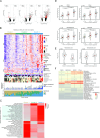
Figure 4. Association of mutational signature-associated clusters with immune parameters (n=43). (A) Volcano plot of differently expressed genes between clusters. Genes from CIBERSORT lm22 immune signatures are highlighted. (B) Heatmap of differentially expressed genes deregulated between clusters. (C) GSEA analysis results, pathways with absolute normalised enrichment score (NES)>1.9 and p.adj<0.05 in at least one pairwise comparison between clusters. Mean expression of all genes in leading edge for each pathway is taken to represent the direction of deregulation between clusters, z-scored. (D) CTLA-4, PDCD1, PDCD1LG1, PDCD1LG2 gene expression across clusters. (E) Boxplot on TIDE exclusion and dysfunction scores estimation. Only samples with clear morphology were considered and normalisation procedure were made to each morphological group average. C1 and 2 were joined because of samples size. (F) Pearson correlation values between immune parameters and AMPAC clusters. Immune cell type absolute estimation was performed using CIBERSORT. AMPAC, ampullary carcinoma; DEG, differentially expressed genes; MSI, microsatellite instability; GSEA, Gene Set Enrichment Analysis; MSS, microsatellite stable; NK, natural killer; PDCD1, programmed cell death protein 1; PDCD1LG2, programmed cell death 1 ligand 2; TIDE, Tumor Immune Dysfunction and Exclusion.
Figure 5
Figure 5. DNA methylation alterations in AMPAC genomes (n=16). (A) Principal component analysis on 10 000 methylation probes with highest variance in the data set. Ellipses are drawn on groupings of samples in hierarchical clustering based on 1000 regions and 10 000 most variable probes. (B) Log2 Ratios between numbers of probes with higher methylation in C3 compared with C1 and 2 to probes with lower methylation in C3 compared with C1 and 2. Only probes with p value for difference between clusters were considered. Positive value in Log2 scale indicates increased methylation in corresponding regions in C3. (C) Loss of global methylation in C1 and 2 compared with C3 plotted as roll mean statistics on 1000 5 kb tiling windows. Normal bile duct data is used as control. (D) Proportion of mutated cytosines, normalised by CG content and plotted as roll mean statistics on 1000 5 kb tiling windows. (E) Separation of C1 and 2 and C3 based on the normalised fraction of (C->T)G mutations (signature 1) to all mutations and the hypermethylated cytosine fraction across genes. (F) Normalised fraction of (C->T)G mutations to all mutations across clusters. (G) Normalised fraction of hypermethylated cytosine fraction in gene regions across C1 and 2 versus C3. (H) (C->T)G mutations across genes. Genes with recurrent hotspots are coloured in red. Wnt pathway (C->T)G alterations are over-represented in clusters 1 and 2. AMPAC, ampullary carcinoma; ANOVA, analysis of variance; APC, adenomatous polyposis coli; AQP7, Aquaporin 7; ARID2, AT-rich interactive domain 2; ATM, serine-protein kinase; DNAH5, dynein axonemal heavy chain 5; ELF3, E74-like ETS transcription factor 3; KRAS, Kristen rat sarcoma viral oncogene homolog; MSI, microsatellite instability; MSS, microsatellite stable; PDZD2, activated in prostate cancer protein; PIK3CA, phosphatidylinositol-4,5-bisphosphate 3-kinase catalytic subunit alpha; SMAD4, SMAD family member 4; TP53, Tumour protein P53.
Figure 6
Figure 6. DNA methylation alterations associated with gene expression in AMPAC (n=16). (A) Venn diagram on the numbers of genes with variance of expression above median and with significant association to promoters, enhancers and gene body methylation. (B) Top differently methylated regions (DMR) between C1 and 2 and C3 (402 regions). (C) Expression of genes associated with methylation changes in regions between C1 and 2 and C3. (D) Over-representation analysis of genes regulated by methylation changes. (E) Volcano plot of genes regulated by methylation between C1 and 2 and C3. Genes with absolute logFC>1.5 and adjusted p<0.01 are labelled, oncogenes and tumour suppressor genes are labelled in bold. AMPAC, ampullary carcinoma; APC, adenomatous polyposis coli; ELF3, E74-like ETS transcription factor 3; MSI, microsatellite instability; MSS, microsatellite stable; SMAD4, SMAD family member 4; SNV, single-nucleotide variant.

References

    1. Adsay V, Ohike N, Tajiri T, et al. Ampullary region carcinomas: definition and site specific classification with delineation of four clinicopathologically and prognostically distinct subsets in an analysis of 249 cases. Am J Surg Pathol. 2012;36:1592–608. doi: 10.1097/PAS.0b013e31826399d8. - DOI - PubMed
    1. Pea A, Riva G, Bernasconi R, et al. Ampulla of Vater carcinoma: Molecular landscape and clinical implications. World J Gastrointest Oncol. 2018;10:370–80. doi: 10.4251/wjgo.v10.i11.370. - DOI - PMC - PubMed
    1. Rostain F, Hamza S, Drouillard A, et al. Trends in incidence and management of cancer of the ampulla of Vater. World J Gastroenterol. 2014;20:10144–50. doi: 10.3748/wjg.v20.i29.10144. - DOI - PMC - PubMed
    1. Shoji H, Morizane C, Hiraoka N, et al. Twenty-six cases of advanced ampullary adenocarcinoma treated with systemic chemotherapy. Jpn J Clin Oncol. 2014;44:324–30. doi: 10.1093/jjco/hyt237. - DOI - PubMed
    1. Kim RD, Kundhal PS, McGilvray ID, et al. Predictors of failure after pancreaticoduodenectomy for ampullary carcinoma. J Am Coll Surg. 2006;202:112–9. doi: 10.1016/j.jamcollsurg.2005.08.002. - DOI - PubMed

LinkOut - more resources