. 2025 Jan 10;12(1):ENEURO.0418-24.2024.
doi: 10.1523/ENEURO.0418-24.2024. Print 2025 Jan.

Functional Regrowth of Norepinephrine Axons in the Adult Mouse Brain Following Injury

Affiliations

Functional Regrowth of Norepinephrine Axons in the Adult Mouse Brain Following Injury

Patrick Cooke et al. eNeuro. .

Abstract

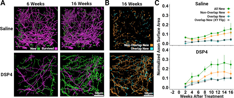
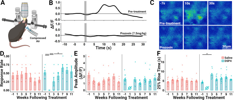
It is widely believed that axons in the central nervous system of adult mammals do not regrow following injury. This failure is thought, at least in part, to underlie the limited recovery of function following injury to the brain or spinal cord. Some studies of fixed tissue have suggested that, counter to dogma, norepinephrine (NE) axons regrow following brain injury. Here, we have used in vivo two-photon microscopy in layer 1 of the primary somatosensory cortex in transgenic mice harboring a fluorophore selectively expressed in NE neurons. This protocol allowed us to explore the dynamic nature of NE axons following injury with the selective NE axon toxin N-(2-chloroethyl)-N-ethyl-2-bromobenzylamine (DSP4). Following DSP4, NE axons were massively depleted and then slowly and partially recovered their density over a period of weeks. This regrowth was dominated by new axons entering the imaged volume. There was almost no contribution from local sprouting from spared NE axons. Regrown axons did not appear to use either the paths of previously lesioned NE axons or NE axons that were spared and survived DSP4 as a guide. To measure NE release, GCaMP8s was selectively expressed in neocortical astrocytes and startle-evoked, NE receptor-mediated Ca2+ transients were measured. These Ca2+ transients were abolished soon after DSP4 lesion but returned to pre-lesion values after 3-5 weeks, roughly coincident with NE axon regrowth, suggesting that the regrown NE axons are competent to release NE in response to a physiological stimulus in the awake mouse.

Keywords: axon regeneration; in vivo microscopy; norepinephrine.


Conflict of interest statement

The authors declare no competing financial interests.

Figures

Figure 1.
Figure 1.
NE axons innervating the adult mouse primary somatosensory cortex regrow following a chemical lesion with the NE-specific neurotoxin DSP4 (50 mg/kg). A, Schematic diagram of a sagittal section of an adult mouse brain depicting the general projection of axons from NE cell bodies located within the locus ceruleus (LC). The red box indicates the region where this histological analysis was conducted: the primary somatosensory cortex (1–1). *Other abbreviations: ob, olfactory bulb; stri, striatum; ac, nucleus accumbens; amg, amygdala; hyp, hypothalamus; th, thalamus; cer, cerebellum. B, Representative maximum projected confocal stack images of layer 1 of the primary somatosensory cortex in mice killed 1, 4, 12, and 24 weeks following DSP4 or saline delivery and sliced in the sagittal plane. Dopamine-β-hydroxylase (DBH)-cre × mTmG mice were used to selectively label NE axons, neuronal tissue was sliced along the sagittal plane, and the fluorescent signal was amplified through processing with antibodies raised against GFP. C, IMARIS software was used to quantify the total axon surface area within 3-D reconstructed z-stacks (z = 30 µm) of layer 1. Each plot symbol represents the total axon surface area of a single sagittal section of an individual mouse (n = 5/group) and vertical bars show the standard error. *p < 0.05; ns, not significant.
Figure 2.
Figure 2.
Long-term in vivo two-photon imaging shows the regrowth of NE axons innervating the adult mouse neocortex after DSP4 challenge. A, Representative 108-μm-thick maximum projected stack images of layer 1 of the primary somatosensory cortex show the widespread loss and slow recovery of NE density over 16 weeks in a representative DSP4-treated mouse. In a representative saline-treated mouse, the overall axon density and individual axon location and morphology was quite stable. Specific labeling of NE axons was achieved using DBHcre × Ai14 transgenic mice. The dark, sinuous patches in these images are the shadows created by large surface blood vessels. B, Top, Population time-course data measuring total axon surface area are normalized to the value measured 1 d prior to saline or DSP4 delivery. Recovery of axon density following DSP4 is dominated by new axon growth. There is almost no contribution from collateral sprouting originating from survived axons. Axons that initially survived DSP4 continued to survive at a rate similar to those that were saline-treated. Vertical bars show the standard error. The number of mice imaged at each time point, shown in the bottom panel, varied due to the deterioration of the imaging window’s clarity over time and downtime due to repair of the imaging rig from week to week. This change in the number of mice assessed at each time point, coupled with the variation in lesion magnitude across DSP4 animals (SE of Survived axons = 5.0% at Week 2), led to some apparent instability across time in the population measures. However, examining survived axons within individual animals shows consistent stability over 16 weeks (Extended Data Fig. 2-3).
Figure 3.
Figure 3.
Imaging the regrowth trajectory of NE axons innervating the adult mouse primary somatosensory cortex after DSP4 administration reveals that they do not appear to be contact-guided by survived axons, nor do they preferentially regrow where axons were present before the DSP4 lesion. A, Representative 3-D images of segmented NE axons innervating the primary somatosensory cortex 6 and 16 weeks following DSP4 or saline delivery. Two weeks later, axon segments were classified as Survived if they lay within 5 µm of the location of an axon segment present 1 d prior to DSP4 or saline delivery. All other segments are labeled New as they are likely the result of growth originating from outside of the analyzed volume. In all subsequent weeks, segments are categorized as Survived if they lay within 5 μm of those classified as Survived at the 2 week time point and New if they are 5 μm or further. New axons do not appear to be regenerating along Survived axons and thus are unlikely to use surviving axons as a scaffold guiding their regrowth. B, New axon segments, shown in A, were isolated and further subcategorized according to their location relative to the location of axon segments present 1 d prior to injury. New axon segments that lie within 5 µm of a segment present 1 d prior to injury are categorized as Overlap new (cyan) while those that lie 5 µm or further are categorized as Non-Overlap New (orange). C, Population time-course data measuring the total surface area of segments categorized as All New (green; labeled New in Fig. 2B), Non-Overlap New (orange), Overlap New (cyan), and Overlap New (XY flip; black) are normalized to the total surface area of all segments 1 d prior to saline or DSP4 delivery. The black line depicts the normalized surface area of axon segments that lie within 5 μm of the relative location of axon segments present 1 d following saline or DSP4 when that volume is flipped on its x- and y-axis. This provides an index of the expected proportion of Overlap New axons due to chance crossings. Note that the Overlap New and Overlap New (XY Flip) measurements are very similar, suggesting that nearly all of the Overlap New scoring can be attributed to chance crossings rather than regrowth within the trajectories of previously lesioned axons. Vertical bars depict the standard error of the mean.
Figure 4.
Figure 4.
Regrown NE axons are competent to release NE as measured using started-evoked NE-mediated Ca2+ transients in astrocytes. A, Schematic of the functional imaging configuration. The primary somatosensory cortex of DBHcre × Ai14 mice was infected with AAV5-gfaABC1D-jGCaMP8s (designed to drive GCaMP8s expression in astrocytes) 5–7 weeks prior to recording. The awake mouse was placed on a treadmill (not depicted) and head fixed. Compressed air (30 PSI) was delivered through a tube to the back of the neck to elicit a startle response and, following a short delay, NE release. B, Representative single trial showing a Ca2+ transient measured in a group of adjacent astrocytes evoked by a 1-s-long air puff (gray bars) to the back of the mouse’s neck. Administration of the selective α1-adrenoreceptor antagonist prazosin (7.5 mg/kg) 15 min prior to the trial abolishes the startle-evoked astrocytic Ca2+ transient, indicating that it is mediated by NE release. C, Representative individual (nonaveraged) false color thermal-coded false color images of GCaMP8s fluorescence before and after delivery of 7.5 mg/kg Prazosin. D–F, Select response characteristics before and after administration of the NE-specific neurotoxin DSP4 (50 mg/kg) or saline. During each assessment week, animals underwent four individual trials with a 5 min intertrial interval. Individual trials were excluded from analysis using a baseline stability criterion (see methods). Each plot point represents an individual mouse and vertical bars show the standard error. D, The proportion of trials each assessment week that elicited a startle-evoked Ca2+ response in neocortical astrocytes. E, The peak amplitude of all GCaMP8s response traces each week. F, The latency from stimulus onset to 25% of the maximum amplitude of all GCaMP8s response traces each week. *p < 0.05; ***p < 0.001.

Similar articles

References

    1. Akassoglou K, et al. (2017) In vivo imaging of CNS injury and disease. J Neurosci 37:10808–10816. 10.1523/JNEUROSCI.1826-17.2017 - DOI - PMC - PubMed
    1. Alilain WJ, Horn KP, Hu H, Dick TE, Silver J (2011) Functional regeneration of respiratory pathways after spinal cord injury. Nature 475:196–200. 10.1038/nature10199 - DOI - PMC - PubMed
    1. Aston-Jones G, Bloom FE (1981a) Norepinephrine-containing locus coeruleus neurons in behaving rats exhibit pronounced responses to non-noxious environmental stimuli. J Neurosci 1:887–900. 10.1523/JNEUROSCI.01-08-00887.1981 - DOI - PMC - PubMed
    1. Aston-Jones G, Bloom FE (1981b) Activity of norepinephrine-containing locus coeruleus neurons in behaving rats anticipates fluctuations in the sleep-waking cycle. J Neurosci 1:876–886. 10.1523/JNEUROSCI.01-08-00876.1981 - DOI - PMC - PubMed
    1. Berridge CW, Waterhouse BD (2003) The locus coeruleus-noradrenergic system: modulation of behavioral state and state-dependent cognitive processes. Brain Res Brain Res Rev 42:33–84. 10.1016/S0165-0173(03)00143-7 - DOI - PubMed

LinkOut - more resources