. 2025 Feb 5;45(6):e0298242024.
doi: 10.1523/JNEUROSCI.0298-24.2024.

Targeting Tiam1 Enhances Hippocampal-Dependent Learning and Memory in the Adult Brain and Promotes NMDA Receptor-Mediated Synaptic Plasticity and Function

Affiliations

Targeting Tiam1 Enhances Hippocampal-Dependent Learning and Memory in the Adult Brain and Promotes NMDA Receptor-Mediated Synaptic Plasticity and Function

Francisco A Blanco et al. J Neurosci. .

Abstract

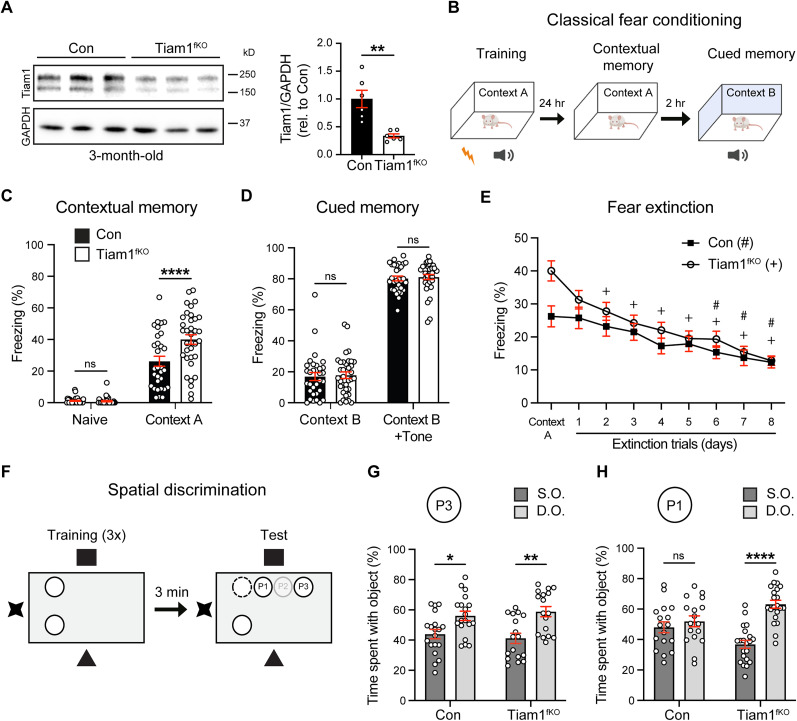
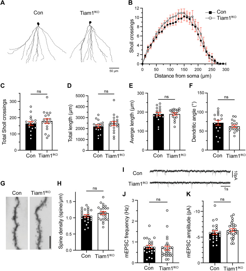
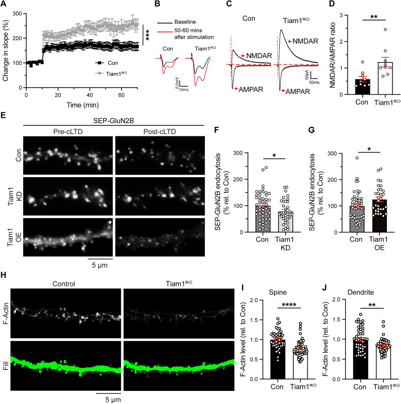
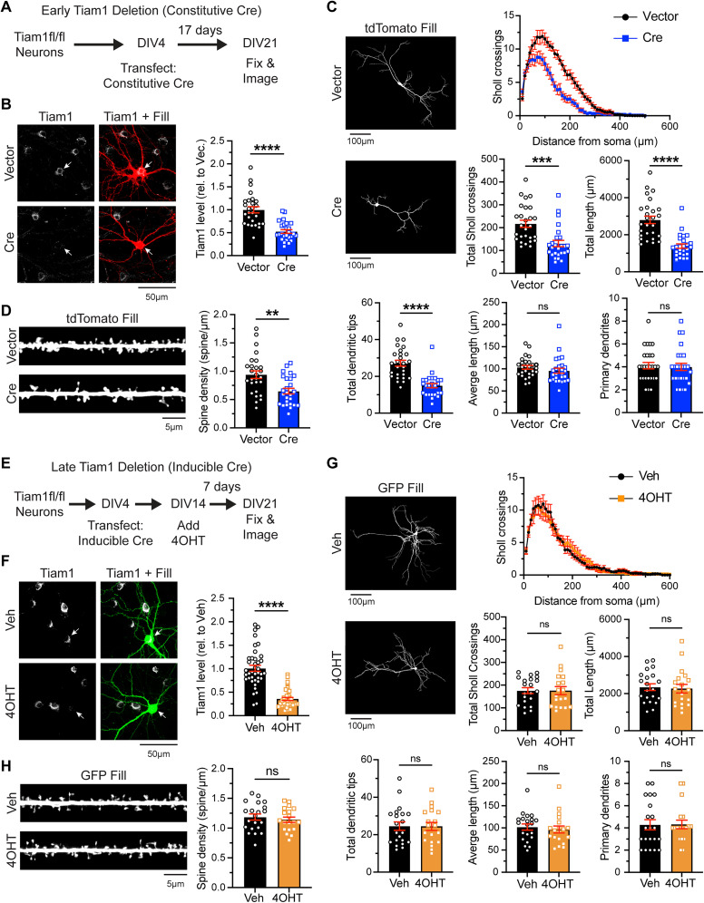
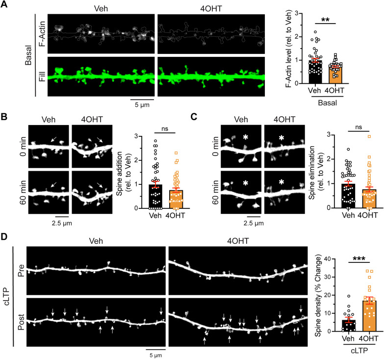
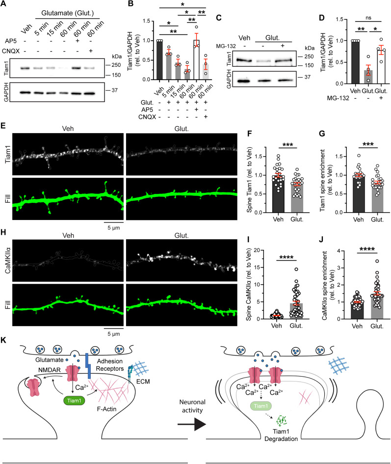
Excitatory synapses and the actin-rich dendritic spines on which they reside are indispensable for information processing and storage in the brain. In the adult hippocampus, excitatory synapses must balance plasticity and stability to support learning and memory. However, the mechanisms governing this balance remain poorly understood. Tiam1 is an actin cytoskeleton regulator prominently expressed in the dentate gyrus (DG) throughout life. Previously, we showed that Tiam1 promotes dentate granule cell synapse and spine stabilization during development, but its role in the adult hippocampus remains unclear. Here, we deleted Tiam1 from adult forebrain excitatory neurons (Tiam1fKO ) and assessed the effects on hippocampal-dependent behaviors. Adult male and female Tiam1fKO mice displayed enhanced contextual fear memory, fear extinction, and spatial discrimination. Investigation into underlying mechanisms revealed that dentate granule cells from Tiam1fKO brain slices exhibited augmented synaptic plasticity and N-methyl-D-aspartate-type glutamate receptor (NMDAR) function. Additionally, Tiam1 loss in primary hippocampal neurons blocked agonist-induced NMDAR internalization, reduced filamentous actin levels, and promoted activity-dependent spine remodeling. Notably, strong NMDAR activation in wild-type hippocampal neurons triggered Tiam1 loss from spines. Our results suggest that Tiam1 normally constrains hippocampal-dependent learning and memory in the adult brain by restricting NMDAR-mediated synaptic plasticity in the DG. We propose that Tiam1 achieves this by limiting NMDAR availability at synaptic membranes and stabilizing spine actin cytoskeleton and that these constraints can be alleviated by activity-dependent degradation of Tiam1. These findings reveal a previously unknown mechanism restricting hippocampal synaptic plasticity and highlight Tiam1 as a therapeutic target for enhancing cognitive function.

Keywords: NMDAR; Tiam1; actin cytoskeleton; dendritic spines; hippocampus; learning and memory.


Conflict of interest statement

The authors declare no competing financial interests.

Figures

Figure 1.
Figure 1.
Late postnatal deletion of Tiam1 from forebrain excitatory neurons enhances contextual fear memory, contextual fear extinction, and spatial discrimination. Crossing Tiam1fl/fl mice with CaMKIIα-Cre mice results in deletion of Tiam1 from forebrain excitatory neurons. For all figures, mice are abbreviated as Tiam1fl/fl (control or Con) or Tiam1fl/fl;Cre (Tiam1fKO). A, Immunoblots and quantification of hippocampal lysates from 3-month-old control and Tiam1fKO mice showing Tiam1 loss. Remaining Tiam1 protein is likely due to Tiam1 expression in other cell types, including astrocytes (N = 6 mice per genotype for quantification). Two-tailed Student's t test (t(10) = 4.165; p = 0.0019). B, Adult control and Tiam1fKO mice were subjected to classical fear conditioning (pairing of tone with footshock in Context A), and then tested for (C) contextual fear memory (exposure to Context A 24 h after training) and (D) cued fear memory (exposure to tone in Context B 26 h after training) by recording freezing behavior. Freezing behavior in (C) Context A prior to fear conditioning (naive) and (D) Context B before tone serve as controls for contextual and cued fear memory, respectively. Tiam1fKO mice displayed enhanced contextual fear memory and normal cued fear memory compared with control mice. (control, N = 31; Tiam1fKO, N = 35). Two-way RM ANOVA (contextual memory: main effect genotype, F(1,64) = 8.715; p = 0.0044; main effect training, F(1,64) = 224.5; p < 0.0001; genotype × training interaction, F(1,64) = 10.78; p = 0.0017; cued memory: main effect genotype, F(1,64) = 0.1613; p = 0.6893; main effect training, F(1,64) = 1250; p < 0.0001; genotype × training interaction, F(1,64) = 0.0005; p = 0.9825). Tukey's post hoc test showed a significant difference between Con and Tiam1fKO in Context A; p < 0001. E, Adult control and Tiam1fKO mice were subjected to extinction trials after fear conditioning (i.e., repetitive exposure to Context A without footshock). Fear memory extinction was defined as freezing less than during the initial test 24 h after training (Context A). Tiam1fKO mice extinguished the fear memory by Day 2 (+), whereas control animals did so starting on Day 6 (#; control, N = 31; Tiam1fKO, N = 35). Two-way RM ANOVA main effect genotype F(1,64) = 2.404; p = 0.1260; main effect extinction, F(8,512) = 35.30; p < 0.0001; genotype × extinction interaction, F(8,512) = 3.033; p = 0.0024. F, Control and Tiam1fKO mice were assessed in a spatial discrimination task where mice were exposed to two identical objects and then one object was moved to a new position (P1 or P3). Mice typically spend more time with the D.O. than the S.O. G, This was true for both control and Tiam1fKO mice following a large displacement to P3. H, However, only Tiam1fKO mice spent more time with the displaced object following a small displacement to P1, suggesting Tiam1fKO mice have enhanced spatial discrimination (P3, control N = 19; Tiam1fKO N = 17; P1, control N = 17; Tiam1fKO N = 21). Two-way ANOVA (P3, main effect genotype, F(1,68) = 0.000; p > 0.999; main effect object set, F(1,68) = 21.33; p < 0.0001; genotype × object set interaction, F(1,68) = 0.7587; p = 0.3868; P1, main effect genotype, F(1,72) = 0.000; p > 0.9999; main effect object set, F(1,72) = 24.23; p < 0.0001; genotype × object set interaction, F(1,72) = 13.38; p = 0.0005. Tukey's post hoc test showed a significant difference between S.O. and D.O. for Con (p = 0.0397) and Tiam1fKO (p = 0.0019) mice in P3 and Tiam1fKO mice in P1 (p < 0.0001). Data are ±SEM. *p < 0.05; **p < 0.01; ****p < 0.0001. Not significant (ns), p > 0.05. For additional details, see Extended Data Figure 1-1.
Figure 2.
Figure 2.
DG granule cell dendrites, spines, and excitatory synapses appear normal in adult Tiam1fKO mice. A, Reconstructed morphologies of biocytin-filled DG granule cells from 3.5- to 4-month-old control and Tiam1fKO mice. B, Summary of Sholl crossings, (C) total Sholl intersections, (D) total length, (E) average length, and (F) dendritic angle measurements of DG granule cell dendrites (N = 3 mice per genotype; control n = 17 cells; Tiam1fKO n = 18 cells). Two-tailed Student's t test (total Sholl, t(33) = 0.8260; p = 0.4147; total length, t(33) = 1.077; p = 0.2892; average length, t(33) = 0.0096; p = 0.9924; dendritic angle, t(33) = 1.340; p = 0.1894) (G), Representative images of dendritic spines on biocytin-labeled DG granule cells from 3.5- to 4- month-old control and Tiam1fKO mice and (H) quantification of dendritic spine density (scale bar, 5 μm; N = 3–4 mice per genotype; control n = 26 segments; Tiam1fKO n = 25 segments). Two-tailed Student's t test (t(49) = 1.462; p = 0.1500). I, Representative traces and summary graphs of mEPSC (J) frequency and (K) amplitude recorded from DG granule cells from brain slices of 3.5- to 4-month-old control and Tiam1fKO mice (N = 3–4 mice per genotype; control n = 24 cells; Tiam1fKO n = 27 cells). Two-tailed Student's t test (frequency, t(49) = 0.5107; p = 0.6119; amplitude, t(49) = 1.014; p = 0.3158). No significant difference between control and Tiam1fKO mice is seen for any reported measure. Data are ±SEM. Not significant (ns), p > 0.05.
Figure 3.
Figure 3.
Adult loss of Tiam1 does not affect the production or survival of newborn DG granule cells. The production of adult-born granule cells was monitored by labeling with BrdU and the immature neuronal marker DCX 14 d after BrdU injection. A, Representative immunohistochemistry images of adult-born granule cells from the DG of control and Tiam1fKO mice and (B) quantification of neurons labeled with BrdU with or without DCX showing no change in newborn neuron production (N = 3 mice per genotype, 16 hippocampal sections were analyzed per mouse). Two-tailed Student's t test (BrdU in SGZ: t(4) = 1.066, p = 0.3465; BrdU and DCX in SGZ: t(4) = 0.9163, p = 0.4113; BrdU in GCL: t(4) = 0.5692, p = 0.5997; BrdU and DCX in GCL: t(4) = 1.1260, p = 0.0.3232). Adult-born neuron survival was determined by labeling with BrdU and the mature neuronal marker NeuN 28 d after BrdU injection. C, Representative images of adult-born granule cells from the DG of control and Tiam1fKO mice and (D) quantification of neurons labeled with BrdU with or without NeuN showing no change in newborn neuron survival (N = 3 mice per genotype, 16 hippocampal sections were analyzed per mouse). Two-tailed Student's t test (BrdU in SGZ: t(4) = 1.3420, p = 0.2508; BrdU and NeuN in SGZ: t(4) = 1.681, p = 0.1681; BrdU in GCL: t(4) = 1.037, p = 0.3582; BrdU and NeuN in GCL: t(4) = 1.109, p = 0.3298). SGZ, Subgranule zone; GCL, granule cell layer. Data are ± SEM. Not significant (ns), p > 0.05.
Figure 4.
Figure 4.
Tiam1 restricts NMDAR-dependent synaptic plasticity in the DG, facilitates activity-dependent NMDAR internalization in primary hippocampal neurons, and promotes F-actin assembly/stabilization in the spines and dendrites of DG granule cells. A, High-frequency stimulation (HFS)-induced LTP in the DG from Tiam1fKO hippocampal slices is enhanced compared with control slices (N = 5 mice; n = 8 slices per genotype). Two-way RM ANOVA main effect genotype, F(1,14) = 10.59; p = 0.0058; main effect time, F(4.306,60.29) = 2.924; p = 0.0252; genotype × time interaction, F(59,826) = 1.817; p = 0.0003. (B) Traces of fEPSPs at the baseline and 50–60 mins after HFS. C, Traces of DG granule cell NMDAR- and AMPAR-mediated EPSCs evoked by PP stimulation in slices from adult control and Tiam1fKO mice. D, Quantification indicated an increase in the NMDA/AMPA ratio in Tiam1fKO mice (N = 3 mice; n = 9 cells per genotype). Two-tailed Student's t test (t(16) = 03.133; p = 0.0064). E, Surface fluorescence of SEP-tagged GluN2B before and 5 min after a cLTD treatment (30 µM NMDA, 3 min) in control, Tiam1 knockdown (KD), and Tiam1 overexpressing (OE) rat primary hippocampal neurons at DIV 17–18. Quantification of SEP-GluN2B internalization [expressed as SEP-GluN2B endocytosis (% rel. to Con)] showed that (F) Tiam1 KD reduced (N = 3 independent sets; control n = 47 cells; KD n = 35 cells) and (G) Tiam1 OE increased (N = 3 independent sets; control n = 49 cells; OE n = 31 cells) the internalization of GluN2B-containing NMDARs relative to control neurons. Two-tailed Student's t test (KD, t(76) = 2.342; p = 0.0218; OE, t(78) = 2.137; p = 0.0357). H, Phalloidin staining showing basal F-actin in dendritic segments from brain sections of eGFP-expressing adult control and Tiam1fKO mice (N = 3 mice; 45 segments per genotype). Quantification showed lower levels of F-actin in (I) spines and (J) dendrites of Tiam1fKO animals compared with control mice. Two-tailed Student's t test (spines, t(88) = 4.822; p < 0.0001; dendrite, t(88) = 3.159; p = 0.0022). Data are ±SEM. *p < 0.05; **p < 0.01; ****p < 0.0001. Not significant (ns), p > 0.05. See Extended Data Figure 4-1 for additional analyses.
Figure 5.
Figure 5.
Late Tiam1 deletion from hippocampal neuron cultures resembles adult Tiam1 loss in vivo. A, Early deletion of Tiam1 from hippocampal cultures was achieved by transfecting Tiam1fl/fl neurons with a constitutive Cre (Cre) construct and tdTomato as fill. B, Immunocytochemistry was used to quantify Tiam1 loss at DIV 21 (N = 3 independent sets; vector n = 27 cells; Cre n = 26). Two-tailed Student's t test (t(51) = 6.026; p < 0.0001). As expected, early deletion of Tiam1 resulted in a decrease in (C) dendrite complexity, as indicated by a reduction in total Sholl crossings, dendritic length, and dendritic tips, and (D) spine density. Two-tailed Student's t test (total Sholl, t(51) = 4.165; p = 0.0001; total length, t(51) = 6.012; p < 0.0001; total dendritic tips, t(51) = 6.055; p < 0.0001; average length, t(51) = 1.069; p = 0.2901; primary dendrites, t(51) = 0.2651; p = 0.7920; spine density, t(51) = 3.464; p = 0.0011). E, Late deletion of Tiam1 in hippocampal cultures from Tiam1fl/fl mice was accomplished by transfecting neurons with a 4OHT-inducible Cre and eGFP (fill) and treating neurons with 4OHT or vehicle (Veh, as control) at DIV 14, a timepoint where dendrites and spines have formed. F, Similar to early deletion, representative immunocytochemistry image and quantification indicated 4OHT-treated neurons showed Tiam1 loss by DIV 21 (N = 3 independent sets; vehicle n = 21 cells; 4OHT n = 19). Two-tailed Student's t test (t(38) = 7.132; p < 0.0001). Late loss of Tiam1 from cultures did not affect (G) dendrite complexity or (H) spine density. Two-tailed Student's t test (total Sholl, t(38) = 0.0054; p = 0.9958; total length, t(38) = 0.2852; p = 0.7770; total dendritic tips, t(38) = 0.0483; p = 0.9617; average length, t(38) = 0.5134; p = 0.6106; primary dendrites, t(38) = 0.0494; p = 0.9608; spine density, t(38) = 0.6415; p = 0.5250). Data are ±SEM. **p < 0.01; ***p < 0.001. Not significant (ns), p > 0.05. Representative dendritic segments in panels D and H were straightened. For early deletion of Tiam1 using the 4OHT-inducible Cre, see Extended Data Figure 5-1.
Figure 6.
Figure 6.
Hippocampal neurons lacking Tiam1 are primed to undergo NMDAR-dependent synaptic remodeling. A, Phalloidin staining showing basal F-actin in eGFP-expressing vehicle and 4OHT-treated hippocampal mouse neurons indicating decreased F-actin in Tiam1-lacking neurons (N = 3 independent sets; vehicle n = 35 cells, 4OHT n = 30). Two-tailed Student's t test (t(63) = 2.898; p = 0.0052). Live cell imaging of (B) spine addition and (C) elimination events in vehicle- and 4OHT-treated neurons expressing inducible Cre and eGFP (fill) show no difference in basal spine remodeling (N = 4 independent sets; vehicle n = 41 cells; 4OHT n = 41). Two-tailed Student's t test (additions, t(80) = 5.18; p = 0.1329; eliminations, t(80) = 1.6963; p = 0.0944). D, Representative images of spines from vehicle- (Veh, control) and 4OHT-treated (Tiam1 null) Tiam1fl/fl mouse hippocampal neurons expressing inducible Cre and eGFP (fill) before (pre) and 60 min after (post) a cLTP stimulation. Quantification shows an augmented increase in spine density in 4OHT-treated neurons, compared with control (vehicle-) treated neurons (N = 3 independent sets; vehicle n = 15 cells; 4OHT n = 19 cells). Two-tailed Student's t test (t(32) = 4.115; p = 0.0003). Data are ±SEM. **p < 0.01; ***p < 0.001. Not significant (ns), p > 0.05. Representative dendritic segments in panel A were straightened. cLTD was also tested in late Tiam1 deletion neurons (Extended Data Fig. 6-1).
Figure 7.
Figure 7.
Robust NMDAR activity can induce Tiam1 degradation. A, Representative immunoblot assessing endogenous Tiam1 levels from rat hippocampal neurons treated with vehicle (Veh) or glutamate (50 µM) for increasing time periods. GAPDH was used as loading control. B, Quantification of Western blots showing significant Tiam1 loss at 15 and 60 min postglutamate treatment. Preincubation with the NMDAR inhibitor AP5 (100 µM) but not the AMPAR inhibitor CNQX (100 µM) blocked Tiam1 loss induced by 60 min glutamate treatment (N = 3 independent sets). ANOVA (F(5,12) = 9.854; p = 0.0006) followed by Tukey's post hoc test. C, D, Similarly, the proteasome inhibitor MG-132 (10 µM) also blocked Tiam1 loss induced by 60 min glutamate treatment (N = 4 independent sets). ANOVA (F(2,9) = 14.34; p = 0.0016). Tukey's post hoc test showed a difference between Veh versus Glut. (p = 0.0014) and Glut. versus Glut. + MG-132 (p = 0.0142). E, Immunocytochemistry of DIV 21 hippocampal neurons expressing eGFP (fill) and Flag-tagged Tiam1 and treated with vehicle (Veh) or glutamate for 15 min. Neurons were fixed and stained with anti-Flag antibodies. F, G, Quantification showed glutamate-induced Tiam1 loss from spines (N = 3 independent sets; Veh n = 24 cells; Glut. N = 25 cells). Quantification measures of (F) relative levels of Tiam1 in spines (Spine Tiam1) and (G) relative levels of Tiam1 in spines versus dendrites (Tiam1 spine enrichment). Two-tailed Student's t test (spine, t(47) = 2.396; p = 0.0206; spine enrichment, t(47) = 3.746; p = 0.0005). H, Representative images and (I, J) quantification showing that 15 min glutamate treatment resulted in the enrichment of endogenous CaMKIIα in spines, in contrast to Tiam1 (N = 3 independent sets; Veh n = 47 cells; Glut. N = 39). Two-tailed Student's t test (spine, t(84) = 7.198; p < 0.0001; spine enrichment, t(84) = 5.442; p < 0.0001). K, Proposed model. Tiam1 normally restrict the plasticity of DG granule cell synapses by limiting the availability of NMDARs at synaptic membranes and promoting actin filament assembly/stabilization at spines. Strong neuronal activity induces Tiam1 degradation, priming synapses for NMDAR-dependent remodeling. Data are ±SEM. *p < 0.05; **p < 0.01; ***p < 0.001; ****p < 0.0001. Not significant (ns), p > 0.05. Model made using BioRender. For additional analyses, see Extended Data Figure 7-1.

Similar articles

References

    1. Abraham WC, Robins A (2005) Memory retention–the synaptic stability versus plasticity dilemma. Trends Neurosci 28:73–78. 10.1016/j.tins.2004.12.003 - DOI - PubMed
    1. Allen TA, Fortin NJ (2013) The evolution of episodic memory. Proc Natl Acad Sci U S A 110:10379–10386. 10.1073/pnas.1301199110 - DOI - PMC - PubMed
    1. Antonelli R, De Filippo R, Middei S, Stancheva S, Pastore B, Ammassari-Teule M, Barberis A, Cherubini E, Zacchi P (2016) Pin1 modulates the synaptic content of NMDA receptors via prolyl-isomerization of PSD-95. J Neurosci 36:5437–5447. 10.1523/JNEUROSCI.3124-15.2016 - DOI - PMC - PubMed
    1. Bavelier D, Levi DM, Li RW, Dan Y, Hensch TK (2010) Removing brakes on adult brain plasticity: from molecular to behavioral interventions. J Neurosci 30:14964–14971. 10.1523/JNEUROSCI.4812-10.2010 - DOI - PMC - PubMed
    1. Bingol B, Schuman EM (2006) Activity-dependent dynamics and sequestration of proteasomes in dendritic spines. Nature 441:1144–1148. 10.1038/nature04769 - DOI - PubMed

Substances

LinkOut - more resources