

[Electroacupuncture improves learning and memory function and promotes hippocampal synaptic regeneration in rats with cerebral ischemia-reperfusion injury]
- PMID: 39725620
- PMCID: PMC11683340
- DOI: 10.12122/j.issn.1673-4254.2024.12.07
[Electroacupuncture improves learning and memory function and promotes hippocampal synaptic regeneration in rats with cerebral ischemia-reperfusion injury]
Abstract
Objectives: To explore the neuroprotective mechanism of electroacupuncture at the acupoints Baihui and Shenting in rats with cerebral ischemia-reperfusion (IR) injury.
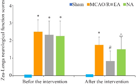
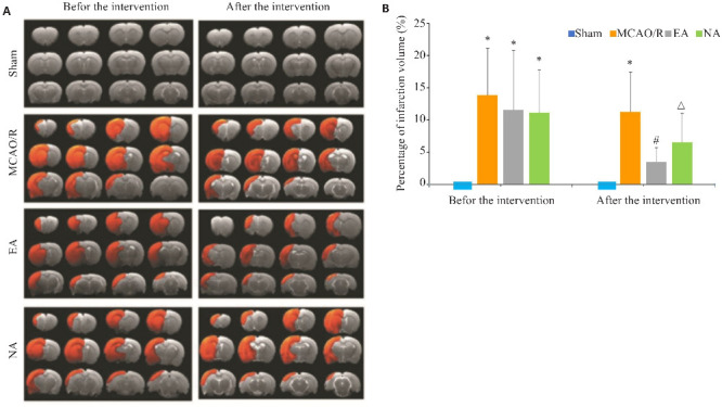
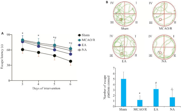
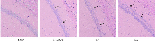
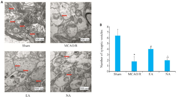
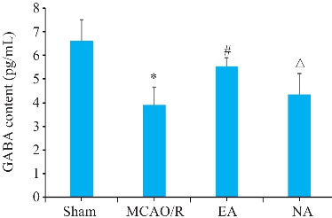
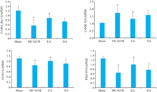
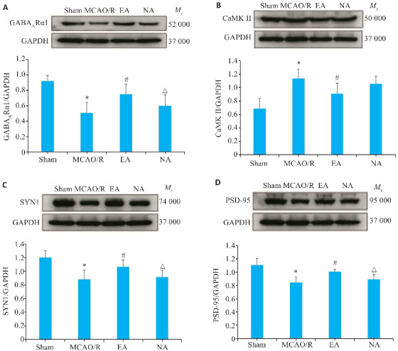
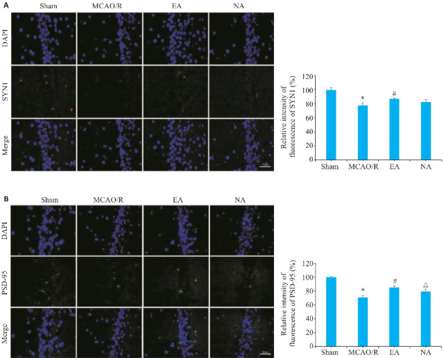
Methods: Forty-eight male SD rats were equally randomized into sham operation group, cerebral IR model group, acupoint electroacupuncture group and non-acupoint acupuncture group. In the latter 3 groups, cerebral focal ischemic injury was induced using the Longa method; in the two electroacupuncture groups, electroacupuncture was performed either at the acupoints Baihui and Shenting or at non-acupoint sites for 7 days. The changes in neurological deficit scores, cerebral infarction volume, learning and memory function, pathologies in hippocampal CA1 area, neuronal and synaptic ultrastructures, and synaptic density of the rats were observed, and serum GABA level and mRNA and protein expressions of GABAAR α1, CaMK II, SYN1 and PSD-95 in the hippocampal tissue were detected.
Results: Compared with those in cerebral IR model group, the rats receiving electroacupuncture at the acupoints, but not those with electroacupuncture at the non-acupoints, showed significantly decreased neurological deficit scores and cerebral infarction volume with shortened escape latency and increased platform crossings. Electroacupuncture at the acupoints significantly increased neuronal cell number, decreased the width of the synaptic gaps and increased density of synaptic bodies in the ischemic hippocampal CA1 area, resulting also in increased serum GABA levels and hippocampal expressions of GABAARα1, SYN1 and PSD-95 and lowered expression level of CaMK II.
Conclusions: Electroacupuncture at Baihui and Shenting improves learning and memory function of rats with cerebral IR injury possibly through a mechanism that promotes synaptic regeneration, upregulates hippocampal expressions of GABAAR α 1, SYN1 and PSD-95 and downregulates the expression of CaMK II.
目的: 探讨电针百会、神庭对脑缺血再灌注损伤大鼠的神经保护作用机制。方法: 将雄性SD大鼠48只随机分为假手术组、模型组、电针组和非穴组,12只/组。采用Longa法制备局灶性缺血损伤大鼠模型。电针干预大鼠百会、神庭穴7 d。观察大鼠神经功能缺损评分、脑梗死体积变化、学习记忆功能、海马CA1区病理变化、神经元及突触超微结构,并计数突触密度、血清血清γ-氨基丁酸(GABA)、海马组织中GABAARα1、钙调蛋白依赖性蛋白激酶II(CaMKⅡ)、突触素1(SYN1)和突触后致密蛋白-95(PSD-95)蛋白及mRNA表达情况、SYN1和PSD-95表达水平。结果: 与模型组比较,电针组大鼠神经功能缺损评分降低(1.75±0.45 vs 1.50±0.67,P<0.05);脑梗死体积降低(11.25±6.22 vs 3.51±2.21,P<0.05);逃避潜伏期降低下降(62.65±5.12 vs 51.83±6.19,P<0.05)及穿越平台次数增加(1.17±0.75 vs 3.17±0.75,P<0.05);缺血侧海马CA1区神经细胞数量增加;突触间隙宽度相对变小,突触小体相对增加(2.00±0.11 vs 4.00±0.25, P<0.05);血清中GABA含量上升(3.90±0.75 vs 5.54±0.35,P<0.05);海马组织中GABAARα1、SYN1和PSD-95 mRNA表达水平升高(P<0.05);CaMKⅡ mRNA表达水平下降(P<0.05)。结论: 电针百会、神庭可改善脑缺血再灌注损伤大鼠学习记忆功能,其机制可能通过促进突触再生,上调GABAARα1、SYN1和PSD-95表达,下调CaMKⅡ表达有关。.
Keywords: cerebral ischemia-reperfusion injury; electroacupuncture; learning and memory function; synaptic plasticity; synaptic regeneration.
Figures
Similar articles
-
[Effects of electroacupuncture on learning-memory function and synaptic plasticity in rats with cerebral ischemia-reperfusion injury based on the BDNF/TrkB pathway].Zhongguo Zhen Jiu. 2024 Sep 12;44(9):1037-45. doi: 10.13703/j.0255-2930.20231208-k0006. Zhongguo Zhen Jiu. 2024. PMID: 39318295 Chinese.
-
[Electroacupuncture improves learning and memory impairment and enhances hippocampal synaptic plasticity through BDNF/TRKB/CREB signaling pathway in cerebral ischemia-reperfusion injury rats].Zhen Ci Yan Jiu. 2023 Sep 25;48(9):843-51. doi: 10.13702/j.1000-0607.20220742. Zhen Ci Yan Jiu. 2023. PMID: 37730254 Chinese.
-
[Effect of electroacupuncture on proBDNF/mBDNF and synaptic plasticity in rats with learning and memory impairment after cerebral ischemia-reperfusion].Zhen Ci Yan Jiu. 2024 Apr 25;49(4):391-397. doi: 10.13702/j.1000-0607.20221313. Zhen Ci Yan Jiu. 2024. PMID: 38649207 Chinese.
-
[Effects of electroacupuncture on mitochondrial autophagy and Sirt1/FOXO3/PINK1/Parkin pathway in rats with learning-memory impairment after cerebral ischemia reperfusion injury].Zhongguo Zhen Jiu. 2025 Feb 12;45(2):193-9. doi: 10.13703/j.0255-2930.20231012-k0005. Zhongguo Zhen Jiu. 2025. PMID: 39943761 Chinese.
-
Electroacupuncture at GB20 improves cognitive ability and synaptic plasticity via the CaM-CaMKII-CREB signaling pathway following cerebral ischemia-reperfusion injury in rats.Acupunct Med. 2024 Feb;42(1):23-31. doi: 10.1177/09645284231202805. Epub 2023 Oct 15. Acupunct Med. 2024. PMID: 38126262
References
-
- 《中国脑卒中防治报告》编写组 . 《中国脑卒中防治报告2020》概要[J]. 中国脑血管病杂志, 2022, 19(2): 136-44.
-
- Mok VC, Lam BY, Wong A, et al. . Early-onset and delayed-onset poststroke dementia-revisiting the mechanisms[J]. Nat Rev Neurol, 2017, 13(3): 148-59. - PubMed
-
- Bettio LEB, Rajendran L, Gil-Mohapel J. The effects of aging in the hippocampus and cognitive decline[J]. Neurosci Biobehav Rev, 2017, 79: 66-86. - PubMed
Publication types
MeSH terms
Substances
Grants and funding
LinkOut - more resources
Full Text Sources
Medical
Miscellaneous