. 2024 Dec 27;82(1):24.
doi: 10.1007/s00018-024-05540-5.

Amiloride sensitizes prostate cancer cells to the reversible tyrosine kinase inhibitor lapatinib by modulating Erbb3 subcellular localization

Affiliations

Amiloride sensitizes prostate cancer cells to the reversible tyrosine kinase inhibitor lapatinib by modulating Erbb3 subcellular localization

Maitreyee K Jathal et al. Cell Mol Life Sci. .

Abstract

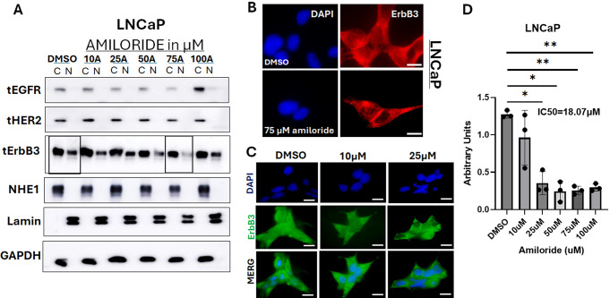
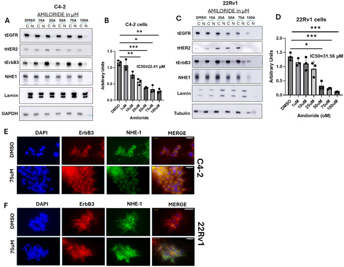
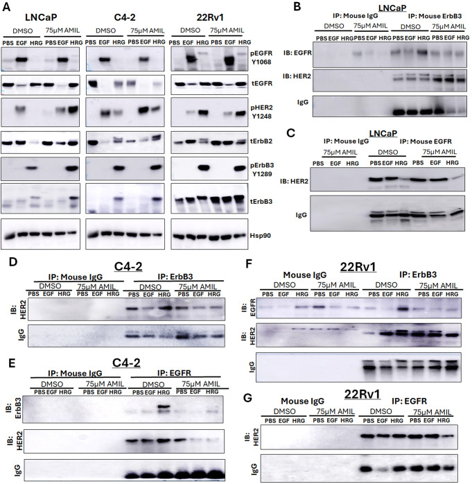
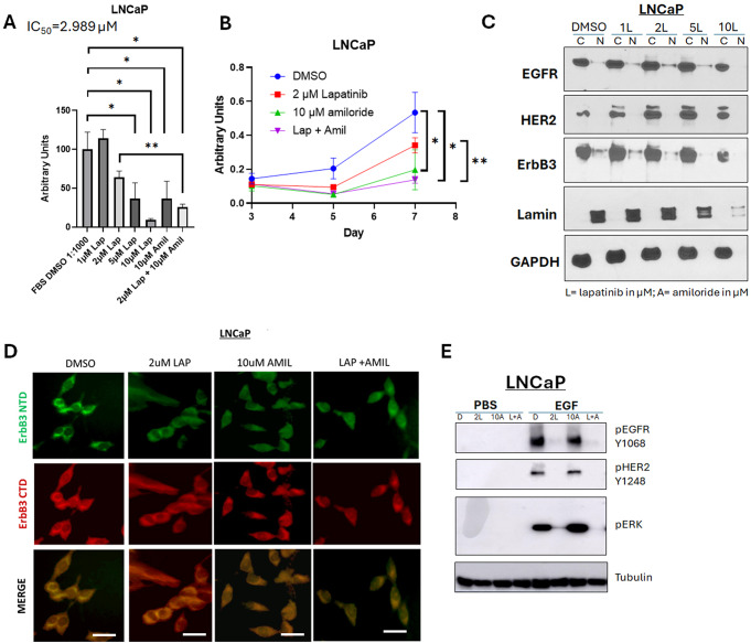
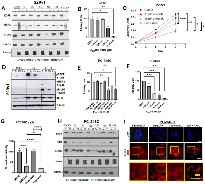
Neoadjuvant therapy (NAT) has been studied in clinically localized prostate cancer (PCa) to improve the outcomes from radical prostatectomy (RP) by 'debulking' of high-risk PCa; however, using androgen deprivation therapy (ADT) at this point risks castration resistant PCa (CRPC) clonal proliferation. Our goal is to identify alternative NAT that reduce hormone sensitive PCa (HSPC) without affecting androgen receptor (AR) transcriptional activity. PCa is associated with increased expression and activation of the epidermal growth factor receptor (EGFR) family, including HER2 and ErbB3. The FDA-approved HER2 inhibitor lapatinib has been tested in PCa but was ineffective due to continued activation of ErbB3. We now demonstrate that this is due to ErbB3 being localized to the nucleus in HSPC and thus protected from lapatinib which affect membrane localized HER2/ErbB3 dimers. Here, we show that the well-established, well-tolerated potassium-sparing diuretic amiloride hydrochloride dose dependently prevented ErbB3 nuclear localization via formation of plasma membrane localized HER2/ErbB3 dimers. This in turn allowed lapatinib inactivation of these dimers via inhibition of its target HER2, which dephosphorylated ERK1/2 and inhibited survival. Amiloride combined with lapatinib significantly increased apoptosis at relatively low doses of both drugs but did not affect AR transcriptional activity. Thus, our data indicate that a combination of amiloride and lapatinib could target HSPC tumors without problems associated with using ADT as NAT in HSPC.

Keywords: Amiloride; Androgen receptor; ErbB3; Heregulin-1β; Lapatinib; Prostate cancer; Subcellular localization.


Conflict of interest statement

Declarations. Ethics approval and consent to participate: Not applicable. Consent for publication: All authors have consented to the contents of the publication. No other consent is necessary as all data presented have been generated by the authors. Conflict of interest: The authors declare that they have no conflicts of interest with the contents of this article.

Figures

Fig. 1
Fig. 1
Amiloride promoted ErbB3 translocation from the nucleus to the cytoplasm and the plasma membrane in HSPC cells (A) Hormone-sensitive LNCaP cells were treated with varying concentrations of amiloride for 72 h before being lysed, fractionated and analyzed by immunoblot. (B) Immunofluorescence microscopy (IF) in LNCaP cells treated with DMSO or 75 µM or and probed with IF specific C-terminal ErbB3 antibodies or (C) 10µM or 25µM amiloride and probed with IF specific N-terminal ErbB3 antibodies for 72 h (scale bars = 30 μm). Note that vehicle treated LNCaP cells expressed nuclear ErbB3 (red) whereas amiloride-treated cells had significantly decreased ErbB3 expression in the nucleus (hollowed out). Location of nuclei are identified by blue DAPI staining. Plasma membrane localization of ErbB3 at cell-cell junction was also noted in amiloride-treated but not in vehicle treated cells. Note that both N- and C-terminal ErbB3 antibodies demonstrate lighter nuclear staining in amiloride-treated cells. (D) Cells were subjected to viability assays using the stated concentrations of amiloride. p-values are calculated with respect to DMSO
Fig. 2
Fig. 2
Amiloride does not promote ErbB3 translocation from the nucleus to the cytoplasm and the plasma membrane in CRPC cells (A) Hormone-insensitive C4-2 or (C) the unrelated cell line 22Rv1 cells were also treated with varying concentrations of amiloride for 72 h before being subjected to viability assays or lysed and fractionated as previously described. For all viability assays, results were obtained from triplicate experiments. Error bars represent standard deviation. Tables show p-values with respect to DMSO for each tested cell line. All densitometry was performed using ImageJ. C = cytoplasmic and N = nuclear. (B, D) Cells were subjected to viability assays using the stated concentrations of amiloride. p-values are calculated with respect to DMSO. (E, F) C4-2 and 22Rv1 cells were treated with IF-specific antibodies to NHE-1 (prototypical target of amiloride) and imaged as described in previous figure legends
Fig. 3
Fig. 3
Differential activation and dimerization of ErbB family members and their downstream targets in HSPC and CRPC cells with high concentrations of amiloride (A) HSPC (LNCaP) and CRPC (C4-2, 22Rv1) cells were treated for 72 h with 75µM amiloride dissolved in 100% sterile DMSO and stimulated with PBS, EGF or HRG for 15 min prior to collection to observe activation of ErbB family members and their downstream targets. Cells were lysed in denaturing lysis buffer before being analysed by immunoblotting. 25 µg of protein were loaded per lane. Hsp90 was used as a loading control. (B,C) LNCaP cells (HSPC) or (D,E) C4-2 cells (CRPC) or (F, G) 22Rv1 cells (unrelated CRPC cells) were treated with 75µM amiloride or 100% DMSO (0.1% v/v) for 72 h and stimulated with PBS, EGF or HRG for 15 min to activate ErbB family dimers just prior to collection. 400ug of whole cell lysate were used in each pulldown lane. Mouse IgG antibody was used as an isotype control. Amiloride increases ErbB3-HER2 dimers and stabilizes ErbB3-EGFR dimers
Fig. 4
Fig. 4
Amiloride efficacy is enhanced by EGFR knockdown in HSPC cells and by HER2 knockdown in CRPC cells. (A) CRPC C4-2 cells were transfected with control (CT) or EGFR or ErbB3 siRNA or (B) HER2 siRNA and treated with or without 75µM amiloride before being analysed for changes in viability with the MTT assay. Error bars represent standard deviation. Experiments were performed in triplicate. (C) Whole cell immunoblot for siRNA efficacy. 20 µg of protein were loaded per lane. Tubulin was used as a loading control. (D) 22Rv1 cells (CRPC) were transiently transfected with empty vector (EV), EGFR (B1), ErbB2 (B2), ErbB3 (B3) or mutant ErbB3 (mB3) as previously described by us in detail [23, 45]. Cells were collected, fractionated and analysed by immunoblot as described in previous figure legends. (E) ErbB3 overexpression was visualized microscopically in 22Rv1 cells using the reagents and procedures as described in previous figure legends
Fig. 5
Fig. 5
Amiloride enhances the sensitivity of HSPC cells to low concentrations of lapatinib (A, B) LNCaP cells were treated with 1–10µM lapatinib and assayed for viability. Lapatinib and amiloride were both dissolved in 100% DMSO. In a head-to-head comparison of lapatinib and amiloride, the combination was additive when cell viability was assayed with the MTT reagent. Experiments were done in triplicate. Error bars represent standard deviation. (C) Cells were treated with varying concentrations of lapatinib for 72 h before being collected and lysed into cytoplasmic and nuclear fractions as described in earlier figure legends. (D) Cells were treated with 2µM lapatinib, 10µM amiloride or a combination of the two for 72 h before being collected, fixed and processed for indirect immunofluorescent microscopy using immunofluorescent-specific antibodies to the C- and N- termini of ErbB3 (‘CTD’ and ‘NTD’ respectively) as previously described. Scale bars = 7.5 μm. Co-administration of lapatinib and amiloride increases the accumulation of ErbB3 compared to 2µM lapatinib alone. (E) LNCaP cells were treated for 72 h with lapatinib, amiloride or the combination or 100% sterile DMSO and stimulated with PBS, EGF or HRG for 15 min prior to collection to observe activation of ErbB family members and their downstream targets. Cells were lysed in denaturing lysis buffer before being analysed by immunoblotting. 25 µg of protein were loaded per lane. Tubulin was used as a loading control
Fig. 6
Fig. 6
Amiloride enhances the sensitivity of HSPC and CRPC cell lines to low concentrations of lapatinib. (A) 22Rv1 cells were treated with varying concentrations of lapatinib, 10µM amiloride or a combination of the two for 72 h before being collected and lysed into cytoplasmic and nuclear fractions as described in earlier figure legends. Co-administration of lapatinib and amiloride does not increase the accumulation of ErbB3 in the cytoplasmic fraction. (B) 22Rv1 cells were treated with 1–10µM lapatinib and assayed for viability. Lapatinib was dissolved in 100% DMSO. Experiments were done in triplicate. Error bars represent standard deviation. (C) In a head-to-head comparison of lapatinib and amiloride, the combination was additive when cell viability was assayed with the MTT reagent. Experiments were done in triplicate. Error bars represent standard deviation. Coloured dotted line estimates 50% viability. Table shows p-values with respect to FBS DMSO. (D) 22Rv1 (CRPC) cells were treated for 72 h with lapatinib, amiloride or the combination or 100% sterile DMSO and stimulated with PBS, EGF or HRG for 15 min prior to collection to observe activation of ErbB family members and their downstream targets. Cells were lysed in denaturing lysis buffer before being analysed by immunoblotting. 25 µg of protein were loaded per lane. Tubulin was used as a loading control. Tables show p-values with respect to PBS DMSO in each cell line tested. (E) PC-346 C cells were treated with 10–60 µM of amiloride and assayed for viability. Amiloride was dissolved in 100% DMSO. Experiments were done in triplicate. Error bars represent standard deviation. (F) PC-346 C cells were treated with 1–10µM lapatinib and assayed for viability. Lapatinib was dissolved in 100% DMSO. Experiments were done in triplicate. Error bars represent standard deviation. (G) PC-346 C cells were treated with lapatinib, amiloride or the combination, which is shown to be additive when cell viability was assayed with the MTT reagent. Experiments were done in triplicate. Error bars represent standard deviation. (H) PC-346 C cells were treated with varying concentrations of lapatinib, 10µM amiloride or a combination of the two for 72 h before being collected and lysed into cytoplasmic and nuclear fractions as described in earlier figure legends. (I) Cells were treated with 2µM lapatinib, 10µM amiloride or a combination of the two for 72 h before being collected, fixed and processed for indirect immunofluorescent microscopy using immunofluorescent-specific antibodies to the C-terminal domain of ErbB3 as previously described. Yellow boxes (inset) and bold yellow arrow depict area of negligible ErbB3 nuclear staining. Scale bars = 7.5 μm
Fig. 7
Fig. 7
Amiloride and lapatinib synergize to increase apoptosis in HSPC and CRPC cell lines. (A-C) HSPC and CRPC cell lines were treated with 100% DMSO, lapatinib, amiloride or the combination (in µM) for 72 h before being processed for cell death analysis using annexin V and propidium iodide staining. The percentage of cells undergoing early or late apoptosis with DMSO treatment was set to 100% and values for the various treatment conditions calculated accordingly. Experiments were performed in triplicate. Error bars represent standard deviation. (D) Schematic with proposed molecular mechanism of lapatinib-amiloride efficacy. (a) EGFR, HER2 and ErbB3 exist at the cell membrane and signal via pathways such as ERK and AKT. (b,c) ErbB3 monomers cycle between the nucleus and cell membrane. (EGFR and HER2 behave similarly but have been omitted for clarity). (d) Lapatinib is a dual-kinase TKI (tyrosine kinase inhibitor) of HER2 and EGFR dimers but will also inhibit HER2 in HER2-ErbB3 dimers. Lapatinib is unlikely able to inhibit ErbB3 if it is in the nucleus and not at the cell surface. (e) Amiloride is a macropinocytosis inhibitor that prevents internalization of ErbB3 and retains it at the cell surface. As a result, nuclear ErbB3 decreases and cytoplasmic surface ErbB3 increases. (f) Amiloride-induced ErbB3 retention enables its dimerization with HER2, enabling the formation of ErbB3-HER2 dimers which are now inhibited by the addition of low concentrations of lapatinib

Update of

Similar articles

Cited by

References

    1. Eifler JB, Humphreys EB, Agro M, Partin AW, Trock BJ, Han M (2012) Causes of death after radical prostatectomy at a large tertiary center. J Urol 188(3):798–801 - PubMed
    1. Shore ND, Moul JW, Pienta KJ, Czernin J, King MT, Freedland SJ (2024) Biochemical recurrence in patients with prostate cancer after primary definitive therapy: treatment based on risk stratification. Prostate Cancer Prostatic Dis 27(2):192–201 - PMC - PubMed
    1. Koskas Y, Lannes F, Branger N, Giusiano S, Guibert N, Pignot G et al (2019) Extent of positive surgical margins following radical prostatectomy: impact on biochemical recurrence with long-term follow-up. BMC Urol 19(1):37 - PMC - PubMed
    1. Eastham JA, Heller G, Halabi S, Monk JP 3rd, Beltran H, Gleave M et al (2020) Cancer and Leukemia Group B 90203 (Alliance): radical prostatectomy with or without Neoadjuvant Chemohormonal Therapy in Localized, high-risk prostate Cancer. J Clin Oncol 38(26):3042–3050 - PMC - PubMed
    1. Ryan ST, Patel DN, Parsons JK, McKay RR (2020) Neoadjuvant approaches prior to radical prostatectomy. Cancer J 26(1):2–12 - PubMed

MeSH terms