. 2024 Dec 27;82(1):30.
doi: 10.1007/s00018-024-05548-x.

Endothelial CD38-induced endothelial-to-mesenchymal transition is a pivotal driver in pulmonary fibrosis

Affiliations

Endothelial CD38-induced endothelial-to-mesenchymal transition is a pivotal driver in pulmonary fibrosis

Min Hu et al. Cell Mol Life Sci. .

Abstract

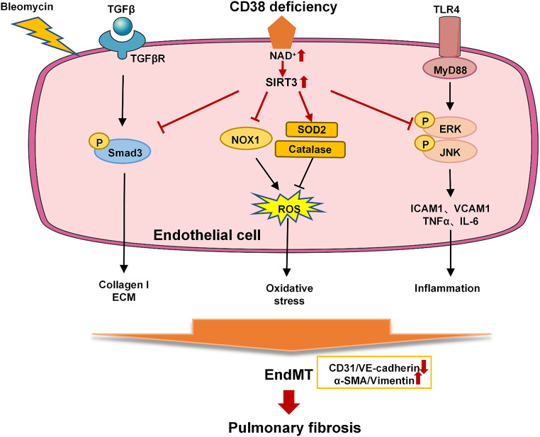
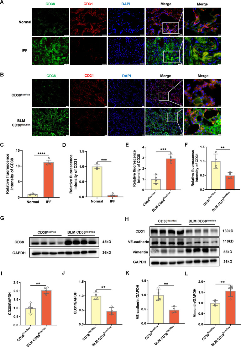
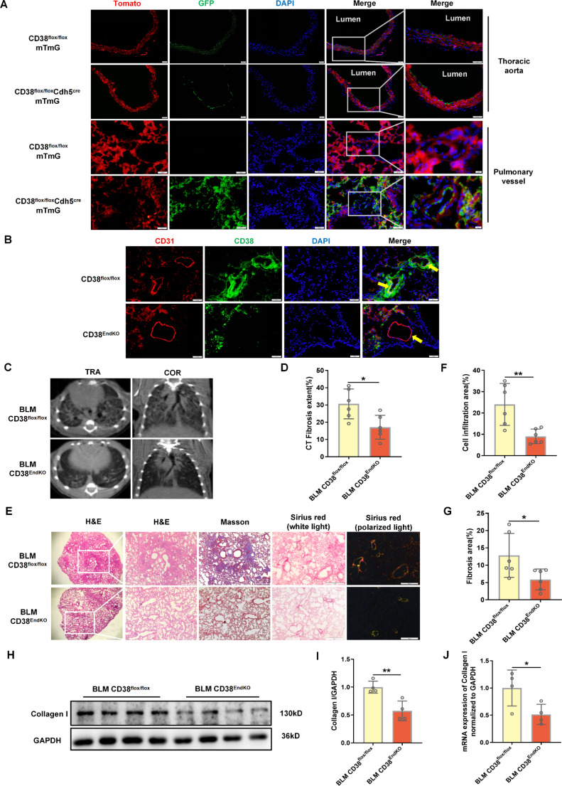
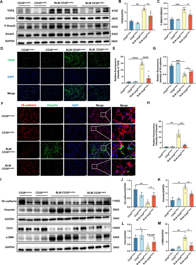
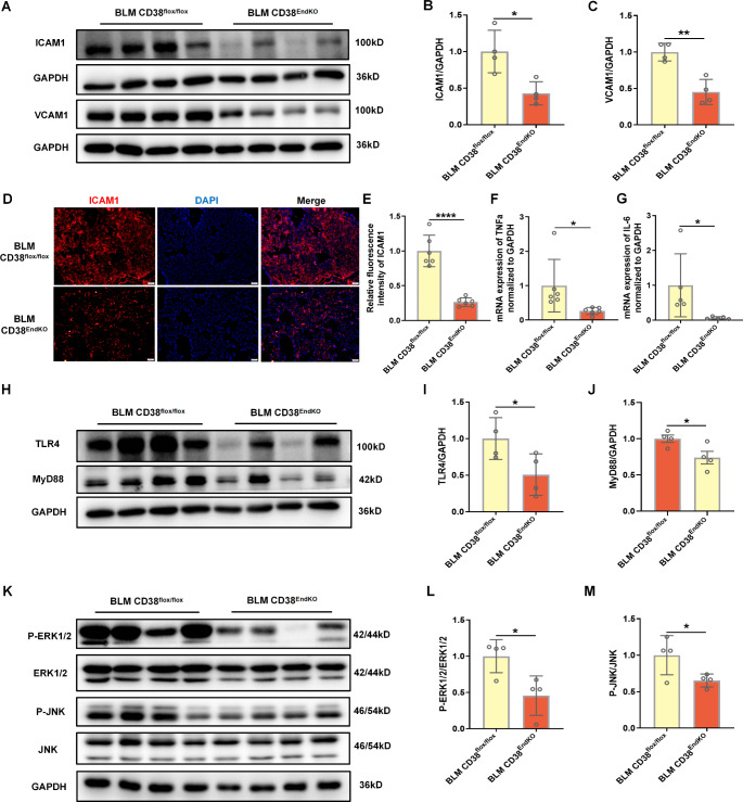
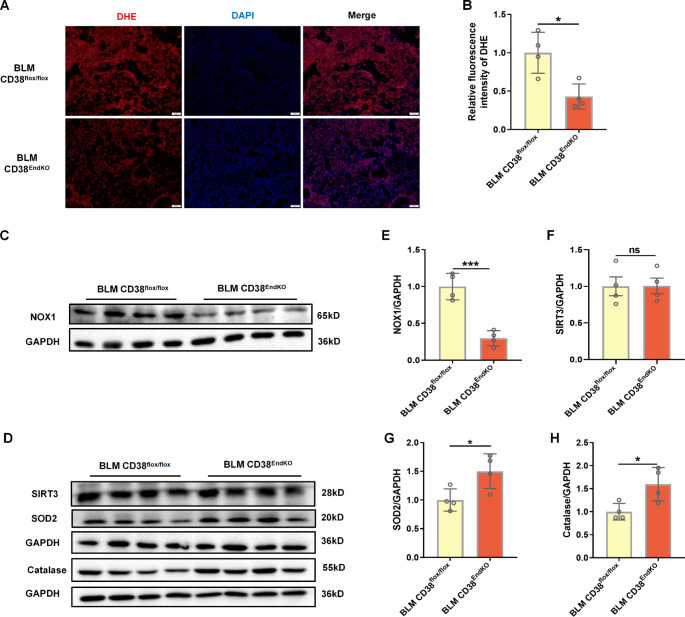
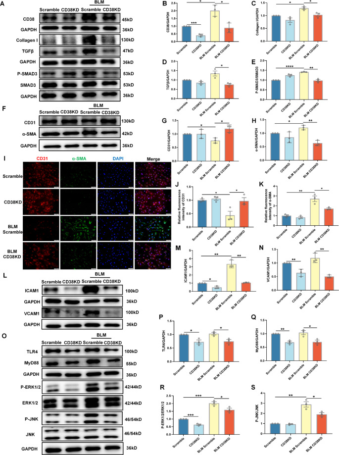
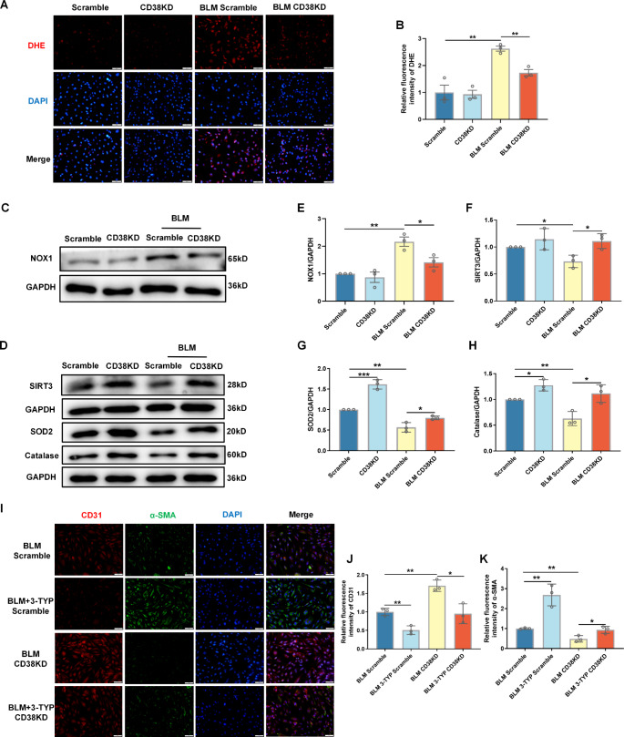
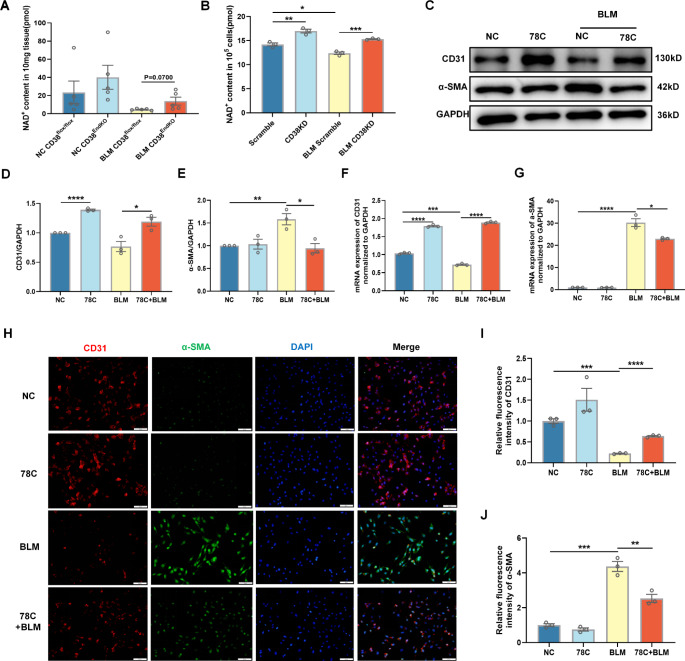
Idiopathic pulmonary fibrosis (IPF) is a prevalent interstitial lung disease with high mortality. CD38 is a main enzyme for intracellular nicotinamide adenine dinucleotide (NAD+) degradation in mammals. It has been reported that CD38 participated in pulmonary fibrosis through promoting alveolar epithelial cells senescence. However, the roles of endothelial CD38 in pulmonary fibrosis remain unknown. In the present study, we observed that the elevated expression of CD38 was related to endothelial-to-mesenchymal transition (EndMT) of lung tissues in IPF patients and bleomycin (BLM)-induced pulmonary fibrosis mice and also in human umbilical vein endothelial cells (HUVECs) treated with BLM. Micro-computed tomography (MCT) and histopathological staining showed that endothelial cell-specific CD38 knockout (CD38EndKO) remarkably attenuated BLM-induced pulmonary fibrosis. In addition, CD38EndKO significantly inhibited TGFβ-Smad3 pathway-mediated excessive extracellular matrix (ECM), reduced Toll-like receptor4-Myeloid differentiation factor88-Mitogen-activated protein kinases (TLR4-MyD88-MAPK) pathway-mediated endothelial inflammation and suppressed nicotinamide adenine dinucleotide phosphate oxidases1 (NOX1)-mediated oxidative stress. Furthermore, we demonstrated that 3-TYP, a SIRT3-specific inhibitor, markedly reversed the protective effect of HUVECsCD38KD cells and 78 C, a CD38-specific inhibitor, on BLM-induced EndMT in HUVECs. Therefore, we concluded that CD38EndKO significantly ameliorated BLM-induced pulmonary fibrosis through inhibiting ECM, endothelial inflammation and oxidative stress, further alleviating EndMT in mice. Our findings suggest that endothelial CD38 may be a new therapeutic target for the prevention and treatment of pulmonary fibrosis clinically.

Keywords: CD38; Endothelial-to-mesenchymal transition; Inflammation; Oxidative stress; Pulmonary fibrosis.


Conflict of interest statement

Declarations. Ethical approval: All the experimental procedures were approved by Nanchang University Institutional Animal Research Committee (Date: 20230822/No: 23 × 007) and were carried out in accordance with Jiangxi Province Laboratory Animal Care Guidelines for the use of animals in research. Consent to participate: Not applicable. Consent for publication: All authors have read the manuscript and agree with submission for publication. Competing interests: The authors have no relevant financial or non-financial interests to disclose.

Figures

None
Protective role and the underlying mechanism of endothelial CD38 in bleomycin-induced pulmonary fibrosis in mice. Endothelial cells-specific CD38 knockout (CD38EndKO) inhibited TGFβ-Smad3-mediated ECM, ROS-mediated oxidative stress and TLR4-mediated inflammation, and in turn, suppressed endothelial-to-mesenchymal transition (EndMT), eventually, alleviated pulmonary fibrosis induced by bleomycin in mice, suggesting endothelial CD38 may be a therapeutic target for the prevention and the treatment of pulmonary fibrosis clinically
Fig. 1
Fig. 1
Analysis of CD38 expression and EndMT related markers in IPF patients and BLM-induced pulmonary fibrosis mice. (A) Co-immunostaining of CD38 and CD31 in lung tissues of normal and IPF patients. (B) Co-immunostaining of CD38 and CD31 in lung tissues of control and BLM-induced mice. (C-D) Quantitative analysis of CD38 and CD31. (E-F) Quantitative analysis of CD38 and CD31. (G) The expression level of CD38 was determined by western blot analysis in lung tissues of control and BLM-induced pulmonary fibrosis mice. (H) The expression level of CD31, VE-cadherin, and Vimentin was determined by western blot analysis in lung tissues of control and BLM-treated mice. (I) Quantitative analysis of CD38 expression. (J-L) Quantitative analysis of CD31, VE-cadherin and Vimentin, respectively. Data were represented as mean ± SEM, **p < 0.01, ***p < 0.001, n = 4 per group
Fig. 2
Fig. 2
Endothelial cell-specific CD38 deficiency (CD38EndKO) ameliorated pulmonary injury and fibrosis in mice. (A) Represent diagram of thoracic aorta and pulmonary vessel slices of CD38flox/floxmTmG and CD38flox/floxCdh5cremTmG mice (B) Co-immunostaining of CD31 and α-SMA of primary vessels of CD38flox/flox and CD38flox/floxCdh5cre mice. (C) Micro CT for lungs of CD38flox/flox and CD38EndKO mice treated with BLM for 21 days. (D) Quantitative analysis of fibrosis extent. (E) H&E, Masson and Sirius red staining of lungs from CD38flox/flox and CD38EndKO mice treated with BLM. (F-G) Quantitative analysis of cell infiltration and fibrosis area. (H) The expression of Collagen I was determined by western blot analysis in lung tissues of CD38flox/flox and CD38EndKO mice treated with BLM. (I) Quantitative analysis of Collagen I. (J) The expression of Collagen I was determined by RT-qPCR in lung tissues of CD38flox/flox and CD38EndKO mice treated with BLM. Data were represented as mean ± SEM, *p < 0.05, **p < 0.01, n = 4–6 per group
Fig. 3
Fig. 3
CD38EndKO ameliorated BLM-induced pulmonary fibrosis by inhibiting TGFβ-Smad3 signal pathway and EndMT in mice. (A) The expressions of TGFβ, phosphorylated and total Smad3 were determined by western blot analysis in lung tissues of CD38flox/flox, CD38flox/flox and CD38EndKO mice treated with saline or BLM. (B-C) Quantitative analysis of TGFβ and phosphorylated Smad3 respectively. (D) The expression of TGFβ was determined by immunofluorescence assay in lung tissues of CD38flox/flox, CD38flox/flox and CD38EndKO mice treated with saline or BLM. (E) Quantitative analysis of TGFβ. (F) Co-immunostaining of VE-cadherin and Vimentin in lung tissues of CD38flox/flox, CD38flox/flox and CD38EndKO mice treated with saline or BLM. (G-H) Quantitative analysis of VE-cadherin and Vimentin. (I) The expressions of VE-cadherin, Vimentin, CD31 and α-SMA were determined by western blot analysis in lung tissues of CD38flox/flox, CD38flox/flox and CD38EndKO mice treated with saline or BLM. (J-M) Quantitative analysis of VE-cadherin, Vimentin, CD31 and α-SMA. Data were represented as mean ± SEM, *p < 0.05, **p < 0.01, ***p < 0.001 ****p < 0.0001, n = 4–6 per group
Fig. 4
Fig. 4
CD38EndKO alleviated BLM-induced pulmonary fibrosis by modulating inflammation in mice. (A) The expressions of ICAM1 and VCAM1 were determined by western blot analysis in lung tissues of CD38flox/flox and CD38EndKO mice treated with BLM. (B-C) Quantitative analysis of ICAM1 and VCAM1. (D) Immunostaining of ICAM1 in lung tissues of CD38flox/flox and CD38EndKO mice treated with bleomycin. (E) Quantitative analysis of ICAM1. (F-G) The expressions of TNFα and IL-6 were determined by RT-qPCR in lung tissues of CD38flox/flox and CD38EndKO mice treated with BLM. (H) The expressions of TLR4 and MyD88 was determined by western blot analysis in lung tissues of CD38flox/flox and CD38EndKO mice treated with BLM. (I-J) Quantitative analysis of TLR4 and MyD88. (K) The expressions of phosphorylated and total ERK1/2 and JNK were determined by western blot analysis in lung tissues of CD38flox/flox and CD38EndKO mice treated with BLM. (L-M) Quantitative analysis of phosphorylated ERK1/2 and JNK. Data were represented as mean ± SEM, *p < 0.05, **p < 0.01, ****p < 0.0001, n = 4–6 per group
Fig. 5
Fig. 5
CD38EndKO decreased BLM-induced pulmonary oxidative stress in mice. (A) DHE staining of lung from CD38flox/flox and CD38EndKO mice treated with BLM. (B) Quantitative analysis of DHE staining. (C) The expression of NOX1 was determined by western blot analysis in lung tissues of CD38flox/flox and CD38EndKO mice treated with BLM. (D) The expressions of SIRT3, SOD2 and Catalase were determined by western blot analysis in lung tissues of CD38flox/flox and CD38EndKO mice treated with BLM. (E-H) Quantitative analysis of NOX1, SIRT3, SOD2 and Catalase. Data were represented as mean ± SEM, *p < 0.05, ***p < 0.001, n = 4 per group
Fig. 6
Fig. 6
CD38 knockdown inhibited BLM-induced excessive ECM, EndMT and inflammation in HUVECs. (A) The expressions of CD38, Collagen I, TGFβ, phosphorylated and total SMAD3 were determined by western blot analysis in scramble and CD38KD HUVECs treated with 10 µg/mL BLM for 48 h. (B-E) Quantitative analysis of CD38, Collagen I, TGFβ and phosphorylated SMAD3, respectively. (F) The expressions of CD31 and α-SMA were determined by western blot analysis in scramble and CD38KD HUVECs treated with 10 µg/mL BLM for 48 h. (G-H) Quantitative analysis of CD31 and α-SMA. (I) Co-immunostaining of CD31 and α-SMA in scramble and CD38KD HUVECs treated with 10 µg/mL BLM for 48 h. (J-K) Quantitative analysis of CD31 and α-SMA. (L) The expressions of ICAM1 and VCAM1 were determined by western blot analysis in scramble and CD38KD HUVECs treated with 10 µg/mL BLM for 48 h. (M-N) Quantitative analysis of ICAM1 and VCAM1. (O) The expressions of TLR4, MyD88, phosphorylated and total ERK1/2 and JNK were determined by western blot analysis in scramble and CD38KD HUVECs treated with 10 µg/mL BLM for 48 h. (P-S) Quantitative analysis of TLR4, MyD88, phosphorylated ERK1/2 and JNK. Data were represented as mean ± SEM, *p < 0.05, **p < 0.01, ***p < 0.001, ****p < 0.0001, n = 3 per group
Fig. 7
Fig. 7
CD38 knockdown attenuated BLM-induced oxidative stress and further EndMT in HUVECs. (A) DHE staining of BLM-induced (10 µg/mL) scramble and CD38KD HUVECs. (B) Quantitative analysis of DHE staining. (C-D) The expressions of NOX1, SIRT3, SOD2 and Catalase were determined by western blot analysis in scramble and CD38KD HUVECs treated with 10 µg/mL BLM for 48 h. (E-H) Quantitative analysis of NOX1, SIRT3, SOD2 and Catalase, respectively. (I) Co-immunostaining of CD31 and α-SMA in BLM-induced (10 µg/mL) scramble and CD38KD HUVECs treated with 3-TYP (50µM) for 48 h. (J-K) Quantitative analysis of CD31 and α-SMA. Data were represented as mean ± SEM, *p < 0.05, **p < 0.01, ***p < 0.001, n = 3 per group
Fig. 8
Fig. 8
Inhibition of CD38 activities attenuated BLM-induced EndMT in HUVECs. (A-B) The content of NAD+ in lung tissues of mice and HUVECs (C) The expressions of CD31 and α-SMA were determined by western blot analysis in HUVECs treated with 78 C (2µM) and BLM (10 µg/mL) for 48 h. (D-E) Quantitative analysis of CD31 and α-SMA. (F-G) The expressions of CD31 and α-SMA were determined by RT-qPCR analysis in HUVECs treated with 78 C (2µM) and BLM (10 µg/mL) for 48 h. (H) Co-immunostaining of CD31 and α-SMA in HUVECs treated with 78 C (2µM) and BLM (10 µg/mL) for 48 h. (I-J) Quantitative analysis of CD31 and α-SMA. Data were represented as mean ± SEM, *p < 0.05, **p < 0.01, ***p < 0.001, ****p < 0.0001, n = 3 per group

Similar articles

References

    1. Bagnato G, Harari S (2015) Cellular interactions in the pathogenesis of interstitial lung diseases. Eur Respir Rev 24:102–114 - PMC - PubMed
    1. Wang X, Wang Y, Rong S, Ma H (2014) [Early treatment with hepatocyte growth factor improves pulmonary artery and right ventricular remodeling in rats with pulmonary artery hypertension by modulating cytokines expression]. Zhonghua Jie He He Hu Xi Za Zhi 37:427–432 - PubMed
    1. Spagnolo P, Cottin V (2017) Genetics of idiopathic pulmonary fibrosis: from mechanistic pathways to personalised medicine. J Med Genet 54:93–99 - PubMed
    1. Wolters PJ, Collard HR, Jones KD (2014) Pathogenesis of idiopathic pulmonary fibrosis. Annu Rev Pathol 9:157–179 - PMC - PubMed
    1. Piera-Velazquez S, Li Z, Jimenez SA (2011) Role of endothelial-mesenchymal transition (EndoMT) in the pathogenesis of fibrotic disorders. Am J Pathol 179:1074–1080 - PMC - PubMed

MeSH terms

LinkOut - more resources