. 2024 Dec 26;82(1):15.
doi: 10.1007/s00018-024-05496-6.

Characterisation and hierarchy of the spermatogonial stem cell compartment in human spermatogenesis by spectral cytometry using a 16-colors panel

Affiliations

Characterisation and hierarchy of the spermatogonial stem cell compartment in human spermatogenesis by spectral cytometry using a 16-colors panel

C Lapoujade et al. Cell Mol Life Sci. .

Abstract

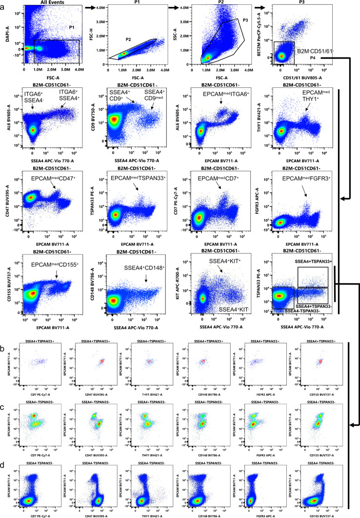
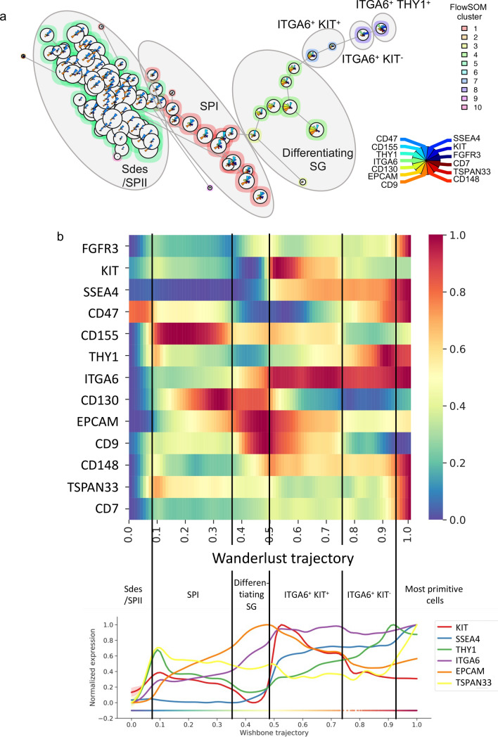
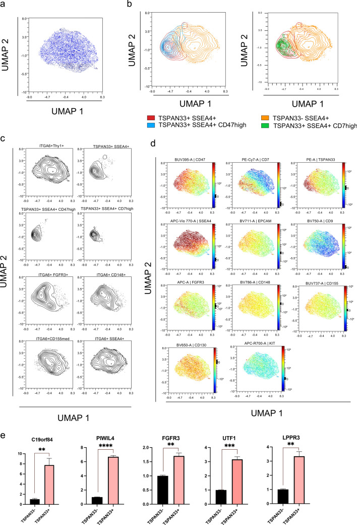
About one in six couples experience fertility problems, and male infertility accounts for about half of these cases. Spermatogenesis originates from a small pool of spermatogonial stem cells (SSCs), which are of interest for the treatment of infertility but remain poorly characterised in humans. Using multiparametric spectral flow cytometric analysis with a 16-colours (16-C) panel of cell markers, we identify novel markers of SSCs and provide insights into unravelling and resolving the heterogeneity of the human spermatogonial cells. This 16-C panel of markers allowed the identification of a primitive SSCs state with the β2M-CD51/61-ITGA6+SSEA4+TSPAN33+THY1+CD9medEPCAMmedCD155+CD148+CD47highCD7high phenotype, with a profile close to the most primitive SSCs states 0 and SSC1-B previously defined by sc-RNAseq approach. The hierarchy of events in the spermatogonial stem cell and progenitor compartment of human spermatogenesis can be delineated. This highlights the importance of a multi-parametric and spectral cytometry approach. The in-depth characterisation of testicular cells should help to overcome the lack of stem cell knowledge, that hinders the understanding of the regenerative potential of SSCs, and is a critical parameter for the successful development of new SSCs-based cell therapies.

Keywords: Human; Spectral cytometry; Spermatogenesis; Spermatogonial stem cell.


Conflict of interest statement

Declarations. Conflict of interest: The authors have no relevant financial or non-financial interests to disclose. Ethical approval and consent: All procedures performed in this protocol were approved by the French Institutional Review Board-Comité de Protection des Personnes, Ile de France IV (IRB 00003835; 2012/40ICB). The participants signed a written informed consent form.

Figures

Fig. 1
Fig. 1
Characterisation of SSCs and spermatogonial progenitors using 8-C fluorescence panel. a Representative flow cytometry plots of the 8-C panel analysis on the human spermatogonial progenitors according to forward (FSC) and side scatter (SSC), β2 microglobulin, ITGA6, SSEA4, KIT, THY1, FGFR3, and TSPAN33 parameters. Viable propidium iodide (PI)-negative cells were gated based on morphology to remove cellular debris and doublets. Population frequencies are shown in the graph. b Expression of SSEA4, FGFR3, or TSPAN33 in the β2MITGA6+THY1+ population. c Flow cytometric cytogram of TSPAN33 and SSEA4 markers in the β2M germinal population, and THY1 and KIT expression in SSEA4+TSPAN33+, SSEA4+TSPAN33, SSEA4TSPAN33 germinal populations. Arrows indicate the strategy of gating and analysis
Fig. 2
Fig. 2
Flow cytometry profiling of adult testicular cell samples using the 16-C panel. a Representative flow cytometry plots of the spectral flow analysis on the human spermatogonial progenitors according to FSC, SSC, β2M, CD51/CD61, ITGA6, THY1, SSEA4, and TSPAN33 parameters. Testicular cells were gated based on morphology to remove cellular debris, sperm and doublets. bd Expression of CD7, CD155, CD148, CD47, and KIT markers in the β2M/CD51/CD61SSEA4+TSPAN33+ (b), the β2M/CD51/CD61SSEA4+TSPAN33 (c), and the β2M/CD51/CD61SSEA4TSPAN33 (d) germinal populations. Arrows indicate the strategy of gating and analysis. Figure S4 shows the most relevant fluorescence minus one (FMO) controls. ITGA6 is marked AL6 in the cytogram axis
Fig. 3
Fig. 3
Comprehensive analysis of multidimensional 16-C panel dataset of human testicular cells. a High-dimensional UMAP analysis of human testicular cells, (b) Testicular cell subsets defined in Fig. 2 and Fig. S3 are indicated on the UMAP. Spermatocyte I (SPI), spermatocyte II (SPII), spermatids (Sd), and the “S phase” subpopulation (differentiating spermatogonia) were assigned according to the expression of CD148, CD47, CD155 and ITGA6 as previously defined in Figure S2. c Expression patterns of individual spermatogonial markers overlaid onto the UMAP representation of human testis cells. The colour indicates the fluorescence intensity of the marker (red: high to blue: neg)
Fig. 4
Fig. 4
Trajectory analysis of the human germ cell differentiation dataset. a FlowSOM tree inferred from unsupervised clustering of β2M+CD51/61 germinal cells using FlowSOM analysis. Germinal subsets are indicated on the FlowSOM tree plot. b Heatmap and Wanderlust trace of the expression of the germinal markers after Wanderlust trajectory inference. The different differentiation stages are indicated on the heatmap and Wanderlust trace to show the expression patterns of each differentiation state
Fig. 5
Fig. 5
Comprehensive analysis of multidimensional 16-C panel dataset of ITGA6+ spermatogonia. a High-dimensional UMAP analysis of human ITGA6+ spermatogonia, (b) Testicular cell subsets defined in Fig. 2, Fig. S2 and Fig. S3 are indicated on the UMAP. c Expression patterns of individual spermatogonial markers overlaid onto the UMAP. Colour indicates fluorescence intensity. d Heatmap of differentially expressed markers in the spermatogonial populations
Fig. 6
Fig. 6
Comprehensive analysis of multidimensional 16-C panel dataset on the ITGA6+THY1+ spermatogonial population. a High-dimensional UMAP analysis of β2M/CD51/CD61ITGA6+THY1+ cells (UMAP zoom to main cluster, full UMAP is shown in Figure S6A). b Overlay of SSEA4+TSPAN33+, SSEA4+TSPAN33, SSEA4+TSPAN33+CD47high and SSEA4+TSPAN33+CD7high populations as defined in Fig. 2 and Fig. S3. c Spermatogonial cell subsets defined in Fig. 2 and Fig. S3 are shown on the UMAP. d Expression patterns of individual spermatogonial markers overlaid onto the UMAP. Colour indicates fluorescence intensity (red: high to blue: neg) (e) Expression of C19orf84, PIWIL4, FGFR3, UTF1 and LPPR3 markers in sorted viable β2MSSEA4+TSPAN33+ (TSPAN33 +) and β2MSSEA4+TSPAN33 (TSPAN33-) populations (n = 3)

References

    1. Hermann BP, Sukhwani M, Winkler F, Pascarella JN, Peters KA, Sheng Y, Valli H, Rodriguez M, Ezzelarab M, Dargo G et al (2012) Spermatogonial stem cell transplantation into rhesus testes regenerates spermatogenesis producing functional sperm. Cell Stem Cell 11:715–726. 10.1016/j.stem.2012.07.017 - PMC - PubMed
    1. Fayomi AP, Peters K, Sukhwani M, Valli-Pulaski H, Shetty G, Meistrich ML, Houser L, Robertson N, Roberts V, Ramsey C et al (2019) Autologous grafting of cryopreserved prepubertal rhesus testis produces sperm and offspring. Science 363:1314–1319. 10.1126/science.aav2914 - PMC - PubMed
    1. Oatley JM, Brinster RL (2012) The germinal stem cell niche unit in mammalian testes. Physiol Rev 92:577–595. 10.1152/physrev.00025.2011 - PMC - PubMed
    1. Hara K, Nakagawa T, Enomoto H, Suzuki M, Yamamoto M, Simons BD, Yoshida S (2014) Mouse spermatogenic stem cells continually interconvert between equipotent singly isolated and syncytial states. Cell Stem Cell 14:658–672. 10.1016/j.stem.2014.01.019 - PMC - PubMed
    1. de Rooij DG (2017) The nature and dynamics of spermatogonial stem cells. Development 144:3022–3030. 10.1242/dev.146571 - PubMed

LinkOut - more resources