. 2024 Dec 27;82(1):28.
doi: 10.1007/s00018-024-05541-4.

HFM1 is essential for the germ cell intercellular bridge transport in primordial follicle formation in mice

Affiliations

HFM1 is essential for the germ cell intercellular bridge transport in primordial follicle formation in mice

Yuheng He et al. Cell Mol Life Sci. .

Abstract

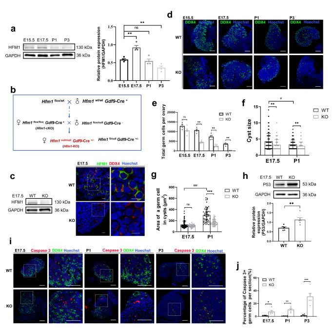
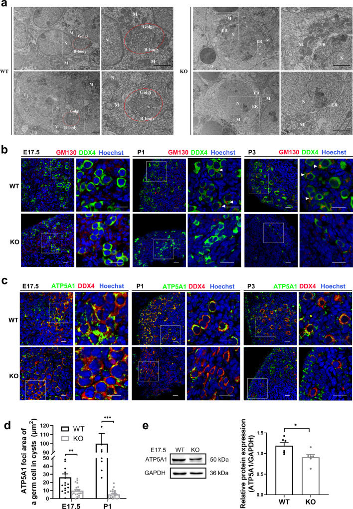
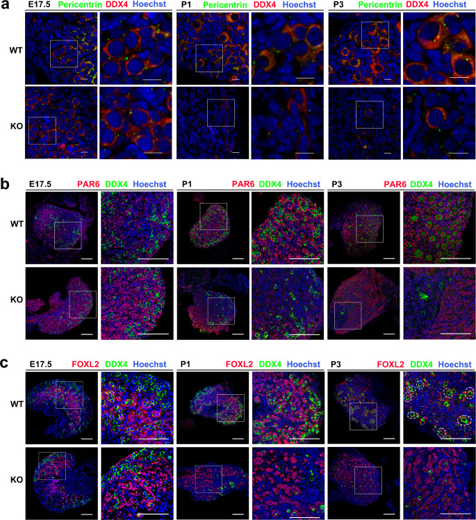
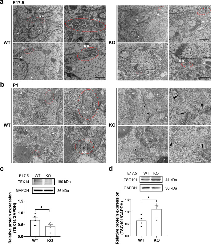
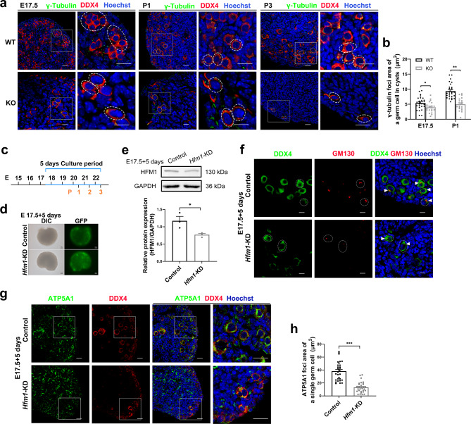
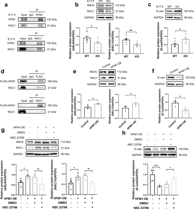
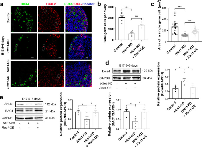
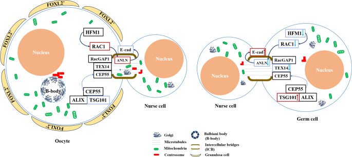
The reproductive lifespan of female mammals is determined by the size of the primordial follicle pool, which comprises oocytes enclosed by a layer of flattened pre-granulosa cells. Oocyte differentiation needs acquiring organelles and cytoplasm from sister germ cells in cysts, but the mechanisms regulating this process remain unknown. Previously helicase for meiosis 1 (HFM1) is reported to be related to the development of premature ovarian insufficiency. Here, it is found that HFM1 is involved in oocyte differentiation through organelle enrichment from sister germ cells. Further study indicates that HFM1 is involved in intercellular directional transport through intercellular bridges via the RAC1/ANLN/E-cad signaling pathway, which is indispensable for oocyte differentiation and primordial follicle formation. These findings shed light on the critical role of HFM1 in intercellular bridge transport, which is essential for the establishment of the primordial follicle pool and presenting new horizons for female fertility protection.

Keywords: HFM1; Intercellular bridge; Oocyte differentiation; POI; Primordial follicle formation.


Conflict of interest statement

Declarations. Ethics approval: Mouse breeding conditions and breeding methods in this experiment strictly complied with the Committee on the Ethics of Animal Experiments of Nanjing Medical University. Consent to publish: All authors read and approved the submission and final publication. Conflict of interest: The authors have no relevant financial or non-financial interests to disclose.

Figures

Fig. 1
Fig. 1
HFM1 was indispensable for primordial follicle formation. a Western blotting analysis of HFM1 expression patterns in the mouse ovaries from E15.5 to P3 (unpaired Student’s t-test, n = 4 independent experiments, ** P = 0.001 or ** P = 0.005 vs. E15.5). GAPDH served as a loading control. The protein level of HFM1 increased markedly at E17.5. b Breeding scheme to achieve Hfm1-KO female mice. c Western blotting analysis showed the complete deletion of HFM1 protein in E17.5 Hfm1-KO ovaries. d Immunofluorescence staining with germ cell marker DDX4 (green) in E15.5, E17.5, P1 and P3 ovaries. Hoechst (blue) was used to identify the nuclear DNA. Scale bars: 100 μm. e Quantitative analysis of germ cell count is shown for WT and Hfm1-KO ovaries at E15.5, E17.5, P1, and P3 (unpaired Student’s t-test, n = 5 animals for each genotype, *** P < 0.001 vs. WT). f The number of germ cells (cyst size) in cysts in E17.5 (n = 118 for WT and n = 71 for KO) and P1 ovaries (n = 91 for WT and n = 65 for KO) (Mann-Whitney U test, ** P = 0.003 or 0.009 vs. WT, #P = 0.015 vs. E17.5). g The area of the germ cell in cysts at E17.5 (n = 81 for WT and n = 41 for KO) and P1 (n = 53 for WT and n = 46 for KO) (Mann-Whitney U test, *** P < 0.001 vs. WT, ###P < 0.001 vs. E17.5). h Western blotting was performed to examine the level of P53 in E17.5 ovaries. The level of P53 was higher in the Hfm1-KO than in the WT ovaries (unpaired Student’s t-test, n = 6 independent experiments, ** P = 0.004 vs. WT). i E15.5, E17.5, P1, and P3 ovaries were stained with germ cell marker DDX4 (green), apoptotic signal Caspase 3 (red). Hoechst (blue) was used to identify the nuclear DNA. Scale bars: 100 μm. j The number of apoptotic cells (Caspase3-positive signals) per section increased significantly in Hfm1-KO ovaries (unpaired Student’s t-test, n = 6, * P = 0.015, ** P = 0.001, *** P < 0.001 vs. WT). Data are presented as mean ± SEM. ns: no significant difference
Fig. 2
Fig. 2
Depletion of HFM1 affected organelle enrichment in germline cysts. a Transmission electron micrograph of Balbiani body (B-body, red circle) in P1 WT and Hfm1-KO mice. Nucleus (N), Mitochondria (M), Endoplasmic reticulum (ER). Scale bars: 2 μm. b Immunofluorescence staining for GM130 (red) and DDX4 (green) at E17.5, P1, and P3. The nucleus was stained by Hoechst (blue). GM130 labeled B-body (a large Golgi sphere, white triangle). Scale bars: 20 μm. c E17.5, P1 and P3 ovaries were stained with mitochondria marker ATP5A1 (green) and DDX4 (red). Hoechst (blue) was used to identify the nuclear DNA. Scale bars: 20 μm. d The ATP5A1 foci area of a germ cell in cysts was generated by ImageJ software at E17.5 and P1 (unpaired Student’s t-test, n = 20, ** P = 0.001, *** P < 0.001 vs. WT). Hfm1-KO resulted in less distribution of the ATP5A1 foci area. e Western blotting analysis of ATP5A1 protein level in E17.5 wild-type and Hfm1-KO ovaries (unpaired Student’s t-test, n = 6 independent experiments, * P = 0.022 vs. WT). Data are presented as mean ± SEM
Fig. 3
Fig. 3
Depletion of HFM1 affected primordial follicle formation. a E17.5, P1, and P3 ovaries stained with Pericentrin (green) to reveal centrosomes. Hoechst (blue) was used to identify the nuclear DNA. Scale bars: 10 μm. b Ovaries were stained for DDX4 (green) and PAR6 (red), a potential marker of germ cells for the primordial follicle formation at E17.5, P1, and P3. The nucleus was stained by Hoechst (blue). Scale bars: 100 μm. c Immunostaining of WT and Hfm1-KO ovaries at E17.5, P1, and P3 using antibodies against DDX4 (green), granulosa cell marker FOXL2 (red), and Hoechst (blue). Primordial follicle (dashed line). Scale bars: 100 μm
Fig. 4
Fig. 4
The formation and stabilization of intercellular bridges were damaged in Hfm1-KO mice. a, b Transmission electron micrograph of the intercellular bridge (ICB, red circle) between germ cells and the plasma membrane gaps (black triangle) in ovaries at E17.5 (a) and P1 (b). Scale bars: 1 μm. c, d Ovaries in E17.5 WT and Hfm1-KO mice. Western blotting was performed to examine the intercellular bridge marker TEX14 (c, unpaired Student’s t-test, n = 6 independent experiments, * P = 0.036 vs. WT) and the membrane abscission protein TSG101 (d, unpaired Student’s t-test, n = 4 independent experiments, * P = 0.047 vs. WT). Data are presented as mean ± SEM
Fig. 5
Fig. 5
HFM1 knockdown disturbed the directional organelles transport of intercellular bridge. a Immunofluorescence staining for γ-tubulin (green) and DDX4 (red, dash line) at E17.5, P1 and P3. The nucleus was stained by Hoechst (blue). Scale bars: 20 μm. b The γ-tubulin foci area of a germ cell in cysts was generated by ImageJ software at E17.5 (n = 26 for WT and n = 21 for KO) and P1(n = 27 for WT and n = 20 for KO) (unpaired Student’s t-test, * P = 0.021, *** P < 0.001 vs. WT). c Timeline showing the 5-day culture scheme. d Fluorescence microscope analyses of adenovirus infection efficiency. Strong green fluorescence was observed under the fluorescent microscope following 5 days of adenovirus infection. Scale bars: 100px. e Western blotting analysis of HFM1 knockdown efficiency. HFM1 protein expression was down-regulated in Ad-shRNA-Hfm1 transfected ovaries after 5 days of culture (unpaired Student’s t-test, n = 3 independent experiments, * P = 0.014 vs. Control). f After 5 days of culture, ovaries transfected with Ad‐shRNA-Hfm1 stained with GM130 (red, white triangle) marking Golgi. DDX4 (green) is a germ cell marker. Hoechst (blue) was used to identify the nuclear DNA. Scale bars: 10 μm. g E17.5 ovaries were cultured with or without Ad‐shRNA-Hfm1 for 5 days, staining with ATP5A1 (green) and germ cell marker DDX4 (Red). Hoechst (blue) was used to identify the nuclear DNA. Scale bars: 20 μm. h The ATP5A1 foci area of a single germ cell was measured by ImageJ software (unpaired Student’s t-test, n = 30, *** P < 0.001 vs. Control). Data are presented as mean ± SEM
Fig. 6
Fig. 6
RAC1/ANLN/E-cad signaling pathway was regulated after knockout or overexpression of HFM1. a Co-IP assays depicted that HFM1 interacts with RAC1 in E17.5 ovaries (n = 3 independent experiments). b, c Western blotting analysis of RAC1, ANLN, and E-cad protein levels in E17.5 WT and Hfm1-KO ovaries. GAPDH served as a loading control (unpaired Student’s t-test, n = 8 independent experiments, * P = 0.029 or 0.011, ** P = 0.002 vs. WT). d The interaction between HFM1 and RAC1 in HEK293T cells. HEK293T cells were transfected with Flag-HFM1 lentiviruses for co-IP assays (n = 3 independent experiments). e, f RAC1, ANLN, and E-cad protein levels were observed and quantified in HEK293T cells. HEK293T cells were transfected with Flag-HFM1 lentiviruses for Western blotting assays (unpaired Student’s t-test, n = 4–5 independent experiments, * P = 0.018 or 0.048 or 0.011 vs. Control). g, h Western blotting analysis showed that the Rac1 inhibitor NSC23766 negatived the influence of HFM1 on ANLN and E-cad protein levels (one-way ANOVA, n = 5 independent experiments, * P = 0.023 or 0.020, *** P < 0.001 vs. Control, #P = 0.029, ##P = 0.009 or 0.010 vs. HFM1-OE + DMSO). Data are presented as mean ± SEM
Fig. 7
Fig. 7
RAC1 overexpression mitigated the effects of HFM1 knockdown on E17.5 ovaries in vitro. E17.5 ovaries were cultured with Ad-shRNA-Hfm1 or Ad‐shRNA-Hfm1 plus LV-Rac1 for 5 days. a Ovaries stained with FOXL2 (red) and germ cell marker DDX4 (green). Hoechst (blue) was used to identify the nuclear DNA. Scale bars: 20 μm. b Statistical analysis of the numbers of germ cells in ovaries (one-way ANOVA, n = 6, ***P < 0.001 vs. Control, ###P < 0.001 vs. Hfm1-KD). c The area of a single germ cell was measured by ImageJ in ovaries treated with Ad‐shRNA-Hfm1 or Ad‐shRNA-Hfm1 plus LV-Rac1 (one-way ANOVA, n = 24, ***P < 0.001 vs. Control, ###P < 0.001 vs. Hfm1-KD). d, e Western blotting analysis of RAC1, ANLN, and E-cad protein levels in ovaries treated with Ad‐shRNA-Hfm1 or Ad‐shRNA-Hfm1 plus LV-Rac1(one-way ANOVA, n = 4–5 independent experiments, * P = 0.012 or 0.021 or 0.015 vs. Control, #P = 0.020 or 0.029 or 0.017 vs. Hfm1-KD). Data are presented as mean ± SEM
Fig. 8
Fig. 8
The schematic illustration of HFM1 regulated germ cell intercellular bridge transport. HFM1 regulated the expression of RAC1 in germ cells, this process impacts ANLN and E-cad expression associated with intercellular bridge and organelle transport, and promotes organelle enrichment and formation of primordial follicles. In Hfm1-KO germ cells, deleted level of HFM1 downregulated RAC1, resulting in the defective organelle enrichment in female germline cysts, and the failure of primordial follicles formation

Similar articles

References

    1. Telfer EE, Grosbois J, Odey YL, Rosario R, Anderson RA (2023) Making a good egg: human oocyte health, aging, and in vitro development. Physiol Rev 103:2623–2677. 10.1152/physrev.00032.2022 - PMC - PubMed
    1. Webber L, Davies M, Anderson R, Bartlett J, Braat D, Cartwright B et al (2016) ESHRE Guideline: management of women with premature ovarian insufficiency. Hum Reprod 31:926–937. 10.1093/humrep/dew027 - PubMed
    1. Panay N, Anderson RA, Nappi RE, Vincent AJ, Vujovic S, Webber L et al (2020) Premature ovarian insufficiency: an International Menopause Society White Paper. Climacteric 23:426–446. 10.1080/13697137.2020.1804547 - PubMed
    1. Huhtaniemi I, Hovatta O, La Marca A, Livera G, Monniaux D, Persani L et al (2018) Advances in the Molecular Pathophysiology, Genetics, and Treatment of Primary Ovarian Insufficiency. Trends Endocrinol Metab 29:400–419. 10.1016/j.tem.2018.03.010 - PubMed
    1. Pu D, Wang C, Cao J, Shen Y, Jiang H, Liu J et al (2016) Association analysis between HFM1 variation and primary ovarian insufficiency in Chinese women. Clin Genet 89:597–602. 10.1111/cge.12718 - PubMed

Substances

LinkOut - more resources