. 2025 Jan;169(1):e16292.
doi: 10.1111/jnc.16292.

Plasticity of Mouse Dorsal Root Ganglion Neurons by Innate Immune Activation Is Influenced by Electrophysiological Activity

Affiliations

Plasticity of Mouse Dorsal Root Ganglion Neurons by Innate Immune Activation Is Influenced by Electrophysiological Activity

Timothy N Friedman et al. J Neurochem. 2025 Jan.

Abstract

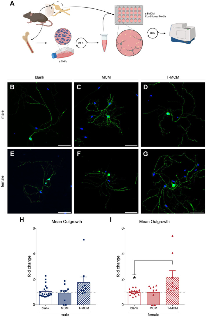
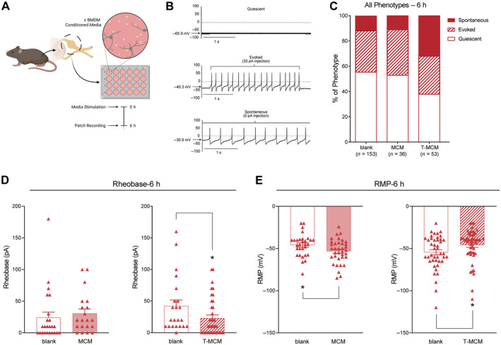
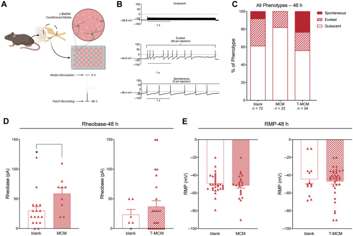
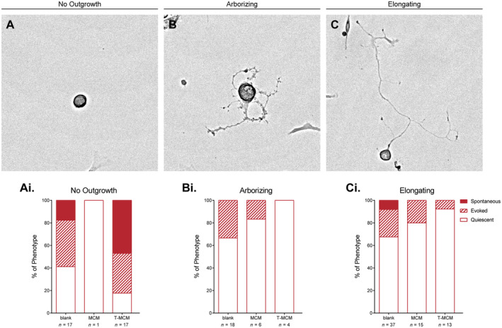
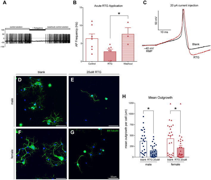
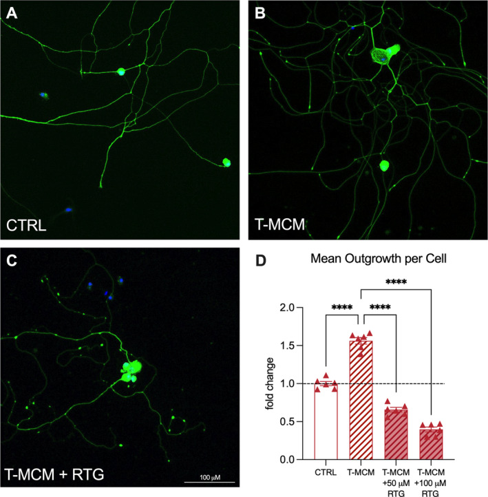
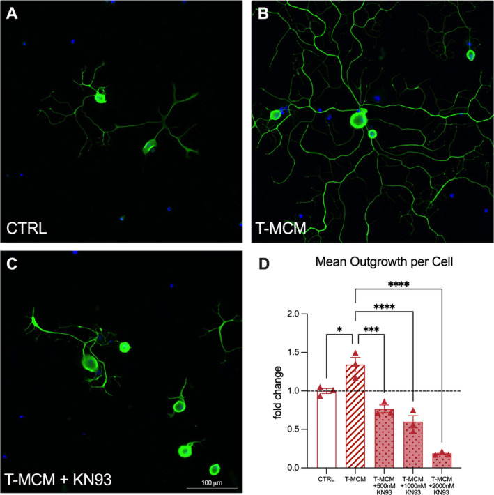
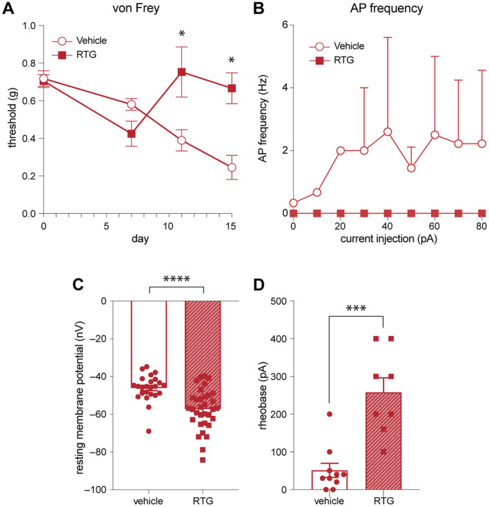
The complex relationship between inflammation, its effects on neuronal excitability and the ensuing plasticity of dorsal root ganglion (DRG) sensory neurons remains to be fully explored. In this study, we have employed a system of experiments assessing the impact of inflammatory conditioned media derived from activated immune cells on the excitability and activity of DRG neurons and how this relates to subsequent growth responses of these cells. We show here that an early phase of increased neuronal activity in response to inflammatory conditioned media is critical for the engagement of plastic processes and that neuronal excitability profiles are linked through time to the structural phenotype of individual neurons. Pharmacological blockade of neuronal activity was able to abolish the growth-promoting effects of inflammatory media. Our results suggest that targeting the activity of DRG neurons may provide a novel therapeutic avenue to manipulate their growth status and potential for plasticity in response to inflammation. Importantly, the same pharmacological blockade in vivo abolished pain responses in a mouse model of multiple sclerosis. While further studies are needed to fully elucidate the underlying mechanisms of the relationship between neural activity and growth status, a more complete understanding of this relationship may ultimately lead to the development of new treatments for neuropathic pain in disorders associated with heightened immune responses such as rheumatoid arthritis and multiple sclerosis.

Keywords: DRG; Kv7 channels; TNFα; electrophysiology; inflammation; neurite extension; pain; plasticity.


Conflict of interest statement

The authors declare no conflicts of interest.

Figures

FIGURE 1
FIGURE 1
Male and female DRG neurons display increased neurite outgrowth in anti‐inflammatory and female‐specific pro‐inflammatory BMDM‐conditioned media. (A) Schematic of DRG neuron cultures and CM stimulation. (B–G) Representative images of neurites stained with βIII tubulin at the experimental endpoint. Quantification of male (H) and female (I) neurites with sex‐matched condition media treatments, normalised to the extent of outgrowth measured in the control (blank) conditions. Scale bar = 50 μm; *p < 0.05. Data is analysed by one‐way ANOVA with Dunnett's multiple comparison test. Bar graphs represent mean ± SEM, with n = 9–21 wells from three independent cell culture preparations.
FIGURE 2
FIGURE 2
Whole‐cell patch clamp recordings of female DRG neurons incubated in BMDM‐conditioned media (CM) for 6 h reveal distinct firing patterns and electrophysiological profiles. (A) Schematic of experimental workflow; neurons were recorded 6 h post‐plating and CM stimulation. (B) Example traces of the three characteristic firing patterns: Quiescent, Evoked and Spontaneous. (C) Quantification of categorical labels for stereotypic firing patterns across CM treatments. Quantification of rheobase (D), resting membrane potential (RMP) (E) across CM treatments. *p < 0.05. Data is analysed by unpaired two‐tailed t‐test. Bar graphs represent mean ± SEM, with n = 20–51 cell recordings per group from three independent cell culture preparations. Categorical data is represented by parts‐of‐whole transformation and analysed by Fisher's exact test on untransformed values.
FIGURE 3
FIGURE 3
Whole‐cell patch clamp recordings of established female DRG neurons incubated in BMDM‐conditioned media (CM) reveal distinct firing patterns and electrophysiological profiles. (A) Schematic of experimental workflow; neurons were recorded 48 h post‐plating and CM stimulation. (B) Example traces of the three characteristic firing patterns: Quiescent, Evoked and Spontaneous. (C) Quantification of categorical labels for stereotypic firing patterns across CM treatments. Quantification of rheobase (D) and resting membrane potential (RMP) (E) across CM treatments. *p < 0.05. Data is analysed by unpaired two‐tailed t‐test. Bar graphs represent mean ± SEM, with n = 22–34 cell recordings per group from three independent cell culture preparations. Categorical data is represented by parts‐of‐whole transformation.
FIGURE 4
FIGURE 4
Whole‐cell patch clamp recordings of established female DRG neurons with distinct structural phenotypes have distinct firing patterns and electrophysiological profiles. (A–C) Representative brightfield images of the distinct neuronal phenotypes (no outgrowth, arborizing, elongating) observed at 48 h post‐plating and CM stimulation. (Ai–Ci) Quantification of categorical labels for stereotypic firing patterns across CM treatments. Categorical data is represented by parts‐of‐whole transformation.
FIGURE 5
FIGURE 5
Neurite outgrowth of DRG neurons is diminished by retigabine (RTG) treatment. (A) Example traces of spontaneously firing DRG neurons that become quiescent under RTG treatment. (B) Quantification of action potential (AP) frequency in binned time domains centred around the period of RTG stimulation. (C) Individual traces of unstimulated and RTG‐stimulated APs. Note the deeper AHP curve and slower return to RMP in the RTG trace. (D–G) Representative images of neurites stained with βIII tubulin at the experimental endpoint after treatment with blank media (D, F) or RTG (E, G). (H) Quantification of male and female neurites after treatment with RTG or control (blank media). Scale bar = 50 μm; *p < 0.05. Data is analysed by an unpaired two‐tailed t‐test. Bar graphs represent mean ± SEM with n = 24 wells from three independent cell culture preparations. Categorical data is represented by parts‐of‐whole transformation.
FIGURE 6
FIGURE 6
Retigabine prevents the growth‐promoting action of inflammatory conditioned media. (A–C) Representative images of neurites from mouse DRG neurons after culture for 48 h with either no treatment, BMDM‐conditioned media treatment, or BMDM‐conditioned media +100 μM RTG treatment. (D) Quantification of mean area of neurite outgrowth. Scale bar = 100 μm, and dashed line represents mean of untreated control; ****p < 0.0001. Data is analysed by one‐way ANOVA. Bar graphs represent mean ± SEM normalised to untreated controls, with n = 6 wells from one cell culture preparation, and the dashed line represents the mean of the untreated control bar.
FIGURE 7
FIGURE 7
KN93 prevents the growth promoting action of inflammatory conditioned media. (A–C) Representative images of neurites from mouse DRG neurons after culture for 48 h with either no treatment, BMDM‐conditioned media treatment, or BMDM‐conditioned media +500 nM KN93 treatment. (D) Quantification of mean area of neurite outgrowth. Scale bar = 100 μm, and dashed line represents mean of untreated control; *p < 0.05, ***p < 0.001, ****p < 0.0001. Data is analysed by one‐way ANOVA. Bar graphs represent mean ± SEM normalised to untreated controls, with n = 3 wells across two independent cell culture preparations, and dashed line represents mean of untreated control bar.
FIGURE 8
FIGURE 8
Retigabine prevents pain hypersensitivity in the EAE model of neuroinflammatory disease. (A) Withdrawal thresholds to von Frey hair stimulation in mice immunised for EAE and treated with vehicle control (left) or RTG (10 mg/kg) (right). Vehicle‐treated mice exhibit stereotypical reductions in withdrawal thresholds indicative of pain hypersensitivity. Mice that begin treatment with RTG on day 7 post‐immunisation exhibit a complete reversal of these behaviours. *p < 0.05, two‐way ANOVA, Tukey host hoc test. (B) Number of action potentials from DRGNs of EAE‐immunised mice treated with retigabine (RTG, 100 μM) versus vehicle control. Frequency of action potentials (Hz) was recorded from 3 s current injections in 10 pA increments. (C) Resting membrane potential (RMP, mV) from DRGNs of EAE‐immunised mice treated with retigabine (RTG, 100 μM) versus vehicle control. ****p < 0.001, two‐tailed unpaired Student's t‐test, DRGNs treated with RTG (100 μM) versus vehicle control. (D) Action potential injection threshold (rheobase, pA) from DRGNs of EAE‐immunised mice treated with retigabine (RTG, 100 μM) versus vehicle control. Line graphs represent mean ± SEM, with n = 8 animals from one in vivo experiment. Bar graphs represent mean ± SEM, with n = 24–33 cell recordings from one cell culture preparation. ***p < 0.001, two‐tailed unpaired Student's t‐test, DRGNs treated with RTG (100 μM) versus vehicle control.

Similar articles

References

    1. Benowitz, L. I. , and Popovich P. G.. 2011. “Inflammation and Axon Regeneration.” Current Opinion in Neurology 24, no. 6: 577–583. - PubMed
    1. Berthézène, C. , Rabiller L., Jourdan G., et al. 2021. “Tissue Regeneration: The Dark Side of Opioids.” International Journal of Molecular Sciences 22, no. 14: 7336. - PMC - PubMed
    1. Boettger, M. K. , Hensellek S., Richter F., et al. 2008. “Antinociceptive Effects of Tumor Necrosis Factor α Neutralization in a Rat Model of Antigen‐Induced Arthritis: Evidence of a Neuronal Target.” Arthritis & Rheumatism 58, no. 8: 2368–2378. - PubMed
    1. Bradke, F. , Fawcett J. W., and Spira M. E.. 2012. “Assembly of a New Growth Cone After Axotomy: The Precursor to Axon Regeneration.” Nature Reviews Neuroscience 13, no. 3: 183–193. - PubMed
    1. Brown, D. A. , and Passmore G. M.. 2009. “Neural KCNQ (Kv7) channels.” British Journal of Pharmacology 156, no. 8: 1185–1195. - PMC - PubMed

Substances

LinkOut - more resources