. 2024 Dec 26;22(1):796.
doi: 10.1186/s12951-024-03076-1.

Unveiling the molecular blueprint of SKP-SCs-mediated tissue engineering-enhanced neuroregeneration

Affiliations

Unveiling the molecular blueprint of SKP-SCs-mediated tissue engineering-enhanced neuroregeneration

Hui Zhu et al. J Nanobiotechnology. .

Abstract

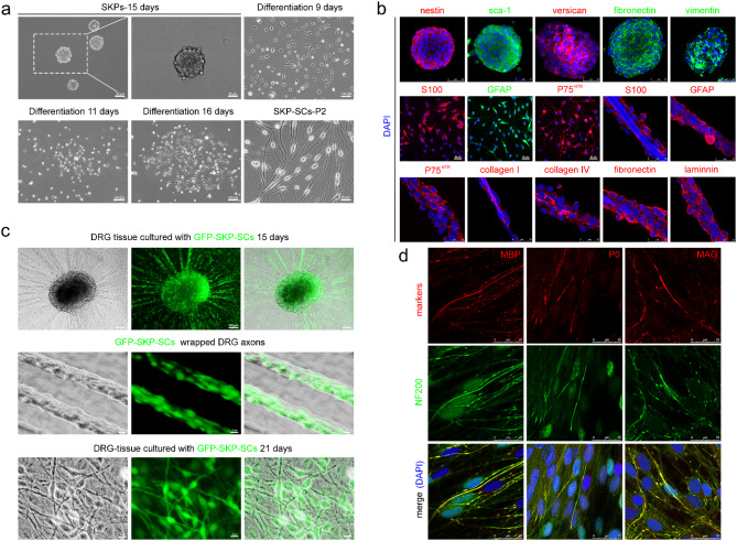
Peripheral nerve injury poses a significant challenge to the nervous system's regenerative capacity. We previously described a novel approach to construct a chitosan/silk fibroin nerve graft with skin-derived precursor-induced Schwann cells (SKP-SCs). This graft has been shown to promote sciatic nerve regeneration and functional restoration to a level comparable to that achieved by autologous nerve grafts, as evidenced by behavioral, histological, and electrophysiological assessments. However, the underlying molecular mechanisms based on SKP-SCs mediated tissue engineering-aid regeneration remain elusive. In the present work, we systematically identified gene modules associated with the differentiation of SKPs into SCs by employing weighted gene co-expression network analysis (WGCNA). By integrating transcriptomic data from the regenerated nerve segment, we constructed a network that delineated the molecular signatures of TENG aid neuroregeneration. Subsequent quantitative PCR (qPCR) validation was performed to substantiate the WGCNA findings. Our WGCNA approach revealed a robust molecular landscape, highlighting hub genes pivotal for tissue engineering-aid regeneration. Notably, the upregulation of specific genes was observed to coincide with the acquisition of SC characteristics. The qPCR validation confirmed the expression patterns of these genes, underscoring their role in promoting neuroregeneration. The current study harnesses the power of WGCNA to elucidate the molecular blueprint governing tissue engineering-aid regeneration. The identified gene modules and validated targets offer novel insights into the cellular and molecular underpinnings of tissue engineering-augmented neuroregeneration. These findings pave the way for developing targeted therapeutics and advanced tissue engineering grafts to enhance peripheral nerve repair.

Keywords: Peripheral nerve regeneration; Schwann cells; Sciatic nerve injury; Skin-derived precursors; Tissue engineering; Weighted gene co-expression network analysis (WGCNA).


Conflict of interest statement

Declarations. Ethics approval and consent to participate: All studies complied with all relevant animal use guidelines and ethical regulations. All animal use and study protocols were approved both by the Institutional Animal Care and by the Administration Committee of Experimental Animals, Jiangsu Province, China, in accordance with the guidelines of the Institutional Animal Care and Use Committee, Nantong University, China (Inspection No: 20190225-004). Consent for publication: Not applicable. Competing interests: The authors declare no competing interests.

Figures

Fig. 1
Fig. 1
Isolation, characterization, and axonal myelination function of SKP-SCs. (a) Phase contrast image showed the SKP spheres were generated from juvenile SKPs after 15 days of culture, the process of induced differentiation 9, 11, 16 days, and the passage two SKP-SCs. Scale bar: 50 μm, 100 μm for low magnification; 25 μm for higher magnification. (b) Characterizations by the immunostainings of nestin, sca-1, versican, fibronectin, and vimentin for SKPs and the immunostainings of S100β, GFAP and p75 NTR for SKP-SCs. SCs markers (S100 β, GFAP and p75 NTR) and ECM molecules (collagen I, collagen IV, fibronectin and laminin) were confirmed by immunostainings on the silk fibroin filaments 7 days post construction of TENG. 50 μm for low magnification; 25 μm for higher magnification. (c) Co-culture of GFP-SKP-SCs and DRG nerve tissues in vitro (15 & 21 days). Most of GFP-SKP-SCs aligned with DRG axons after 7 days in vitro. Some GFP-SKP-SCs initiated wrapping the DRG axons. At 21 days after co-cultures, SKP-SCs were induced to myelinate axons. 100 μm for low magnification; 5 μm for higher magnification. (d) The slides were labeled with the markers of myelin as MBP, P0, and MAG to show myelination, NF-200 to show axons, and DAPI (blue) to show cell nuclear. Scale bar: 25 μm
Fig. 2
Fig. 2
Construction of SKP-SCs-TENG in vitro and gross view post transplantation. Scanning electron microscope images showed cross section of chitosan conduit (a, d), silk fibroin filaments (b), inner surface of chitosan conduit (c), and the micromorphology of SKP-SCs cultured on silk fibroin filaments (e) and chitosan conduit (f) 14 days post construction of TENGs in vitro. The wall of nerve conduit displayed porous structure. Scale bar, 1 mm, 20 μm, 50 μm, 200 μm, 20 μm, 50 μm. The gross view obtained 12 weeks post-surgery in SKP-SCs-TENG (g, h), chitosan/silk fibroin neural scaffold (i, j), autograft (k) and non‐grafted (l) groups. The proximal and the distal coaptations were indicated by an arrow and an arrowhead, respectively. Scale bar: 2000 μm
Fig. 3
Fig. 3
The locomotor function, electrophysiological evaluation, histological observation and morphometric analysis of the regenerated nerve. (a-c) Scatter plots comparing the sciatic function index values among three groups at 4 w, 8 w, and 12 w post-surgery. *p < 0.05 versus scaffold group. (d-g) Representative CMAP recordings at 12 weeks post-surgery, were obtained from the injured side of animals in TENG, autograft, scaffold groups and on the contralateral uninjured side (normal) of animals, respectively. (h-j) Scatter plots showing the proximal and distal CMAP amplitude detected on the injured side of animals, and the motor nerve conduction velocity and in the TENG, autograft, scaffold groups and on the contralateral uninjured side (normal), respectively. *p < 0.05 versus scaffold group and #p < 0.05 versus normal group. Transmission electron micrographs (k) were obtained at 12 weeks post-surgery. Scale bar: 5 μm for TEM images. (l-n) Scatter plots showing the thickness of the regenerated myelin sheath (l), the diameter of regenerated myelinated nerve fibers (m), and g-ratio of the regenerated nerve (n). One-way ANOVA and the post hoc Tukey’s t test are used to analyze the data. *p < 0.05 versus scaffold group and #p < 0.05 versus normal group
Fig. 4
Fig. 4
FluoroGold™ retrograde nerve tracing. Representative fluorescence micrographs following FluoroGold™ (FG) retrograde nerve tracing. (a) FG retrogradely labeled sensory neurons in DRGs and motor neurons in the spinal cord. The high magnifications clearly showed the FG-labeled sensory neurons in DRGs and motor neurons in longitudinal sections of spinal cord, respectively. Scale bar: 100 μm for low magnifications, 50 μm for high magnifications. (b, c) Scatter plots of the number of FG-labeled spinal motor neurons (b) and percentage of FG-labeled sensory DRG neurons (c) were shown and analyzed (n = 3). *p < 0.05 versus scaffold group and #p < 0.05 versus normal group. (d-f) Scatter plots showing the average wet weight ratio of total anterior tibialis and gastrocnemius muscle. *p < 0.05, **p < 0.01, ****p < 0.0001
Fig. 5
Fig. 5
The weighted gene co-expression network analysis (WGCNA) on various grafts. (a-c) Gene dendrogram showing the co-expression modules defined by the WGCNA labeled by colors. Module band: cluster dendrogram groups genes into distinct modules using all samples of TENG (a), autograft (b), and scaffold (c). The modules were designated numerically based on size, and the 14, 12, 48 largest modules are labeled adjacent to their respective color band, respectively. (d) Scatter plot showing the distribution of local properties of nodes in all samples of TENG, autograft, and scaffold group. The x-axis in scatter plot represents the node degree, and the y-axis in scatter plot represents the local clustering coefficient. High-degree nodes or hub genes represent gene regulatory hubs, whereas high clustering coefficients denote highly collaborative gene association and network robustness. The TENG group dramatically increased the high degree nodes (hubs) and the local clustering coefficient, similar to autograft group, while the scaffold group decreased hubs and the clustering coefficient. Heatmap showing the time effect on various gene co-expression modules in TENG (e) and autograft group (g). The vertical axis represents the different modules, each labeled by color. The horizontal axis shows the strength of the correlation, with red indicating positive correlation and green indicating negative correlation. Numerical values within each box represent the correlation coefficient, with the associated p-value shown in parentheses. In TENG group (e), strong positive correlations are observed for MEpink (0.83, p = 4e-07) and MEtan (0.74, p = 4e-05), while strong negative correlations are found for MEyellow (-0.92, p = 3e-10) and MEsalmon (-0.73, p = 6e-05). In autograft group (g), strong positive correlations are observed for MEbrown (0.89, p = 7e-09) and MEyellow (0.53, p = 0.007). Conversely, strong negative correlations are observed for MEgreen (-0.91, p = 8e-10) and MEred (-0.65, p = 6e-04). Dashed lines separate modules with statistically significant correlations (p < 0.05) from those without. Histogram depicting the gene significance across various gene co-expression modules in TENG (f) and autograft group (h). Notably, the yellow, pink, and turquoise modules show the highest gene significance, while the purple and red modules display lower significance in TENG group. The brown and green modules show the highest gene significance, followed by the red and turquoise modules. In contrast, the black, blue, purple, and pink modules exhibit lower gene significance in autograft group. The dashed horizontal line represents the threshold for significance. (i-l) Module preservation assessed in the microarray dataset of autograft (i) or scaffold (j) in TENG group, and scaffold (k) or TENG (l) in autograft group. The green dashed line (Z-summary = 10) marks the “strongly preserved” threshold and the blue dashed line (Z-summary = 2) marks the “moderately preserved” threshold. Module size represents the number of genes within each module. Circled groups highlight clusters of modules with low correlation coefficient (p > = 0.05)
Fig. 6
Fig. 6
(Functional enriched modules and gene ontology annotations. a, b) The modules were positively correlated with various indicators of blood vessel, apoptosis, axon and neurites, immune response, inflammatory responses, migration, myelination, proliferation, extracellular matrix, and transcript regulation. Module preservation between even partitioning of TENG (a) or autograft (b) in IPA functional genes dataset for Pearson-based WGCNA. P-values are represented by the depth of the red color. The darker the color, the smaller is the p-value and the higher is the overlap. (c-f) The eigengene expression plots showed module eigengene expression and top 5 associated gene ontology terms. These selected plots illustrated the expression levels of module eigengenes across different time points in TENG (c, d) and autograft (e, f) groups. The solid line showed a smoothed trend in eigengene expression over time. These modules were enriched in genes associated with Gene Ontology (GO) terms. (g-p) The average expression profiles (Z-scores) of functional genes involved in apoptosis (g), proliferation (h), blood vessel (i), myelination (j), immune response (k), migration (l), inflammatory responses (m), extracellular matrix (n), transcript regulation (o), axon and neurites (p)
Fig. 7
Fig. 7
The functional analysis of gene modules and conserved hub genes selection. (a) Heatmap showing the time effect on various gene co-expression modules in scaffold group. The vertical axis represents the different modules, each labeled by color. The horizontal axis shows the strength of the correlation, with red indicating positive correlation and green indicating negative correlation. Numerical values within each box represent the correlation coefficient, with the associated p-value shown in parentheses. Dashed lines separate modules with statistically significant correlations (p < 0.05) from those without. (b) Histogram depicting the gene significance across various gene co-expression modules in scaffold group. Notably, the darkgrey and lightcyan modules show the highest gene significance. The dashed horizontal line represents the threshold for significance. (c, d) The eigengene expression plots showed module eigengene expression and top 5 associated gene ontology terms. These selected plots illustrated the expression levels of module eigengenes across different time points in scaffold group. The solid line showed a smoothed trend in eigengene expression over time. These modules were enriched in genes associated with GO terms. (e) Venn diagrams showing conserved or specific differentially expressed gene counts time negative, positive correlated, and common genes in TENG, autograft, and scaffold group post-surgery. (f) Protein-protein interaction (PPI) networks for common hub genes in all grafted group post-surgery. Red circles represent specific proteins (e.g., FOSL1, CC12, FGF2). Green gears represent biological processes such as “Angiogenesis,” “Demyelination,” and “Inflammatory response.” Blue crosses indicate diseases related to apoptosis or cell death (e.g., “Apoptosis of endothelial cells,” “Cell death of immune cells”). (g-i) Heatmap of dynamic gene expression for common hub genes in all grafted group post-surgery. (j-u) The qPCR results verified that the differential expression of the genes, including Birc2 (j), Birc3 (k), Hif1a (l), Tnc (m), Tnf (n), Cxcl17 (o), Mmp9 (p), Agt (q), Serpine1 (r), Tgfb1 (s), Il6 (t), Il10 (u), were essential in affecting myelination post-surgery

Similar articles

Cited by

References

    1. Shlapakova LE, Surmeneva MA, Kholkin AL, Surmenev RA. Revealing an important role of piezoelectric polymers in nervous-tissue regeneration: a review. Mater Today Bio. 2024;25:100950. - PMC - PubMed
    1. Wang S, Lu H, Kang X, Wang Z, Yan S, Zhang X, Shi X. Electroconductive and Immunomodulatory Natural polymer-based hydrogel bandages designed for peripheral nerve regeneration. Adv Funct Mater. 2024;34:2310903.
    1. Zhou W, Rahman MSU, Sun C, Li S, Zhang N, Chen H, Han CC, Xu S, Liu Y. Perspectives on the Novel multifunctional nerve Guidance conduits: from specific regenerative procedures to motor function rebuilding. Adv Mater. 2024;36:2307805. - PubMed
    1. Sun Y, Zhang Y, Guo Y, He D, Xu W, Fang W, Zhang C, Zuo Y, Zhang Z. Electrical aligned polyurethane nerve guidance conduit modulates macrophage polarization and facilitates immunoregulatory peripheral nerve regeneration. J Nanobiotechnol. 2024;22:244. - PMC - PubMed
    1. Haider A, Khan S, Iqbal DN, Khan SU, Haider S, Mohammad K, Mustfa G, Rizwan M, Haider A. Chitosan as a tool for tissue engineering and rehabilitation: recent developments and future perspectives – A review. Int J Biol Macromol. 2024;278:134172. - PubMed

LinkOut - more resources