. 2024 Dec 27;13(1):67.
doi: 10.1186/s40035-024-00456-3.

N-terminus α-synuclein detection reveals new and more diverse aggregate morphologies in multiple system atrophy and Parkinson's disease

Affiliations

N-terminus α-synuclein detection reveals new and more diverse aggregate morphologies in multiple system atrophy and Parkinson's disease

James A Wiseman et al. Transl Neurodegener. .

Abstract

Background: Parkinson's disease (PD) and multiple system atrophy (MSA) are classified as α-synucleinopathies and are primarily differentiated by their clinical phenotypes. Delineating these diseases based on their specific α-synuclein (α-Syn) proteoform pathologies is crucial for accurate antemortem biomarker diagnosis. Newly identified α-Syn pathologies in PD raise questions about whether MSA exhibits a similar diversity. This prompted the need for a comparative study focusing on α-Syn epitope-specific immunoreactivities in both diseases, which could clarify the extent of pathological overlap and diversity, and guide more accurate biomarker development.

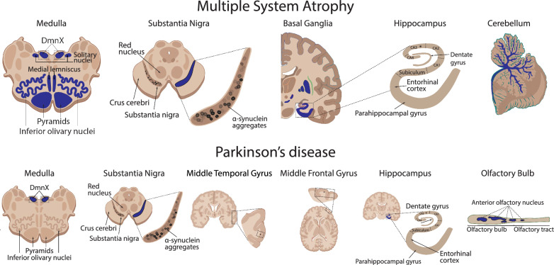
Methods: We utilised a multiplex immunohistochemical approach to detect multiple structural domains of α-Syn proteoforms across multiple regions prone to pathological accumulation in MSA (n = 10) and PD (n = 10). Comparison of epitope-specific α-Syn proteoforms was performed in the MSA medulla, inferior olivary nucleus, substantia nigra, hippocampus, and cerebellum, and in the PD olfactory bulb, medulla, substantia nigra, hippocampus, and entorhinal cortex.

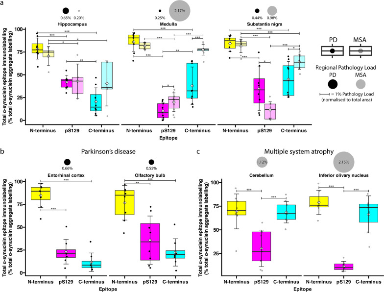
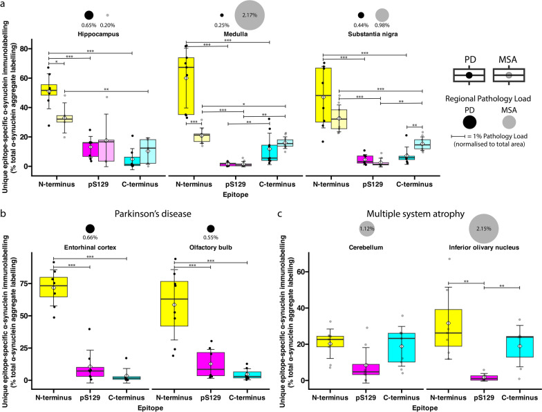
Results: N-terminus and C-terminus antibodies detected significantly more α-Syn pathology in MSA than antibodies for phosphorylated (pS129) α-Syn, which are classically used to detect α-Syn. Importantly, C-terminus immunolabelling is more pronounced in MSA compared to PD. Meanwhile, N-terminus immunolabelling consistently detected the highest percentage of α-Syn across pathologically burdened regions of both diseases, which could be of biological significance. As expected, oligodendroglial involvement distinguished MSA from PD, but in contrast to PD, no substantial astrocytic or microglial α-Syn accumulation in MSA occurred. These data confirm glial-specific changes between these diseases when immunolabelling the N-terminus epitope. In comparison, N-terminus neuronal α-Syn was present in PD and MSA, with most MSA neurons lacking pS129 α-Syn proteoforms. This explains why characterisation of neuronal MSA pathologies is lacking and challenges the reliance on pS129 antibodies for the accurate quantification of α-Syn pathological load across α-synucleinopathies.

Conclusions: These findings underscore the necessity of utilising a multiplex approach to detect α-Syn, most importantly including the N-terminus, to capture the entire spectrum of α-Syn proteoforms in α-synucleinopathies. The data provide novel insights toward the biological differentiation of these α-synucleinopathies and pave the way for more refined antemortem diagnostic methods to facilitate early identification and intervention of these neurodegenerative diseases.

Keywords: Epitope-specific; Lewy body diseases; Multiple system atrophy; Multiplex; N-terminus; Parkinson’s disease; Truncational variants; α-Synuclein.


Conflict of interest statement

Declarations. Ethics approvals and consent to participate: Human post-mortem brain tissues used in this study were received from (1) the Sydney Brain Bank, and (2) the Neurological Foundation Human Brain Bank (New Zealand). All brain tissues were donated with written informed consent from donors and their families prior to brain removal and all protocols were approved by the University of Sydney Human Research Ethics Committee (2019/491) and the University of Auckland Human Participants Ethics Committee (011654). All experiments were conducted in accordance with relevant guidelines and regulations. Consent for publication: Not applicable. Competing Interests: The authors declare no competing financial or non-financial interests.

Figures

Fig. 1
Fig. 1
Comparison of the regional distribution of α-Syn pathology in the human brain with MSA and PD. Schematic diagram depicting the anatomical localisation (blue) of α-Syn pathology in the medulla, substantia nigra, hippocampus, and cerebellum of the human brain with MSA (top), and the medulla, substantia nigra, middle temporal gyrus, middle frontal gyrus, hippocampus, and olfactory bulb of the human brain with PD (bottom)
Fig. 2
Fig. 2
Representative images demonstrating the regional distribution and epitope-specific immunolabelling of α-Syn aggregates in PD (left) and MSA (right). Overview images of the whole regions are displayed in the left column for each disease (Scale bars, 100 μm), and zoomed overview images for each region are displayed in the right column for each disease (Scale bars, 20 μm)
Fig. 3
Fig. 3
Representative confocal images showing different epitope-specific aggregate structures found in PD and MSA. a Perinuclear, glial, and lysosomal α-Syn aggregates with exclusive N-terminus immunoreactivity in the human brain with PD. b Different epitope-specific aggregate structures found in MSA. (1) Punctate aggregates exhibiting exclusive N-terminus immunoreactivity were commonly observed within neuronal cytoplasm. (2) Neuronal inclusions with exclusive N-terminus immunoreactivity were frequently observed in the inferior olivary nucleus. (3, 4) Filamentous neuronal inclusions with exclusive portions of N-terminus immunoreactivity and exclusive portions of C-terminus immunoreactivity were common. (5) Oligodendroglial inclusion exhibiting variable N-terminus, pS129, and C-terminus immunoreactivity. (1–4, 6) pS129 α-Syn immunolabelling was often very weak or, in some instances, completely absent. Yellow and magenta arrowheads label instances of unique N-terminus and C-terminus immunolabelling, respectively. Scale bars, 5 μm
Fig. 4
Fig. 4
Comparison of total individual α-synuclein epitope immunolabelling in PD and MSA. Quantification of total N-terminus (yellow), pS129 (magenta), and C-terminus (cyan) epitope immunolabelling in (a) the hippocampus, medulla, and substantia nigra of PD and MSA cases, (b) the PD entorhinal cortex and olfactory bulb, and (c) the MSA cerebellum and inferior olivary nucleus. Boxplot colours correspond to colours of N-terminus (yellow), pS129 (magenta), and C-terminus (cyan) immunolabelling in representative images. White diamond = mean; crossbar = median; boxplots display interquartile range, and error bars indicate SD. ***P < 0.001, **P < 0.01, *P < 0.05
Fig. 5
Fig. 5
Comparison of unique (epitope-specific) α-synuclein immunolabelling in PD and MSA. Quantification of unique (epitope-specific) N-terminus (yellow), pS129 (magenta), and C-terminus (cyan) immunolabelling in (a) the hippocampus, medulla, and substantia nigra of PD and MSA cases, (b) the PD entorhinal cortex and olfactory bulb, and (c) the MSA cerebellum and inferior olivary nucleus. Boxplot colours correspond to colours of N-terminus (yellow), pS129 (magenta), and C-terminus (cyan) immunolabelling in representative images. White diamond = mean; crossbar = median; boxplots display interquartile range, and error bars indicate SD. ***P < 0.001, **P < 0.01, *P < 0.05
Fig. 6
Fig. 6
Illustration summarising the prevailing α-Syn aggregate structures that are present in PD and MSA. In the human brain with PD, novel perinuclear, microglial, astrocytic, and lysosomal α-Syn aggregate structures that exhibit epitope-specific N-terminus (residues 34–57) immunoreactivity were observed. In the human brain with MSA, microglial and astrocytic inclusions were not observed. Punctate neuronal inclusions with exclusive N-terminus immunoreactivity were often observed; however, these were considerably less frequent than in PD (not included in the illustration). Structurally discrete, whisp-like α-Syn filaments with either exclusive N-terminus or exclusive C-terminus immunoreactivity were frequently observed, which appeared to be interlaced and juxtaposed with one another. Finally, in the MSA ION, several neuronal inclusions with exclusive N-terminus immunoreactivity were observed (not included in the illustration)

References

    1. Schweighauser M, Shi Y, Tarutani A, Kametani F, Murzin AG, Ghetti B, et al. Structures of α-synuclein filaments from multiple system atrophy. Nature. 2020;585(7825):464–9. - PMC - PubMed
    1. Strohäker T, Jung BC, Liou SH, Fernandez CO, Riedel D, Becker S, et al. Structural heterogeneity of α-synuclein fibrils amplified from patient brain extracts. Nat Commun. 2019;10(5535):1–12. - PMC - PubMed
    1. Yang Y, Shi Y, Schweighauser M, Zhang X, Kotecha A, Murzin AG, et al. Structures of α-synuclein filaments from human brains with Lewy pathology. Nature. 2022;610(7933):791–5. - PMC - PubMed
    1. Peelaerts W, Baekelandt V. ɑ-Synuclein strains and the variable pathologies of synucleinopathies. J Neurochem. 2016;139:256–74. - PubMed
    1. Wenning GK, Stankovic I, Vignatelli L, Fanciulli A, Calandra-Buonaura G, Seppi K, et al. The movement disorder society criteria for the diagnosis of multiple system atrophy. Mov Disord. 2022;37(6):1131–48. - PMC - PubMed

Publication types