. 2025 Jan 13;65(1):338-350.
doi: 10.1021/acs.jcim.4c01478. Epub 2024 Dec 26.

Ranking Single Fluorescent Protein-Based Calcium Biosensor Performance by Molecular Dynamics Simulations

Affiliations

Ranking Single Fluorescent Protein-Based Calcium Biosensor Performance by Molecular Dynamics Simulations

Melike Berksoz et al. J Chem Inf Model. .

Abstract

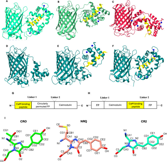
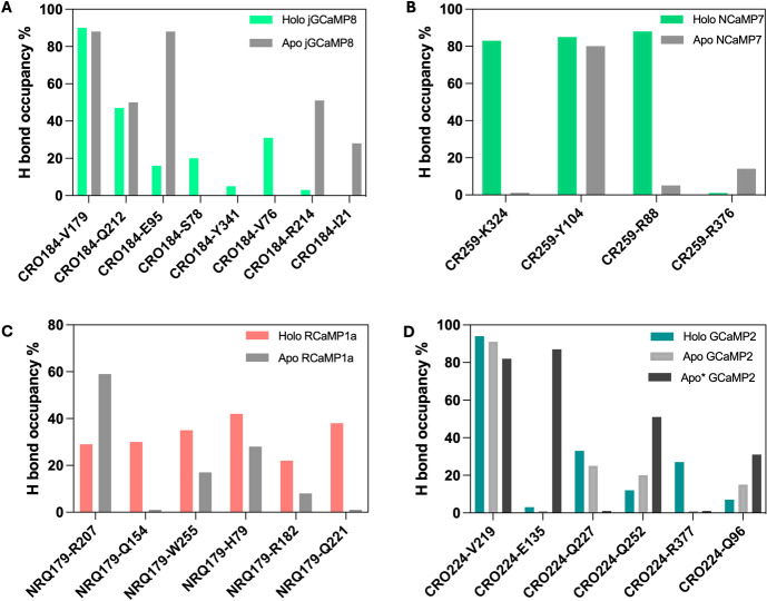
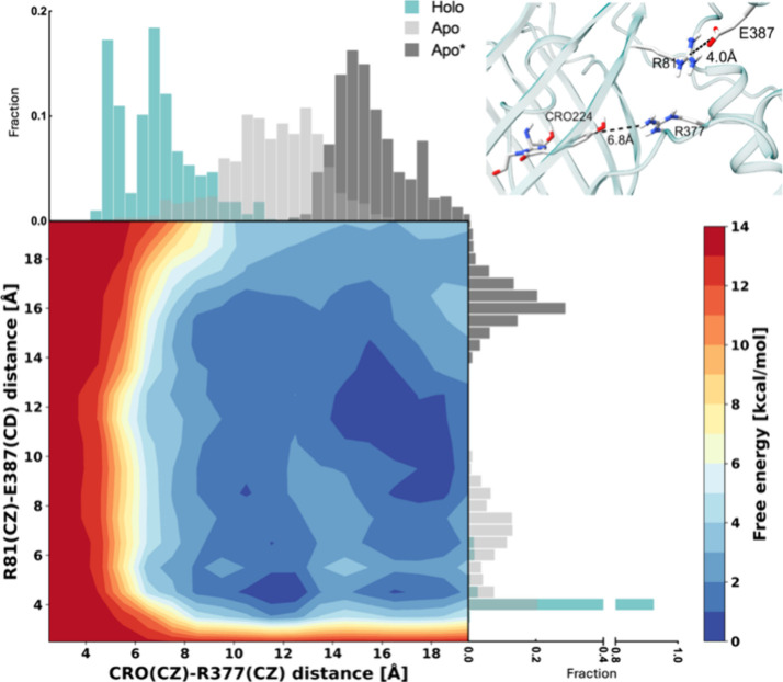
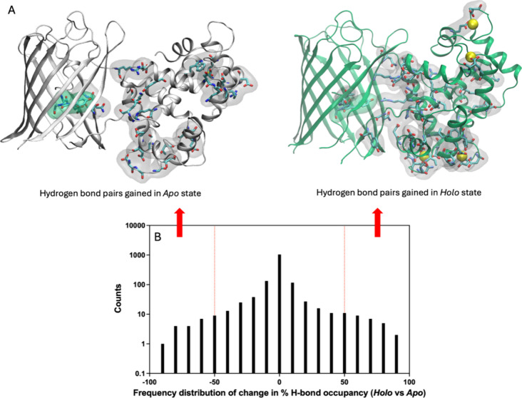
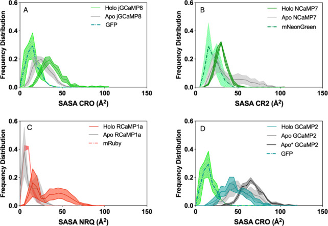
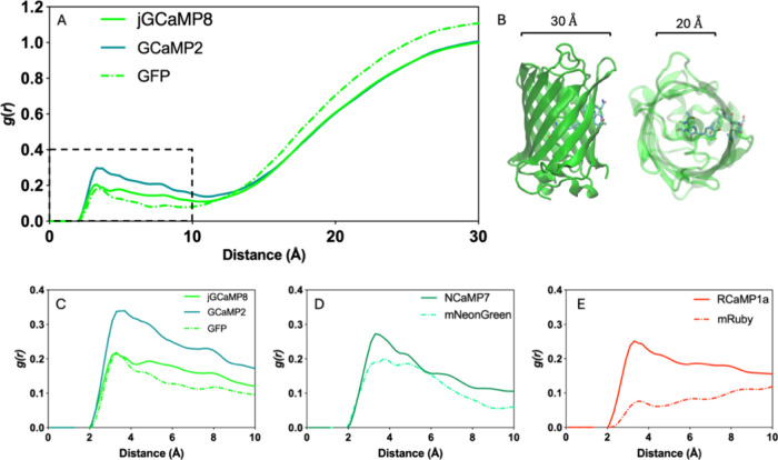
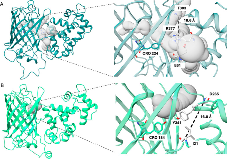
Genetically encoded fluorescent biosensors (GEFBs) have become indispensable tools for visualizing biological processes in vivo. A typical GEFB is composed of a sensory domain (SD) that undergoes a conformational change upon ligand binding or enzymatic reaction; the SD is genetically fused with a fluorescent protein (FP). The changes in the SD allosterically modulate the chromophore environment whose spectral properties are changed. Single fluorescent (FP)-based biosensors, a subclass of GEFBs, offer a simple experimental setup; they are easy to produce in living cells, structurally stable, and simple to use due to their single-wavelength operation. However, they pose a significant challenge for structure optimization, especially concerning the length and residue content of linkers between the FP and SD, which affect how well the chromophore responds to conformational change in the SD. In this work, we use all-atom molecular dynamics simulations to analyze the dynamic properties of a series of calmodulin-based calcium biosensors, all with different FP-SD interaction interfaces and varying degrees of calcium binding-dependent fluorescence change. Our results indicate that biosensor performance can be predicted based on distribution of water molecules around the chromophore and shifts in hydrogen bond occupancies between the ligand-bound and ligand-free sensor structures.


Conflict of interest statement

The authors declare no competing financial interest.

Figures

Figure 1
Figure 1
(A) Four Ca2+-bound jGCaMP8 (PDB: 7ST4). (B) Four Ca2+-bound NCaMP7 (PDB: 6XW2). (C) Four Ca2+-bound RCaMP1a (PDB: 3U0K). (D) Calcium-free GCaMP2 with a partially resolved CaM domain (PDB: 3EKJ). (E) Two Ca2+-bound GCaMP2 (PDB: 3O77). (F) Four Ca2+-bound GCaMP2 (PDB: 3EVR). α-Helical peptide associated with the CaM domain is shown in yellow in all cases. (G) Schematic sequence of jGCaMP8, RCaMP1a, and GCaMP2. (H) Schematic sequence of NCaMP7. (I) Chromophore types. CRO, NRQ, and CR2. Only the heavy atoms provided in the PDB structures are shown; carbons are colored according to the fluorescence color of the respective chromophore. Atom types are taken from CHARMM36 force field topologies.
Figure 2
Figure 2
Hydrogen bond network near the chromophore in the ON and OFF states of each sensor. OFF states are colored gray, and ON states are colored according to the color emitted by the sensor. Images are taken from representative frames of each MD run and are structurally aligned with ChimeraX onto the FP domains. (A) jGCaMP8; (B) NCaMP7; (C) RCaMP1a; (D) GCaMP2. The hydrogen bonds between NRQ179-OH and N25-N and between CR2-OH and C320-SH are mediated by a water molecule. The water/oxygen bond is displayed as a red sphere. Distances are shown only for the holo states; only polar hydrogens of the side chains are explicitly shown.
Figure 3
Figure 3
Occupancies of hydrogen bonds involving the chromophore as either an acceptor or a donor in holo and apo sensors. (A) jGCaMP8, (B) NCaMP7, (C) RCaMP1a, and (D) GCaMP2.
Figure 4
Figure 4
Potential of mean force calculated via well-tempered metadynamics simulations on calcium-removed holo GCaMP2. The frequency distribution of the distances of the selected CVs in holo (green), apo (light gray), and apo* (dark gray) states are shown along the axes. The positions of the selected distances along the interface relative to the chromophore are shown on the upper right.
Figure 5
Figure 5
Shifts in hydrogen bond occupancies between holo and apo states of GCaMP2 differentiate the ON/OFF states. (A) Hydrogen-bonded residue pairs whose occupancy increases (decreases) by more than (less than) 50% in holo (apo) states visualized as licorice and surface representations. (B) Histogram of difference in % hydrogen bond occupancies in holo vs apo states; arrows indicate the hydrogen bond selected to be plotted in (A). For clarity, y-axes are plotted on a logarithmic scale.
Figure 6
Figure 6
Frequency distribution of chromophore SASA grouped according to chromophore type. (A) jGCaMP8 and GFP. (B) NCaMP7 and mNeonGreen. (C) RCaMP1a and mRuby. (D) GCaMP2 and GFP.
Figure 7
Figure 7
Radial distribution function (RDF) of water-oxygen around the chromophore in holo sensors and intact FPs. (A) RDF of water-oxygen up to 30 Å from the chromophore in GFP and GFP-derived sensors. (B) Dimensions of GFP. (C–E) RDF of water-oxygen up to 10 Å in holo sensors and their ON state parental FPs (dashed).
Figure 8
Figure 8
Channels connecting the surface to the chromophore. (A) holo GCaMP2 and (B) holo jGCAMP8. Insets show close-up of channels and distances between gate post residues (T303 and E61 in GCaMP2, I21 and D265 in jGCaMP8) flanking the bulge.

Similar articles

References

    1. Margineanu A.; Chan J. J.; Kelly D. J.; et al. Screening for protein-protein interactions using Förster resonance energy transfer (FRET) and fluorescence lifetime imaging microscopy (FLIM). Sci. Rep. 2016, 6 (June), 28186.10.1038/srep28186. - DOI - PMC - PubMed
    1. Nasu Y.; Shen Y.; Kramer L.; Campbell R. E. Structure- and mechanism-guided design of single fluorescent protein-based biosensors. Nat. Chem. Biol. 2021, 17 (5), 509–518. 10.1038/s41589-020-00718-x. - DOI - PubMed
    1. Oltrogge L. M.; Wang Q.; Boxer S. G. Ground-State Proton Transfer Kinetics in Green Fluorescent Protein. Biochemistry. 2014, 53 (37), 5947–5957. 10.1021/bi500147n. - DOI - PMC - PubMed
    1. Tachibana S. R.; Tang L.; Zhu L.; Liu W.; Wang Y.; Fang C. Watching an Engineered Calcium Biosensor Glow: Altered Reaction Pathways before Emission. J. Phys. Chem. B 2018, 122 (50), 11986–11995. 10.1021/acs.jpcb.8b10587. - DOI - PubMed
    1. Pakhomov A. A.; Martynov V. I. GFP Family: Structural Insights into Spectral Tuning. Chem. Biol. 2008, 15 (8), 755–764. 10.1016/j.chembiol.2008.07.009. - DOI - PubMed

MeSH terms

LinkOut - more resources