

Epstein-Barr Virus-Associated Smooth Muscle Tumor in the Liver Post Kidney Transplant: A Case Report
- PMID: 39726470
- PMCID: PMC11669557
- DOI: 10.7759/cureus.74441
Epstein-Barr Virus-Associated Smooth Muscle Tumor in the Liver Post Kidney Transplant: A Case Report
Abstract
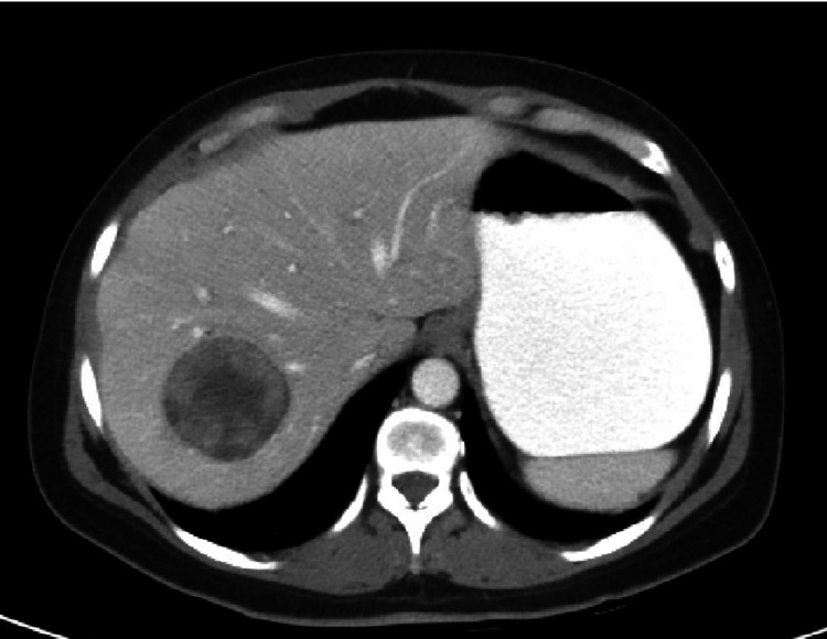
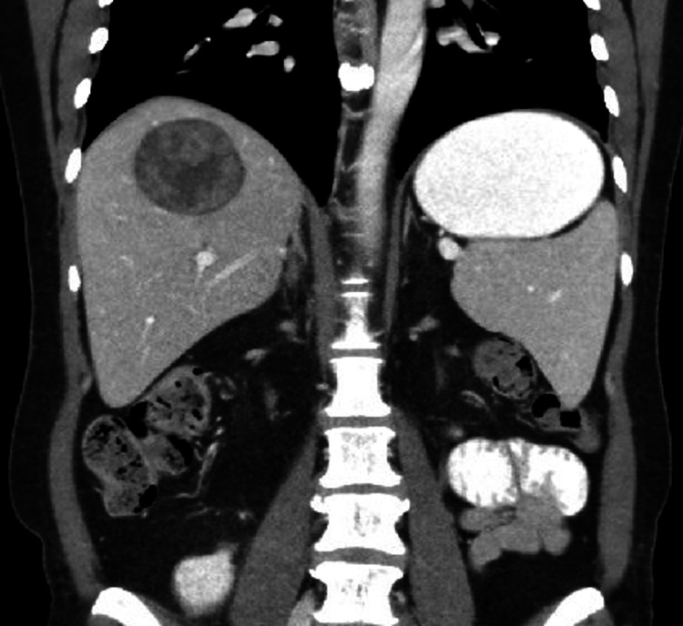
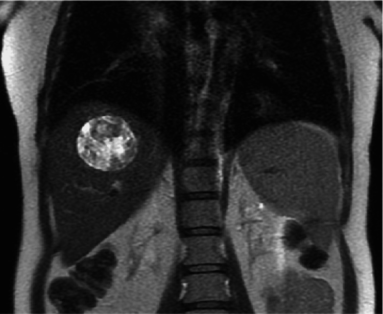
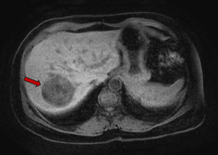
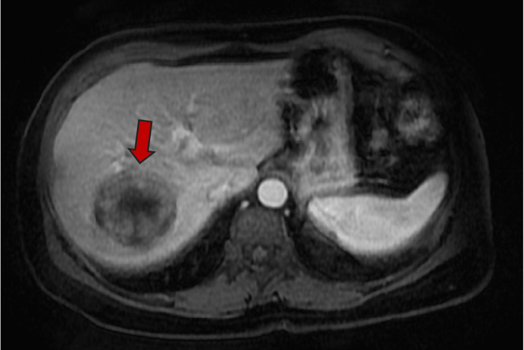
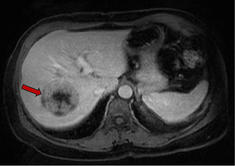
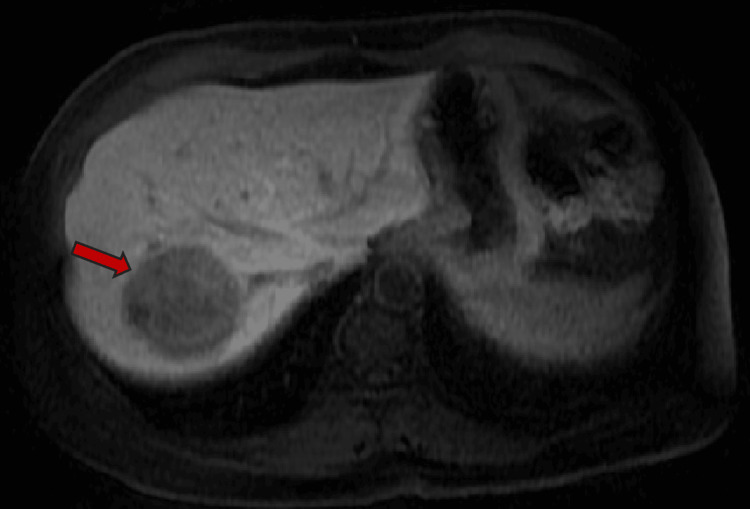
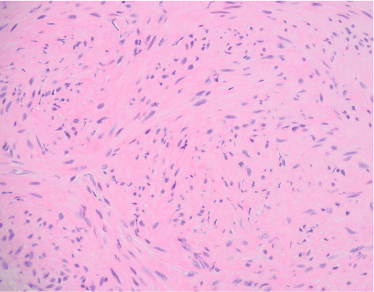
Epstein-Barr virus (EBV) is one of the most common causes of infection from the herpes virus family which also possesses oncogenic potential. EBV-associated smooth muscle tumors (EBV-SMT) are often found in the CNS but here we present the case of a 50-year-old woman with EBV-SMT in the liver. This patient had a kidney transplant in 2009 and had been undergoing immunosuppressive therapy to support her transplant. Subsequent imaging found a liver mass which was not seen on previous imaging. The biopsy revealed an EBV-SMT. The exact pathophysiology of EBV-SMT is not clear though it is believed to involve the reactivation of latent infection through mTOR pathways. Treatment of such masses includes reducing immunomodulating pharmacotherapy though no established management guidelines exist.
Keywords: ebv-smt; immunosuppresion; large liver mass; rare tumors; smooth muscle tumor; transplant kidney.
Copyright © 2024, Ali et al.
Conflict of interest statement
Human subjects: Consent for treatment and open access publication was obtained or waived by all participants in this study. Conflicts of interest: In compliance with the ICMJE uniform disclosure form, all authors declare the following: Payment/services info: All authors have declared that no financial support was received from any organization for the submitted work. Financial relationships: All authors have declared that they have no financial relationships at present or within the previous three years with any organizations that might have an interest in the submitted work. Other relationships: All authors have declared that there are no other relationships or activities that could appear to have influenced the submitted work.
Figures
Similar articles
-
Concurrent Epstein Barr virus-associated smooth muscle tumor and myeloid sarcoma of the liver and acute myeloid leukemia in a patient post kidney transplant: a case report and review of the literature.J Gastrointest Oncol. 2022 Dec;13(6):3329-3335. doi: 10.21037/jgo-21-700. J Gastrointest Oncol. 2022. PMID: 36636068 Free PMC article.
-
Epstein-Barr virus-associated smooth muscle tumor in a kidney transplant recipient: A case-report and review of the literature.Transpl Infect Dis. 2021 Feb;23(1):e13456. doi: 10.1111/tid.13456. Epub 2020 Sep 16. Transpl Infect Dis. 2021. PMID: 32881184
-
Epstein-Barr virus associated smooth muscle tumors in solid organ transplant recipients: Incidence over 31 years at a single institution and review of the literature.Transpl Infect Dis. 2019 Feb;21(1):e13010. doi: 10.1111/tid.13010. Epub 2018 Oct 25. Transpl Infect Dis. 2019. PMID: 30298678 Review.
-
Epstein-Barr virus-associated smooth muscle tumors after lung transplantation.Transpl Infect Dis. 2019 Jun;21(3):e13068. doi: 10.1111/tid.13068. Epub 2019 Mar 27. Transpl Infect Dis. 2019. PMID: 30864272 Review.
-
Epstein-Barr Virus-Associated Smooth Muscle Tumor After Kidney Transplantation: A French Multicenter Retrospective Study.Clin Transplant. 2024 Aug;38(8):e15424. doi: 10.1111/ctr.15424. Clin Transplant. 2024. PMID: 39136236
References
-
- The immunology of Epstein-Barr virus-induced disease. Taylor GS, Long HM, Brooks JM, Rickinson AB, Hislop AD. Annu Rev Immunol. 2015;33:787–821. - PubMed
-
- AIDS-related EBV-associated smooth muscle tumors: a review of 64 published cases. Purgina B, Rao UN, Miettinen M, Pantanowitz L. https://pmc.ncbi.nlm.nih.gov/articles/PMC3062098/ Patholog Res Int. 2011;2011:561548. - PMC - PubMed
-
- Epstein-Barr virus-associated smooth muscle tumor. Dekate J, Chetty R. Arch Pathol Lab Med. 2016;140:718–722. - PubMed
-
- Epstein-Barr virus-associated smooth muscle tumors are distinctive mesenchymal tumors reflecting multiple infection events: a clinicopathologic and molecular analysis of 29 tumors from 19 patients. Deyrup AT, Lee VK, Hill CE, et al. Am J Surg Pathol. 2006;30:75–82. - PubMed
Publication types
LinkOut - more resources
Full Text Sources
Miscellaneous