. 2025 Dec;14(1):2447614.
doi: 10.1080/22221751.2024.2447614. Epub 2025 Jan 8.

Replication kinetics, pathogenicity and virus-induced cellular responses of cattle-origin influenza A(H5N1) isolates from Texas, United States

Affiliations

Replication kinetics, pathogenicity and virus-induced cellular responses of cattle-origin influenza A(H5N1) isolates from Texas, United States

Ahmed Mostafa et al. Emerg Microbes Infect. 2025 Dec.

Abstract

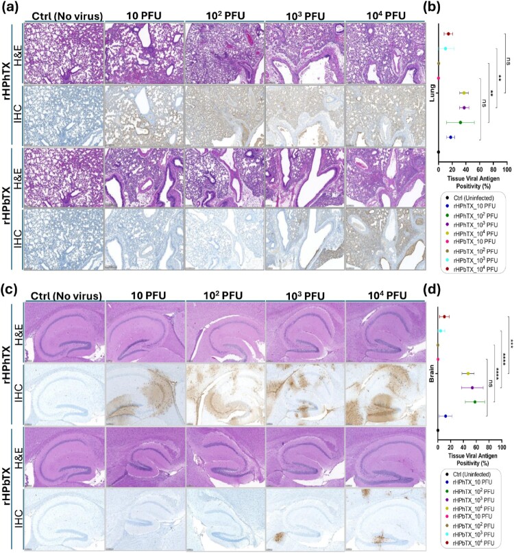
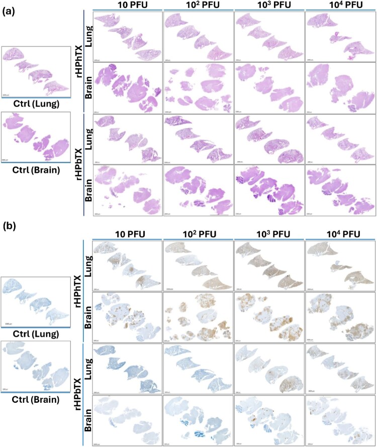
The host range of HPAIV H5N1 was recently expanded to include ruminants, particularly dairy cattle in the United States (US). Shortly after, human H5N1 infection was reported in a dairy worker in Texas following exposure to infected cattle. Herein, we rescued the cattle-origin influenza A/bovine/Texas/24-029328-02/2024(H5N1, rHPbTX) and A/Texas/37/2024(H5N1, rHPhTX) viruses, identified in dairy cattle and human, respectively, and their low pathogenic forms, rLPbTX and rLPhTX, with monobasic HA cleavage sites. Intriguingly, rHPhTX replicated more efficiently than rHPbTX in mammalian and avian cells. Still, variations in the PA and NA proteins didn't affect their antiviral susceptibility to PA and NA inhibitors. Unlike rHPbTX and rLPbTX, both rHPhTX and rLPhTX exhibited higher pathogenicity and efficient replication in infected C57BL/6J mice. The lungs of rHPhTX-infected mice produced higher inflammatory cytokines/chemokines than rHPbTX-infected mice. Our results highlight the potential risk of HPAIV H5N1 virus adaptation in human and/or dairy cattle during the current multistate/multispecies outbreak in the US.

Keywords: Cattle H5N1 virus; HPAIV; adaptation; pathogenicity; zoonosis.


Conflict of interest statement

No potential conflict of interest was reported by the author(s).

Figures

Figure 1.
Figure 1.
Sequence analysis of rHPhTX and rHPbTX, and phenotypic characterization of the rHPhTX and the natural HPhTX isolate. (a) Schematic presentation of the amino acid (aa) variations between rHPhTX (red, top) and rHPbTX (green, bottom) in PB2, PB1, PA, PAX, NA, NS1 proteins (top to bottom). (b) Prevalence rate of the distinct aa residues in HPhTX among recently reported human isolates in the US since April 2024. The graphic was created via a Web-based WebLogo application (http://weblogo.threeplusone.com/create.cgi) [38]. Red asterisks indicate which aa are prevalent among the human H5N1 isolates in the US since the cattle H5N1 outbreak in March 2024. (c) Schematic representation of the rHPhTX (top left), rHPbTX (top right), rLPhTX (bottom left), and rLPbTX (bottom right) viruses used in this study. (d) Plaque morphology of rHPhTX (top) and the natural HPhTX isolate (bottom) using crystal violet (left) and immunostaining (right). (e) Replication kinetics of rHPhTX (red) and HPhTX natural isolate (white) in A549, MDBK, CRFK, and DF-1 cells. Error bars reflect the standard deviation (SD) of three independent replicates. Statistical analysis was performed using two-way ANOVA with Greenhouse-Geisser correction, followed by Dunn-Šídák multiple comparisons post-hoc test. The “ns” indicates a non-significant difference. (f) HPhTX genome coverage by next-generation sequencing (NGS). (g) Clonal populations of the virus with minor nonsynonymous alterations in PA (L655F) and NS1 (P164S) were detected in the adapted natural isolate not previously reported in the published sequences (GenBank accession numbers of PP577940-47). The synonymous mutation T2214A was introduced in the rHPhTX PB2 segment as a genetic tag to differentiate from the natural HPhTX isolate. The figure panel (c) has been created with BioRender.com.
Figure 2.
Figure 2.
In vitro characterization of rHPhTX, rHPbTX, rLPhTX, and rLPbTX. (a) Plaque formation in MDCK monolayers with or without TPCK-treated trypsin. (b) Growth kinetics of rHPhTX (red) and rHPbTX (green) in A549, MDBK, CRFK, and DF-1 cells. (c-f) Susceptibility of rHPhTX and rHPbTX to FDA-approved antivirals. The antiviral activities of amantadine (c), baloxavir (d), oseltamivir (e) and zanamivir (f) against rHPhTX and rHPbTX were determined at 48 hpi by plotting log(agonist) vs. normalized response—variable slope and the connecting line represents a non-linear regression of the underlying data. Error bars reflect the SD of three independent replicates. The EC50 of the indicated antivirals was calculated by plotting log inhibitor versus normalized response (variable slope) and applying the nonlinear regression analyses using GraphPad Prism 9.5.1 software. Error bars reflect the SD of three independent replicates. Statistical analysis was performed using two-way ANOVA with Greenhouse-Geisser correction, followed by Dunn-Šídák multiple comparisons post-hoc test. The significant differences are indicated (* = p < 0.05, ** = p < 0.01, *** = p < 0.001; **** = p < 0.0001; non-significant = ns).
Figure 3.
Figure 3.
Pathogenicity of rHPhTX, rHPbTX, rLPhTX and rLPbTX in C57BL/6J mice. Percentage (%) of body weight changes (top) and survival curves (bottom) of C57BL/6J mice infected with 10–104 PFU of the rHPhTX and rLPhTX (a) or rHPbTX and rLPbTX (b). The mean percent of body weight change (±SEM) is shown. Mice were humanely euthanized when they had lost more than 25% of their initial body weight.
Figure 4.
Figure 4.
Viral loads in different tissues of C57BL/6J mice infected with rHPhTX and rHPbTX. The organs were collected at 2 and 4 dpi, homogenized and were subjected to titration using standard plaque assays. Infectious viral titers in homogenized lung, nasal turbinate, and brain tissues (n = 4) from mice infected with 10–104 PFU of rHPhTX and rLPhTX (a) or rHPbTX and rLPbTX (b) were assessed. Likely, the Infectious viral titers in homogenized heart, spleen, kidney and mammary gland tissues (n = 4) from mice infected with 10–104 PFU of rHPhTX and rLPhTX (c) or rHPbTX and rLPbTX (d) were also evaluated.
Figure 5.
Figure 5.
Histopathologic changes and tissue viral NP antigen positivity (%) in the lungs and brains of infected and control C57BL/6J mice. (a) Histopathologic changes in murine lung tissues infected with rHPhTX and rHPbTX at different infectious doses (10-104 PFU) against the control uninfected C57BL/6J mice. The upper row corresponding to each virus represents the tissue stained with hematoxylin-eosin (H&E). The bottom row represents the immunohistochemistry (IHC) data of the same part of the tissue (immunostained using an influenza A NP rabbit polyclonal antibody). (b) Quantitative assessment of tissue viral antigen positivity (%) in murine lung tissues infected with rHPhTX and rHPbTX at different infectious doses (10-104 PFU) against the control uninfected C57BL/6J mice. (c) Histopathologic changes in murine brain tissues infected with rHPhTX and rHPbTX at different infectious doses (10-104 PFU) against the control uninfected C57BL/6J mice. The upper row corresponding to each virus represents the tissue stained with hematoxylin-eosin, while the bottom row represents the immunohistochemistry data of the same part of the tissue. (d) Quantitative assessment of tissue viral antigen positivity (%) in murine brain tissues infected with rHPhTX and rHPbTX at different infectious doses (10-104 PFU) against the control uninfected C57BL/6J mice. The pathology overall score was determined following virus quantification in lung and brain tissues of infected C57BL/6J mice (n  = 4). Data are presented as mean ± SD. Statistical analyses against indicated groups were performed using one-way ANOVA followed by a Tukey post hoc test. The significant differences are indicated (** = p < 0.01, *** = p < 0.001, **** = p < 0.0001; non-significant = ns). Scale bars represent 200μm.
Figure 6.
Figure 6.
H&E stained and IHC lung and brain tissue sections of control uninfected and rHPhTX- or rHPbTX-infected C57BL/6J mice. (a) H&E staining of control uninfected and rHPhTX- or rHPbTX-infected lung and brain tissues of female C57BL/6J mice. (b) Influenza NP antigen IHC staining of the control uninfected and rHPhTX- or rHPbTX-infected lung and brain tissues of female C57BL/6J mice. Scale bars represent 2000μm.
Figure 7.
Figure 7.
Robust induction of innate immune responses by rHPhTX and rHPbTX in C57BL/6J infected mice. Tissue homogenates from rHPhTX-infected, rHPbTX-infected, or uninfected mice were analyzed at 2 and 4 dpi for secreted cytokines IFN-α, IFN-β, IFN-γ, IL-6, TNF-α, (a) and chemokines CCL5, CXCL2 and CXCL-10 (b) using an 8-plex bead Luminex assay. Data is shown as M ± SEM with n = 4 mice/group. A two-way ANOVA with Holm-Sidak’s multiple comparisons test was used to compare groups within each time-point. The significant differences are indicated (* = p < 0.05, ** = p < 0.01, *** = p < 0.001; **** = p < 0.0001; non-significant = ns).

Similar articles

Cited by

References

    1. Lee SH, Cho AY, Kim TH, et al. . Novel highly pathogenic avian influenza A(H5N1) clade 2.3.4.4b virus in wild birds, South Korea. Emerg Infect Dis. 2023;29:1475–1478. doi:10.3201/eid2907.221893 - DOI - PMC - PubMed
    1. Youk S, Torchetti MK, Lantz K, et al. . H5n1 highly pathogenic avian influenza clade 2.3.4.4b in wild and domestic birds: introductions into the United States and reassortments, December 2021–April 2022. J. Virol. 2023;587:109860. doi:10.1016/j.virol.2023.109860. - DOI - PubMed
    1. Mostafa A, Abdelwhab EM, Mettenleiter TC, et al. . Zoonotic potential of influenza A viruses: A comprehensive overview. Viruses. 2018;10:497. doi:10.3390/v10090497 - DOI - PMC - PubMed
    1. Ngom D, Khoulé A, Faye ET, et al. . Crimean-Congo haemorrhagic fever outbreak in Northern Senegal in 2022: prevalence of the virus in livestock and ticks, associated risk factors and epidemiological implications. Zoonoses Public Health. 2024;71:696–707. - PMC - PubMed
    1. de Glanville WA, Allan KJ, Nyarobi JM, et al. . An outbreak of rift valley fever among peri-urban dairy cattle in Northern Tanzania. Trans R Soc Trop Med Hyg. 2022;116:1082–1090. doi:10.1093/trstmh/trac076 - DOI - PMC - PubMed

MeSH terms

LinkOut - more resources