. 2025 Jan 7;53(1):gkae1263.
doi: 10.1093/nar/gkae1263.

PerR functions as a redox-sensing transcription factor regulating metal homeostasis in the thermoacidophilic archaeon Saccharolobus islandicus REY15A

Affiliations

PerR functions as a redox-sensing transcription factor regulating metal homeostasis in the thermoacidophilic archaeon Saccharolobus islandicus REY15A

Xuemei Zhang et al. Nucleic Acids Res. .

Abstract

Thermoacidophilic archaea thrive in environments with high temperatures and low pH where cells are prone to severe oxidative stress due to elevated levels of reactive oxygen species (ROS). While the oxidative stress responses have been extensively studied in bacteria and eukaryotes, the mechanisms in archaea remain largely unexplored. Here, using a multidisciplinary approach, we reveal that SisPerR, the homolog of bacterial PerR in Saccharolobus islandicus REY15A, is responsible for ROS response of transcriptional regulation. We show that with H2O2 treatment and sisperR deletion, expression of genes encoding proteins predicted to be involved in cellular metal ion homeostasis regulation, Dps, NirD, VIT1/CCC1 and MntH, is significantly upregulated, while expression of ROS-scavenging enzymes remains unaffected. Conversely, the expression of these genes is repressed when SisPerR is overexpressed. Notably, the genes coding for Dps, NirD and MntH are direct targets of SisPerR. Moreover, we identified three novel residues critical for ferrous ion binding and one novel residue for zinc ion binding. In summary, this study has established that SisPerR is a repressive redox-sensing transcription factor regulating intracellular metal ion homeostasis in Sa. islandicus for oxidative stress defense. These findings have shed new light on our understanding of microbial adaptation to extreme environmental conditions.


Figures

Graphical Abstract
Graphical Abstract
Figure 1.
Figure 1.
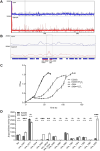
The sisperR knockout strain of Sa. islandicus shows increased tolerance to H2O2 treatment. (A) Growth curves of the WT strain E233S in the presence of different concentrations of H2O2. The cells were inoculated into 30 mL medium to a final estimated OD600 of 0.05. Different concentrations of H2O2 were added in exponentially growing cells. The cells were incubated at 30°C for 1 h before cultured with shaking at 75°C. The growth was monitored using spectrometer. (B) Comparison of the growth of E233S and ΔperR cultured with or without 70 μM H2O2. (C) Survival rates of E233S and ΔperR with or without H2O2 treatment. Exponentially growing cells were treated with different doses of H2O2 at 30°C for 1 h and cultivated at 75°C for 5 h. Cells were taken and plated on STVU plates. The survival rates are calculated as percentages of the number of colonies on plates with treated cultures divided by that on plates with corresponding non-treated cultures. Statistical significance was analyzed by performing a t test with the software GraphPad Prism 9 (*P < 0.05, **P < 0.01, ***P < 0.001, ns, not significant). (D) Growth curves of ΔperR carrying plasmids pSeSD, pSeSD-PerR and pSeSD-PerR(T53A) cultured with or without 70 μM H2O2 in STV medium. (E) Flow cytometry profiles showing the intracellular ROS levels in E233S and ΔperR. E233S and ΔperR were cultured in STVU medium to OD600 0.2–0.3 and then the cells were grown in the presence or absence of 50 μM H2O2. The cells were incubated at 30°C for 1 h and then cultured at 75°C for 2 h. DCFH-DA (5 μM) was added to the cell suspension and the cells were incubated at 37°C for 30 min in the dark. All the samples were analyzed immediately after incubation by flow cytometry using ImageStreamX MarkII with an excitation wavelength of 488 nm. The analysis was performed twice. (F) Examination of the tolerance of ΔperR to different concentrations of H2O2. The cells were treated as in (A) except that the H2O2 concentrations were elevated. For all the experiments, each value was based on data from three independent experiments.
Figure 2.
Figure 2.
Growth of strains with point mutation on the metal binding sites of SisPerR. (A) Superposition of SisPerR to B. subtilis PerR (BsPerR) monomer. A model of SisPerR monomer is predicted by AlphaFold. The structures of SisPerR and BsPerR are colored in orange and gray, respectively. The cysteine/histidine residues of SisPerR and BsPerR are depicted in red and blue, respectively. (B) Growth curves of E233S and the point mutant strains of PerR(C128S), PerR(H32A), PerR(H85A) and PerR(H32A/H85A) in the presence or absence of 70 μM H2O2. (C) Verification of protein expression of the mutant strains by Western blotting. (D) The relative transcription level of sisperR. The transcription levels of sisperR in E233S and PerR(C128S) were measured by RT-qPCR. Data were normalized to the level of 16S rRNA. Error bars represent standard derivations of three replicates.
Figure 3.
Figure 3.
The conserved cysteine residues are critical for the function of PerR. (A) Detection of cysteine mutant proteins in ΔperR/pSeSD-PerR(C90S) and ΔperR/pSeSD-PerR(C90S/C128S) by Western blotting in complementary strains. Anti-TBP was used as the loading control. (B) Verification of protein expression of SisPerR mutants in E. coli by Western blotting. (C) Growth curves ΔperR carrying the plasmids pSeSD, pSeSD-PerR, pSeSD-PerR(C90S) and pSeSD-PerR(C90S/C128S). Cells were cultured in STV medium with or without 70 μM H2O2. The growth was monitored by spectrometry.
Figure 4.
Figure 4.
Four additional residues of PerR are critical for the function of PerR in Sa. islandicus. (A) Superposition of monomeric BsPerR with SisPerR predicted by AlphaFold. The structure of SisPerR and BsPerR are colored in orange and gray, respectively. The residues of SisPerR and BsPerR are depicted in red and blue, respectively. (B) Verification of the expression of mutant SisPerR proteins in ΔperR/pSeSD-PerR(E79A), ΔperR/pSeSD-PerR(N87A), ΔperR/pSeSD-PerR(D98A) and ΔperR/pSeSD-PerR(D92S) by Western blotting. Anti-TBP was used as the loading control. (C) Growth curve of the complementary mutant strains in the presence or absence of 70 μM H2O2. ΔperR carrying the plasmids pSeSD, pSeSD-PerR, pSeSD-PerR(E79A), pSeSD-PerR(N87A), pSeSD-PerR(D98A) and pSeSD-PerR(D92S) were cultured to OD600∼0.2 and cultivated in STV medium with or without 70 μM H2O2 treatment. The growth was monitored by spectrometry.
Figure 5.
Figure 5.
Global transcriptional changes of Sa. islandicus E233S and ΔperR with or without 70 μM H2O2 treatment. (A) Heatmap of genome expression of Sa. islandicus E233S and ΔperR. Both strains were grown in the presence or absence of 70 μM H2O2 at 6 h. The cells were collected for RNA-Seq analysis. Genes are clustered with their log10 (FPKM) values and their expression levels are indicated by different colors. (B) and (C) Volcano plots showing fold changes and levels of significance for the DEGs in E233S (B) and ΔperR (C) in the presence and absence of 70 μM H2O2. The Y-axis (-log10, P-value) represents statistical significance of the fold change, and X-axis (log2, fold change) represents fold change in gene expression.
Figure 6.
Figure 6.
Identification of putative target genes of SisPerR by ChIP-seq. (A) Overview of the genomic binding profile of SisPerR by ChIP-seq. The sample before immunoprecipitation was used as input. (B) A zoom of the profile showing enriched promoter region of sire_0452 (mntH) and sire_0453 (dps). Each rectangle represents a gene sequence. The gene sequences of sire_0452 (mntH) and sire_0453 (dps) were represented by arrows. (C) Growth curves of E233S and Δdps with or without 70 μM H2O2. E233S and Δdps were cultured with or without 70 μM H2O2. (D) In vivo promoter activity assay using lacS as a reporter gene. The promoter of putative PerR-regulated genes sire_0057 (perR), sire_0277 (sucC), sire_0444 (fdhA), sire_0452 (mntH), sire_0453 (dps)/sire_0454 (nirD), sire_0495 (duf973), sire_0980, sire_0982 (duf1641), sire_1083 (tpp), sire_1254 (sam1), sire_1398, sire_1880 (tadC), sire_2636 (vit1/ccc1) and tbp were chosen for the analysis. Sa. islandicus E233S and ΔperR strains carrying each of the reporter gene plasmids were grown in STV medium. The cell mass was collected from which cell extracts were prepared and used for determination of the β-glycosidase activity. The values were obtained based on three biological repeats. Statistical significance was analyzed by performing a t test with the software GraphPad Prism 9 (*P < 0.05, **P < 0.01, ***P < 0.001, ns, not significant).
Figure 7.
Figure 7.
Phylogenetic analysis of PerR homologs. The analyses were performed by aligning 62 amino acid sequences from 42 archaeal and 20 bacterial species using MUSCLE (v. 5.1) (49). The phylogenetic tree was generated using ML-based FastTree (v. 2.1.11) (50). Representative PerR homologs from each archaeal phylum and bacteria were selected. More than one homologs were selected from Crenarchaea and Euryarchaea. The filled circles in different sizes at the nodes indicate different bootstrap values of 1000 replicates. SisPerR is indicated in red.

Similar articles

Cited by

References

    1. Imlay J.A. Pathways of oxidative damage. Annu. Rev. Microbiol. 2003; 57:395–418. - PubMed
    1. Imlay J.A. The molecular mechanisms and physiological consequences of oxidative stress: lessons from a model bacterium. Nat. Rev. Microbiol. 2013; 11:443–454. - PMC - PubMed
    1. Holmström K.M., Finkel T.. Cellular mechanisms and physiological consequences of redox-dependent signalling. Nat. Rev. Mol. Cell Biol. 2014; 15:411–421. - PubMed
    1. Sies H., Belousov V.V., Chandel N.S., Davies M.J., Jones D.P., Mann G.E., Murphy M.P., Yamamoto M., Winterbourn C.. Defining roles of specific reactive oxygen species (ROS) in cell biology and physiology. Nat. Rev. Mol. Cell Biol. 2022; 23:499–515. - PubMed
    1. Johnson L.A., Hug L.A.. Distribution of reactive oxygen species defense mechanisms across domain bacteria. Free Radical Biol. Med. 2019; 140:93–102. - PubMed

MeSH terms