. 2025 Feb 8;53(4):gkae1279.
doi: 10.1093/nar/gkae1279.

Autophosphorylation of the Tousled-like kinases TLK1 and TLK2 regulates recruitment to damaged chromatin via PCNA interaction

Affiliations

Autophosphorylation of the Tousled-like kinases TLK1 and TLK2 regulates recruitment to damaged chromatin via PCNA interaction

Kirk L West et al. Nucleic Acids Res. .

Abstract

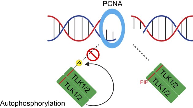
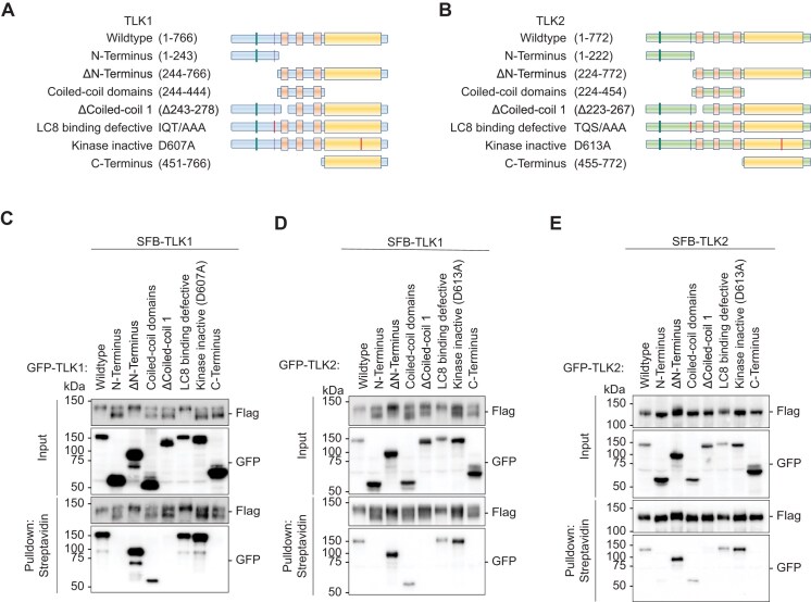
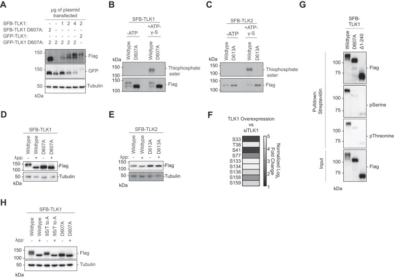
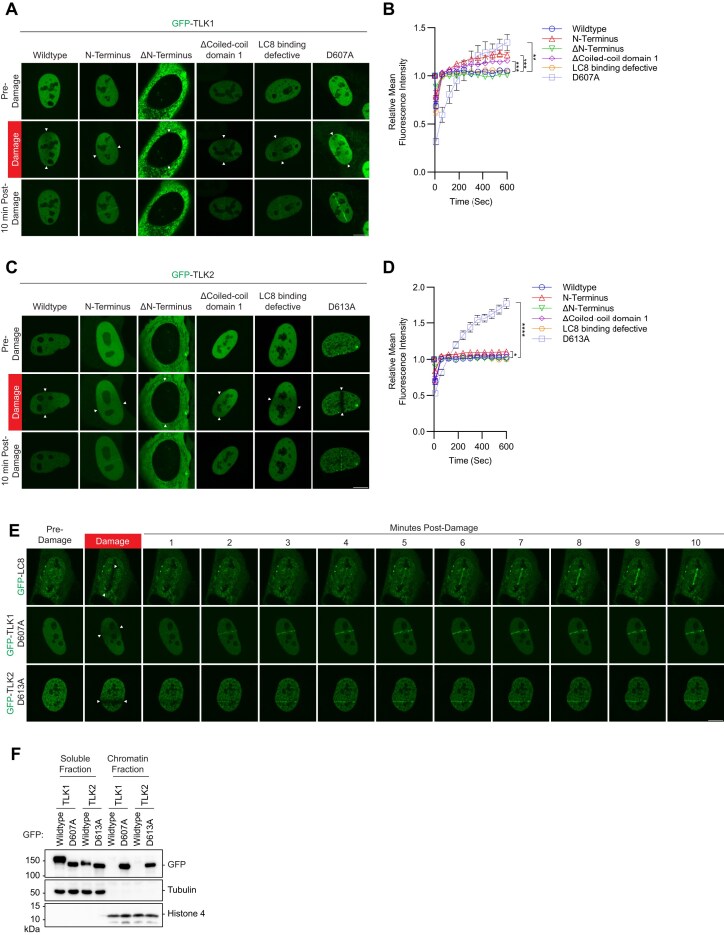
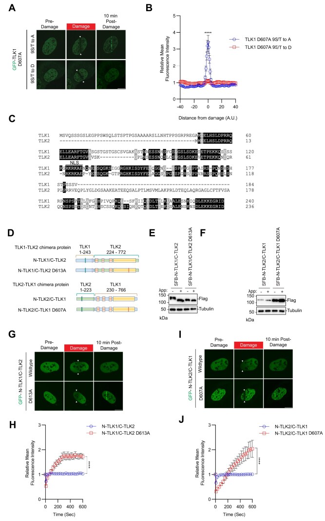
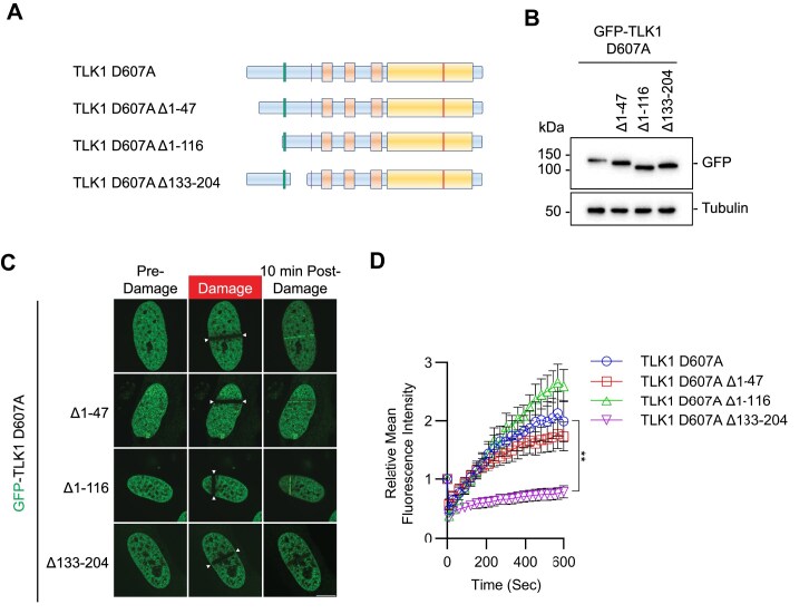
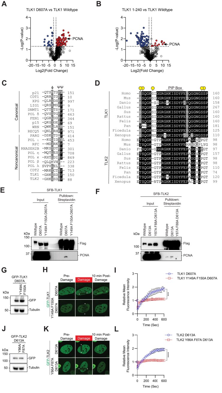
Tousled-like kinases 1 and 2 (TLK1 and 2) are cell cycle-regulated serine/threonine kinases that are involved in multiple biological processes. Mutation of TLK1 and 2 confer neurodegenerative diseases. Recent studies demonstrate that TLK1 and 2 are involved in DNA repair. However, there is no direct evidence that TLK1 and 2 function at DNA damage sites. Here, we show that both TLK1 and TLK2 are hyper-autophosphorylated at their N-termini, at least in part, mediated by their homo- or hetero- dimerization. We found that TLK1 and 2 hyper-autophosphorylation suppresses their recruitment to damaged chromatin. Furthermore, both TLK1 and 2 associate with PCNA specifically through their evolutionarily conserved non-canonical PCNA-interacting protein (PIP) box at the N-terminus, and mutation of the PIP-box abolishes their recruitment to DNA damage sites. Mechanistically, the TLK1 and 2 hyper-autophosphorylation masks the PIP-box and negatively regulates their recruitment to the DNA damage site. Overall, our study dissects the detailed genetic regulation of TLK1 and 2 at damaged chromatin, which provides important insights into their emerging roles in DNA repair.


Figures

Graphical Abstract
Graphical Abstract
Figure 1.
Figure 1.
Tousled-like kinases form dimers and drive autophosphorylation. (AandB) Schematic illustrations of TLK1 and TLK2 constructs for dimerization domain mapping. (CE) TLK dimerization requires an intact coiled-coil domain. HEK293T cells were co-transfected with (C) SFB-TLK1 wildtype and the indicated GFP-TLK1 expression vectors, (D) SFB-TLK1 wildtype and GFP-TLK2 plasmids, (E) SFB-TLK2 and GFP-TLK2 plasmids, and pulldown experiment followed by Western blotting analysis using FLAG and GFP antibodies.
Figure 2.
Figure 2.
Tousled-like kinases are autophosphorylated at their N-terminus. (A) Co-transfection of TLK1 wildtype and catalytic inactive mutant D607A with the indicated amount of plasmids in HEK293T cells followed by Western blotting analysis. (BandC) Kinase assays using ATP-γ-S followed by alkylation by p-Nitrobenzyl mesylate to convert phosphorylated residues into thiophosphate esters. Antibody against thiophosphate ester was used to identify phosphorylation events. (DandE) Lambda protein phosphatase (λpp) assay was performed using SFB-tagged TLK1 wildtype and D607A mutant or SFB-tagged TLK2 wildtype and D613A mutant purified from HEK293T cell lysate. Differential molecular weights were analyzed by Western blotting analysis. (F) TLK1 is autophosphorylated at nine residues within its N-terminus. Phospho-Tandem-Mass-Tag mass spectrometry was performed on phosphopeptides enriched from HEK293T cells with TLK1 overexpression and knockdown using siRNA. (G) TLK1 N-terminus is highly phosphorylated. SFB-tagged TLK1 wildtype, D607A or Δ1–240 (ΔN-terminus) were purified from HEK293T using streptavidin beads followed by Western blotting analysis using phospho-Serine and phospho-threonine antibodies. (H) TLK1 wildtype, N-terminal 9S/T to A and catalytic inactive mutants were treated with λ-phosphatase. Differential molecular weights were analyzed by Western blot.
Figure 3.
Figure 3.
Accumulation of TLK1 and 2 is negatively regulated by their kinase activity and dimerization at damaged chromatin. (AandC) TLK1 and TLK2 recruitment to sites of DNA damage is repressed by dimerization and kinase activity. U2OS cells were transfected with indicated GFP-TLK1 or TLK2 wildtype and mutants. Laser-induced micro-irradiation and live-cell imaging experiments were conducted using a confocal microscope, denoted line between the white arrows. Representative images of each experimental group at 10 min after damage. Scale bar represents 10 μm. (B, D) Quantification of laser-included micro-irradiation as in (A) and (C), respectively. N ≥ 10 cells. Significance was determined by unpaired, two-tailed Student’s t-test and P-values are reported as *P < 0.05, **P < 0.01, ***P < 0.001 and ****P < 0.0001. (E) DNA damage kinetics of LC8, TLK1 and TLK2 catalytic inactive mutants using laser-induced micro-irradiation. Scale bar represents 10 μm. (F) Kinase activity negatively regulates TLK chromatin loading. HEK293T cells were transfected with the indicated GFP-TLK1 wildtype and catalytic inactive mutants. Cells were harvested 24 h after transfection and fractionated to obtain soluble and chromatin fractions.
Figure 4.
Figure 4.
Recruitment of TLK1 and 2 to damaged DNA is N-terminal phosphorylation-dependent. (A) TLK1 autophosphorylation suppresses recruitment to damaged chromatin. The TLK1catalytic mutant (D607A) with N-terminus phospho-defective mutant (9S/T to A) or phospho-mimetic mutant (9S/T to D) were subjected to laser-induced micro-irradiation. Scale bar represents 10 μm. (B) Quantification of the recruitment intensity as shown in A. N = 10. (C) Sequence alignment of the N-terminus of TLK1 and TLK2. (D) Schematic illustration of chimeric proteins generated for TLK1 and TLK2. (EandF) Chimeric N-TLK1/C-TLK2, N-TLK1/C-TLK2 D613A, N-TLK2/C-TLK1 and N-TLK2/C-TLK1D607A were treated with λpp. Apparent molecular weights were analyzed by Western blot. (G) Representative images of GFP- N-TLK1/C-TLK2 and N-TLK1/C-TLK2 D613A chimeras at 10 min after laser-induced micro-irradiation. The damaged area was denoted between the arrows. Scale bar represents 10 μm. (H) Recruitment kinetics quantification of the GFP-chimera proteins after laser-induced micro-irradiation as in (G). N ≥ 10. (I) Representative images of GFP- N-TLK2/C-TLK1 and N-TLK2/C-TLK1 D607A chimeras at 10 min after laser-induced micro-irradiation. The damaged area was denoted between the arrows. Scale bar represents 10 μm. (J) Recruitment kinetics quantification of the GFP-chimera proteins after laser-induced micro-irradiation as in (I). N ≥ 10. Significance was determined by unpaired, two-tailed Student’s t-test and P-values are reported as *P < 0.05, **P< 0.01, ***P< 0.001 and ****P < 0.0001.
Figure 5.
Figure 5.
The N-terminus conserved region of TLK1 is required for its laser-induced micro-irradiation. (A) Schematic illustrations of TLK1 N-terminal deletion mutants for damaged chromatin recruitment region mapping. (B) Western blotting analysis of the mutants as in A and used in C and D. (C) Representative images of TLK1 N-terminal deletion mutants 10 min after laser-induced micro-irradiation. Scale bar represents 10 μm. (D) Recruitment kinetics of the TLK1 N-terminal deletion mutants as in C. N ≥ 10. Significance was determined by unpaired, two-tailed Student’s t-test and P-values are reported as *P < 0.05, **P <0.01, ***P < 0.001 and ****P < 0.0001.
Figure 6.
Figure 6.
PCNA regulates TLK1 and TLK2 DNA damage recruitment via interaction with their PIP-box. (AandB) Volcano plot of proteomic analyses between TLK1 wildtype and catalytic mutant D607A or N-terminal fragment a.a. 1–240. Data was analyzed using Perseus and represents technical duplicate data. (C) Sequence alignment of the canonical and non-canonical PIP-box between TLK, TLK2 and other PCNA interacting proteins. PIP-box QxxϕxxΨΨ, where x represents any amino acid, ϕ represents hydrophobic residues and Ψ represents aromatic residues) or non-canonical sequences (xxxϕxxΨΨ). (D) Sequence alignment of the TLK1 and TLK2 PIP-box across species. Dot labels represent identified TLK1 autophosphorylation sites. (EandF) TLK1 and TLK2 interact with PCNA. HEK293T cells transfected with SFB-TLK1 wildtype, D607A or PIP box mutant Y149A F150A D607A. Cells were pulled down by streptavidin beads and analyzed by western blot using indicated antibodies. (G–I) TLK1 PIP box is required for DNA damage recruitment. (G) GFP-TLK1D607A and D607A PCNA binding defective mutant (Y149A/F150A) protein expression. (H) Representative images of indicated mutants at 10 min after laser-induced micro-irradiation. The irradiated area is induced by the arrows. (I) DNA damage recruitment kinetics quantification as in (H). N = 10. Scale bar represents 10 μm. (J–L) TLK2 PIP box is required for DNA damage recruitment. (J) GFP-TLK2D613A and D613A PCNA binding defective mutant (Y86A/F87A) protein expression. (K) Representative images of indicated mutants at 10 min after laser-induced micro-irradiation. The irradiated area is induced by the arrows. (L) DNA damage recruitment kinetics quantification as in (K). N ≥ 10. Significance was determined by unpaired, two-tailed Student’s t-test and P-values are reported as *P < 0.05, **P < 0.01, ***P < 0.001 and ****P < 0.0001. Scale bar represents 10 μm.

Update of

Similar articles

References

    1. Shalom S., Don J. Tlk, a novel evolutionarily conserved murine serine threonine kinase, encodes multiple testis transcripts. Mol. Reprod. Dev. 1999; 52:392–405. - PubMed
    1. Roe J.L., Rivin C.J., Sessions R.A., Feldmann K.A., Zambryski P.C. The Tousled gene in A. thaliana encodes a protein kinase homolog that is required for leaf and flower development. Cell. 1993; 75:939–950. - PubMed
    1. Lee S.B., Segura-Bayona S., Villamor-Payà M., Saredi G., Todd M.A.M., Attolini C.S.O., Chang T.Y., Stracker T.H., Groth A. Tousled-like kinases stabilize replication forks and show synthetic lethality with checkpoint and PARP inhibitors. Sci. Adv. 2018; 4:eaat4985. - PMC - PubMed
    1. Li Y., DeFatta R., Anthony C., Sunavala G., De Benedetti A. A translationally regulated Tousled kinase phosphorylates histone H3 and confers radioresistance when overexpressed. Oncogene. 2001; 20:726–738. - PubMed
    1. Segura-Bayona S., Stracker T.H. The Tousled-like kinases regulate genome and epigenome stability: implications in development and disease. Cell. Mol. Life Sci. 2019; 76:3827–3841. - PMC - PubMed

MeSH terms