. 2025 Dec;14(1):2447615.
doi: 10.1080/22221751.2024.2447615. Epub 2025 Mar 3.

Enhanced antibody response to the conformational non-RBD region via DNA prime-protein boost elicits broad cross-neutralization against SARS-CoV-2 variants

Affiliations

Enhanced antibody response to the conformational non-RBD region via DNA prime-protein boost elicits broad cross-neutralization against SARS-CoV-2 variants

Yun-Fei Ma et al. Emerg Microbes Infect. 2025 Dec.

Abstract

Preventing immune escape of SARS-CoV-2 variants is crucial in vaccine development to ensure broad protection against the virus. Conformational epitopes beyond the RBD region are vital components of the spike protein but have received limited attention in the development of broadly protective SARS-CoV-2 vaccines. In this study, we used a DNA prime-protein boost regimen to evaluate the broad cross-neutralization potential of immune response targeting conformational non-RBD region against SARS-CoV-2 viruses in mice. Mice with enhanced antibody responses targeting conformational non-RBD region show better performance in cross-neutralization against the Wuhan-01, Delta, and Omicron subvariants. Via analyzing the distribution of conformational epitopes, and quantifying epitope-specific binding antibodies, we verified a positive correlation between the proportion of binding antibodies against the N-terminal domain (NTD) supersite (a conformational non-RBD epitope) and SARS-CoV-2 neutralization potency. The current work highlights the importance of high ratio of conformational non-RBD-specific binding antibodies in mediating viral cross-neutralization and provides new insight into overcoming the immune escape of SARS-CoV-2 variants.

Keywords: Conformational non-RBD epitopes; DNA prime-protein boost; NTD supersite-specific binding antibody; SARS-CoV-2; crossneutralization.


Conflict of interest statement

No potential conflict of interest was reported by the author(s).

Figures
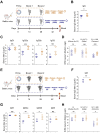
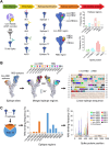
Figure 1.
Figure 1.
Humoral responses of “DNA prime-protein boost” and protein-only immunization strategies. (A) Schematic diagram of RBD antigen immunization. (B) RBD-specific IgG was assessed by ELISA. (C) RBD-specific IgG subtypes were assessed by ELISA. (D) Live virus neutralization titers were determined by in vitro neutralization assays. (E) Schematic diagram of Spike protein immunization. (F) S-specific IgG was assessed by ELISA. (G) S-specific IgG subtypes were assessed by ELISA. (H) Live virus neutralization titers were determined by in vitro neutralization assays. The orange and blue rhombuses represent the RBD “DNA prime-protein boost” group and the RBD protein group, respectively (B)–(D). The orange and blue circles represent the S “DNA prime-protein boost” group and the S protein group, respectively (F)–(H). Each rhombus (B)–(D) or circle (F)–(H) represents one sample, n = 10. *P < 0.05, **P < 0.01, ***P < 0.001 (two-tailed Student t-test). NS, not significant.
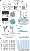
Figure 2.
Figure 2.
Humoral responses of conformational non-RBD and RBD epitopes. (A-B) Structure analysis of conformational non-RBD and RBD subunits. (C) Schematic diagram of immunization. (D) Spike-specific IgG was assessed by ELISA. (E) Spike-specific IgG subtypes were assessed by ELISA. (F) Live virus neutralization titers were determined by in vitro neutralization assays. The orange and blue triangles represent the SDNA-RBD-RBD group and the SDNA-S-S group, respectively (D)–(F). Each triangle (D)–(F) represents one sample, n = 10. *P < 0.05, **P < 0.01, ***P < 0.001 (two-tailed Student t-test). NS, not significant.
Figure 3.
Figure 3.
Non-RBD and RBD epitopes are identified within the spike protein of SARS-CoV-2. (A) Non-RBD and RBD epitope identification flow chart. (B) Left, identified non-RBD and RBD epitopes. Middle, non-RBD, and RBD epitope regions are projected onto protein 3D structures. Right, linear sequence of 12 epitope regions. (C) Left, the ratio of non-RBD and RBD epitope sites. Middle, nAb binding frequency of the non-RBD and RBD epitope regions. Right, Ab binding frequency of epitope residues within the different epitope regions. The gray vertical line represents epitope residues overlapped by at least two epitope regions.
Figure 4.
Figure 4.
Ns-EMPEM analysis of polyclonal Fabs from vaccinated mouse sera complexed with SARS-CoV-2 spikes. (A) Schematic diagram of ns-EM polyclonal epitope mapping (EMPEM) analysis. (B) Representative 2D classes, side and top views of the 3D reconstructions from ns-EMPEM analysis of SARS-CoV-2 spikes complexed with polyclonal Fabs isolated from DNA(S)-RBD-RBD and DNA(S)-S-S vaccination groups. Pie charts show the proportion of Abs targeting the SARS-CoV-2 nsEM-nonRBD (sea green), nsEM-RBD (blue), and spike (w/o Ab) (gray). (C) Surface representation of each antibody epitope on the SARS-CoV-2 spike. The spike trimer with three “down” RBDs is adapted from PDB ID 6XR8. The epitope residues involved in NTD binding are labelled with their respective color. Interaction residues in NTD are defined by a 5 Å distance cut-off. (D) Comparison between identified and ns-EMPEM epitopes. (E) The percentage of ns-EMPEM non-RBD epitopes overlapped with the identified non-RBD epitope regions. (F) Residue sequences of the non-RBD4 epitope region in 17 SARS-CoV-2 variants. Mutated residues are highlighted with a red background and residue deletions are indicated by “-.” (G) Conservative frequency of the non-RBD4 epitope residues across 17 SARS-CoV-2 variants.
Figure 5.
Figure 5.
Contribution of non-RBD antibodies to neutralization. (A) Schematic diagram of the experiment to evaluate the influence of NTD-specific Abs on neutralization against pseudotyped virus. (B)–(C) Pseudotyped virus neutralization titers of serum with or without NTD protein incubation. (D) Fitting analysis of pseudotyped virus neutralization titers of serum with different NTD protein concentrations. *P < 0.05 (two-tailed Student t-test). NS, not significant.

Similar articles

Cited by

References

    1. Cao Y, Yisimayi A, Jian F, et al. . BA.2.12.1, BA.4 and BA.5 escape antibodies elicited by omicron infection. Nature. 2022 Aug;608(7923):593–602. doi:10.1038/s41586-022-04980-y - DOI - PMC - PubMed
    1. Liu C, Ginn HM, Dejnirattisai W, et al. . Reduced neutralization of SARS-CoV-2 B.1.617 by vaccine and convalescent serum. Cell. 2021 Jun 17;184(16):4220–4236.e13. doi:10.1016/j.cell.2021.06.020 - DOI - PMC - PubMed
    1. Lan J, Ge J, Yu J, et al. . Structure of the SARS-CoV-2 spike receptor-binding domain bound to the ACE2 receptor. Nature. 2020 May;581(7807):215–220. doi:10.1038/s41586-020-2180-5 - DOI - PubMed
    1. Valdes-Balbin Y, Santana-Mederos D, Paquet F, et al. . Molecular aspects concerning the Use of the SARS-CoV-2 receptor binding domain as a target for preventive vaccines. ACS Cent Sci. 2021 May 26;7(5):757–767. doi:10.1021/acscentsci.1c00216 - DOI - PMC - PubMed
    1. Yan R, Wang R, Ju B, et al. . Structural basis for bivalent binding and inhibition of SARS-CoV-2 infection by human potent neutralizing antibodies. Cell Res. 2021 Mar 17;31(5):517–525. doi:10.1038/s41422-021-00487-9 - DOI - PMC - PubMed

MeSH terms

Supplementary concepts