. 2025 Feb 25;99(2):e0150724.
doi: 10.1128/jvi.01507-24. Epub 2024 Dec 27.

Loss of mucin 2 and MHC II molecules causes rare resistance to murine RV infection

Affiliations

Loss of mucin 2 and MHC II molecules causes rare resistance to murine RV infection

Carolyn Bomidi et al. J Virol. .

Abstract

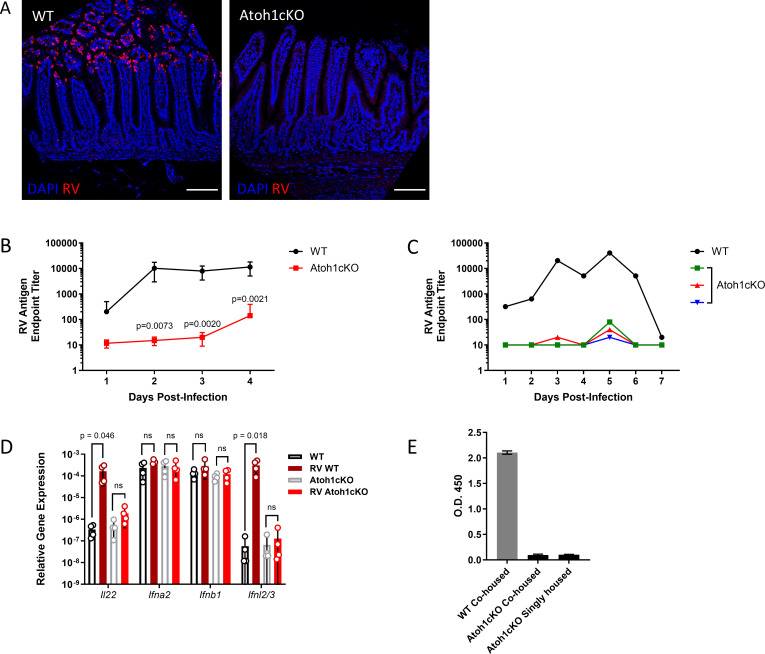
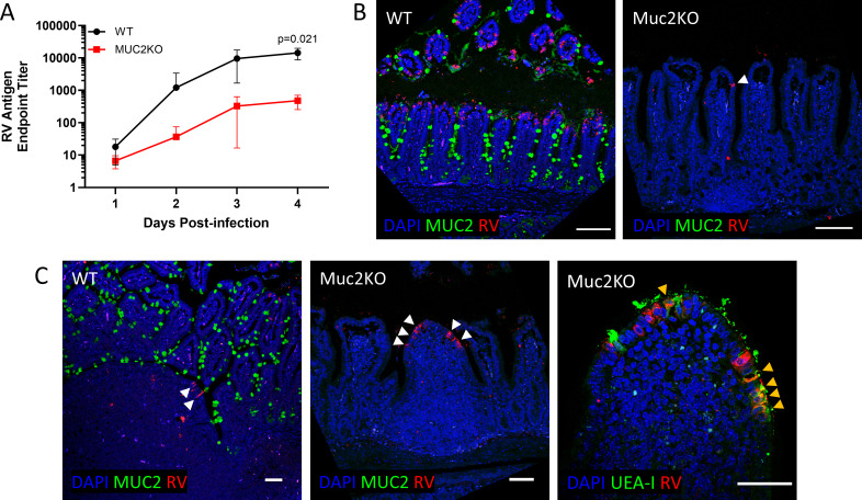
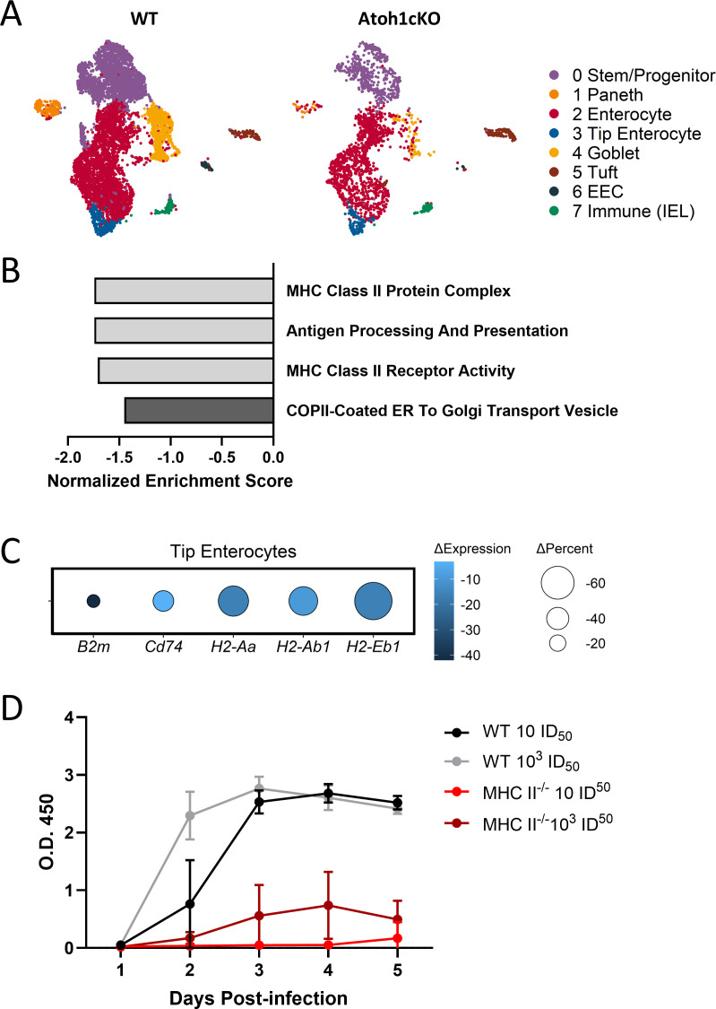
Enteric pathogen rotavirus (RV) primarily infects mature enterocytes at the tips of the intestinal villi; however, the role of secretory Paneth and goblet cells in RV pathogenesis remains unappreciated. Atoh1 knockout mice (Atoh1cKO) were used to conditionally delete Paneth, goblet, and enteroendocrine cells in the epithelium to investigate the role of secretory cells in RV infection. Unexpectedly, the number of infected enterocytes and the amount of RV shedding in the stool were greatly decreased following secretory cell deletion. Resistance to RV infection persisted for 7 days after virus inoculation, and Atoh1 knockout mice co-housed with infected wild-type mice were uninfected, based on lack of shedding virus, despite the highly infectious nature of RV. This response was directly proportional to the extent of secretory cell deletion, with infection predominantly occurring in areas containing intact secretory cells. RV infection of Muc2 knockout mice recapitulated the secretory cell deletion phenotype, indicating that goblet cell loss is responsible for attenuated infection. Transcriptome analysis of Atoh1cKO intestine via single-cell RNA sequencing revealed downregulation of MHC II molecules specifically in tip enterocytes, and MHC II-/- mice were likewise resistant to RV infection. These data suggest a previously unknown role for both MUC2 and MHC II expression in susceptibility to RV infection.IMPORTANCERotavirus (RV) is a highly contagious pathogen that primarily infects mature intestinal enterocytes. Murine rotavirus readily infects infant and adult mice, enabling evaluation of RV infection and immunity. We report that mice lacking secretory cells are one of the few genetically modified mouse lines not susceptible to murine rotavirus. Further investigation revealed loss of mucin 2 (MUC2) expression or major histocompatibility complex II (MCH II) expression recapitulated this rare resistance to rotavirus infection, suggesting a previously unrecognized link between secretory cell products and major histocompatibility complex II expression. Furthermore, these mouse models provide a platform to investigate rotavirus pathogenesis.

Keywords: Atoh1; MHC II; Muc2; RV; pathogenesis; receptor; resistance; secretory cell; tropism.


Conflict of interest statement

The authors declare no conflict of interest.

Figures

Fig 1
Fig 1
Mice lacking intestinal secretory cells are resistant to murine rotavirus infection. (A) Immunofluorescence of Atoh1cKO (right) or littermate control (WT, left) intestine at 4 days post-infection (dpi) with rotavirus ECwt (RV). Scale bar: 100 µm. Red: RV, blue: nuclei. (B) Fecal ELISA monitoring RV shedding in WT or Atoh1cKO stool for 4 dpi. Unpaired t-test results are indicated by P value; all other comparisons are not significant (P value > 0.05). n = 4–6 mice per group; symbols indicate mean ± SD. (C) Fecal ELISA monitoring RV shedding in representative WT or Atoh1cKO stool for 7 dpi. Each line indicates shedding from a single mouse. (D) RNA was isolated from the jejunum of WT and Atoh1cKO mice, and qRT-PCR was used to quantify various immune response transcripts at 4 dpi. n = 4 mice per group; bars show mean ± SD. Unpaired t-test results are indicated by brackets. ns: not significant (P value > 0.05). (E) A WT mouse was infected with RV and co-housed with uninfected Atoh1cKO mice and WT littermate controls. Two additional Atoh1cKO mice were infected and singly housed for comparison. RV shedding was assessed via fecal ELISA 4 days after exposure to RV or the infected WT mouse.
Fig 2
Fig 2
Resistance to murine RV is titratable and localized. (A) Fecal shedding of Vil1Cre/ERT2+; Atoh1fl/fl mice treated with one to three doses of either 1 mg (left) or 0.1 mg (right) tamoxifen and infected with RV. Significant unpaired t-test results between no tamoxifen (0 mg) and one to three doses of tamoxifen are indicated by asterisks; all other comparisons are not significant (P value > 0.05). n = 3–4 mice per condition; symbols indicate mean ± SD. (B) The average number of goblet (left) and Paneth (right) cells was assessed via PAS stain at 4 dpi. Goblet and Paneth cells from at least 20 well-oriented crypts or villi were counted per segment per animal. n = 3 animals averaged per condition. (C) Immunofluorescence of region-specific Atoh1KO (F-Atoh1KO) intestine at 4 dpi. FITC-conjugated LEA lectin (green) marks Paneth and goblet cells as well as surface glycans. Red: RV, blue: nuclei. White arrowheads indicate infected cells. Red signal below the crypts indicates nonspecific staining of red blood cells. Scale bar = 100 µm. (D) Number of infected cells per villus in the proximal or distal intestine of F-Atoh1KO and littermate controls (WT) from (C). >25 well-oriented villi from infected animals were selected for quantification. Dotted line indicates median. (E) Fecal shedding of Fabp1Cre; Atoh1fl/fl (F-Atoh1KO) mice and littermate controls (WT) infected with RV. n = 3–4 animals per condition; symbols indicate mean ± SD.
Fig 3
Fig 3
Tip enterocytes are resistant to RV infection in Muc2KO mice, but the follicle-associated epithelium is susceptible. (A) Fecal RV ELISA from Muc2KO mice and littermate controls (WT). n = 3–4 animals per line; symbols indicate mean ± SD. Unpaired t-test results are indicated by P value; all other comparisons are not significant (P value > 0.05). (B) Immunofluorescent staining for MUC2 (green), RV (red), and nuclei (blue) at 4 days post-infection (dpi). White arrowhead indicates rare infected enterocyte. Scale bar = 100 µm. (C) Infected WT or Muc2KO intestine stained with MUC2 antibody (left) or FITC-conjugated lectin Ulex europaeus agglutinin (UEA-I, right) and anti-RV at 4 dpi. White arrowheads indicate infected cells in the follicle-associated epithelium (left). Yellow arrowheads indicate cells double-positive for UEA-I (green) and RV (red) (right). Scale bar = 50 µm.
Fig 4
Fig 4
Enterocyte MHC II expression is decreased in Atoh1cKO mice and MHC II-/- mice are resistant to RV infection (A) Tip enterocytes (blue) were identified in a previously published Atoh1cKO scRNAseq dataset (29). IEL: Intraepithelial lymphocytes. (B) Select de-enriched Gene Ontology (GO) pathways in Atoh1cKO tip enterocytes versus WT tip enterocytes. p value < 0.05. (C) Differentially expressed genes associated with the GO pathways in (B). Adjusted p value < 0.05. ∆Percent indicates change in percent of cells expressing the gene (Atoh1cKO versus WT), ∆Expression indicates change in mean expression level. (D) MHC II-/- mice were infected with low (10 ID50) or high (1E3 ID50) dose of rotavirus (RV). Stool was collected daily to quantify viral load via RV ELISA. n=4-6 mice per group; symbols indicate mean ± SD.

Similar articles

References

    1. Bishop RF, Davidson GP, Holmes IH, Ruck BJ. 1973. Virus particles in epithelial cells of duodenal mucosa from children with acute non-bacterial gastroenteritis. Lancet 2:1281–1283. doi:10.1016/s0140-6736(73)92867-5 - DOI - PubMed
    1. Crawford SE, Ramani S, Tate JE, Parashar UD, Svensson L, Hagbom M, Franco MA, Greenberg HB, O’Ryan M, Kang G, Desselberger U, Estes MK. 2017. Rotavirus infection. Nat Rev Dis Primers 3:17083. doi:10.1038/nrdp.2017.83 - DOI - PMC - PubMed
    1. Ramig RF. 2004. Pathogenesis of intestinal and systemic rotavirus infection. J Virol 78:10213–10220. doi:10.1128/JVI.78.19.10213-10220.2004 - DOI - PMC - PubMed
    1. Estes M, Greenberg HB. 2013. Edited by Knipe D. and Howley P. M.. Field’s virology, p 1347–1401. Lippincott Williams & Wilkins.
    1. Echeverria P, Blacklow NR, Cukor GG, Vibulbandhitkit S, Changchawalit S, Boonthai P. 1983. Rotavirus as a cause of severe gastroenteritis in adults. J Clin Microbiol 18:663–667. doi:10.1128/jcm.18.3.663-667.1983 - DOI - PMC - PubMed

MeSH terms

Substances