. 2025 Feb 5;16(2):e0351124.
doi: 10.1128/mbio.03511-24. Epub 2024 Dec 27.

Inorganic polyphosphate and the stringent response coordinately control cell division and cell morphology in Escherichia coli

Affiliations

Inorganic polyphosphate and the stringent response coordinately control cell division and cell morphology in Escherichia coli

Christopher W Hamm et al. mBio. .

Abstract

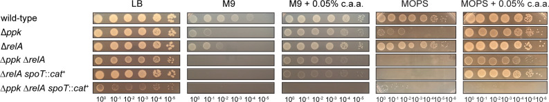
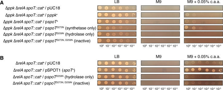
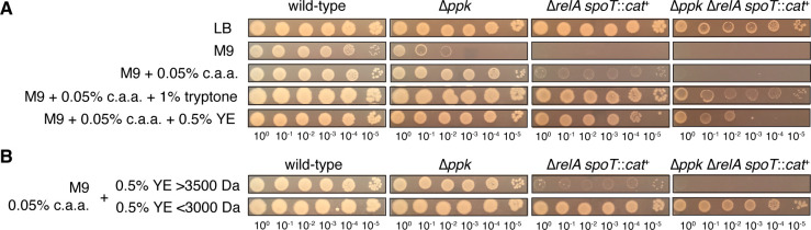
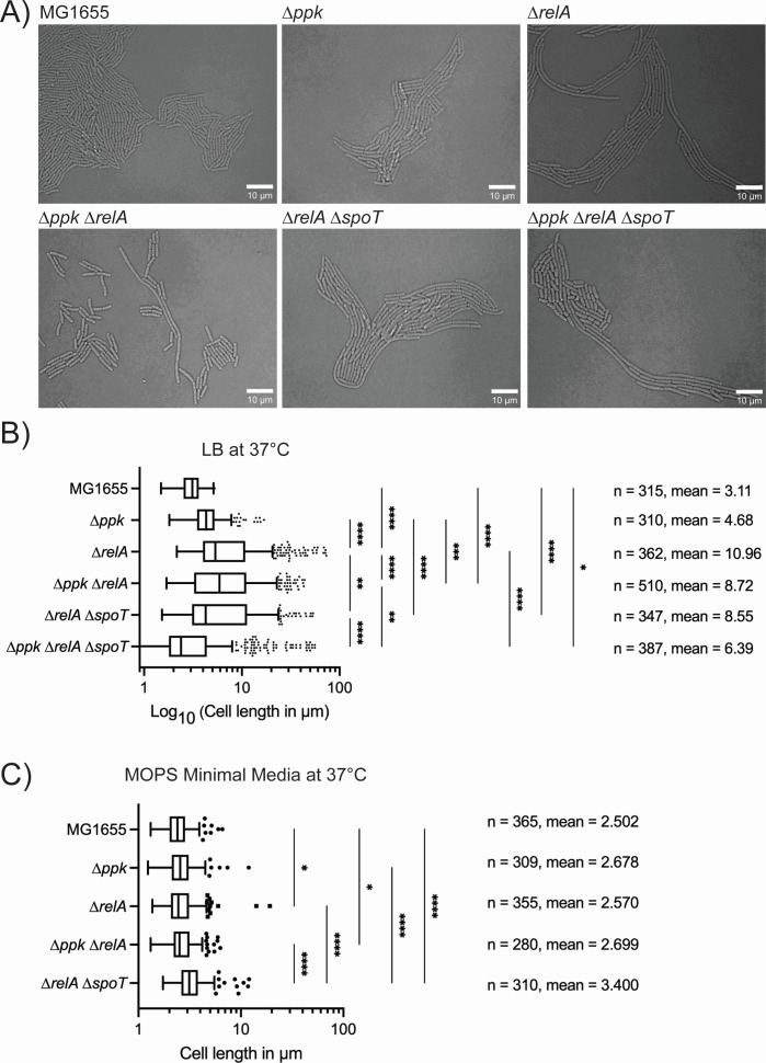
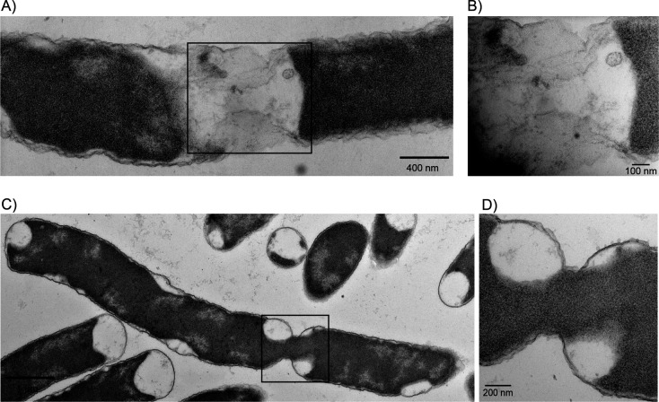
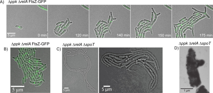
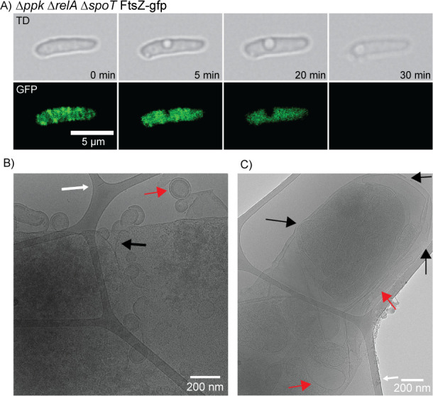
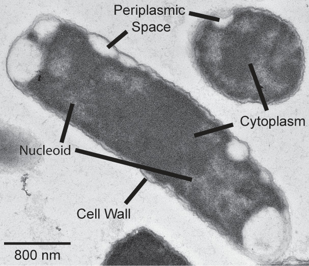
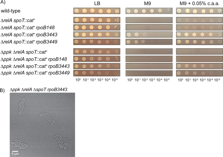
Bacteria encounter numerous stressors in their constantly changing environments and have evolved many methods to deal with stressors quickly and effectively. One well-known and broadly conserved stress response in bacteria is the stringent response, mediated by the alarmone (p)ppGpp. (p)ppGpp is produced in response to amino acid starvation and other nutrient limitations and stresses and regulates both the activity of proteins and expression of genes. Escherichia coli also makes inorganic polyphosphate (polyP), an ancient molecule evolutionary conserved across most bacteria and other cells, in response to a variety of stress conditions, including amino acid starvation. PolyP can act as an energy and phosphate storage pool, metal chelator, regulatory signal, and chaperone, among other functions. Here we report that E. coli lacking both (p)ppGpp and polyP have a complex phenotype indicating previously unknown overlapping roles for (p)ppGpp and polyP in regulating cell division, cell morphology, and metabolism. Disruption of either (p)ppGpp or polyP synthesis led to the formation of filamentous cells, but simultaneous disruption of both pathways resulted in cells with heterogenous cell morphologies, including highly branched cells, severely mislocalized Z-rings, and cells containing substantial void spaces. These mutants also failed to grow when nutrients were limited, even when amino acids were added. These results provide new insights into the relationship between polyP synthesis and the stringent response in bacteria and point toward their having a joint role in controlling metabolism, cell division, and cell growth.IMPORTANCECell division is a fundamental biological process, and the mechanisms that control it in Escherichia coli have been the subject of intense research scrutiny for many decades. Similarly, both the (p)ppGpp-dependent stringent response and inorganic polyphosphate (polyP) synthesis are well-studied, evolutionarily ancient, and widely conserved pathways in diverse bacteria. Our results indicate that these systems, normally studied as stress-response mechanisms, play a coordinated and novel role in regulating cell division, morphology, and metabolism even under non-stress conditions.

Keywords: (p)ppGpp; cell division; cell morphology; polyphosphate; stress response; stringent response.


Conflict of interest statement

The authors declare no conflict of interest.

Figures

Fig 1
Fig 1
Triple mutants lacking ppk, relA, and spoT have a growth defect on a minimal medium that cannot be rescued with casamino acids. E. coli strains MG1655 (wild type), MJG0224 (MG1655 ∆ppk-749), MJG0226 (MG1655 ∆relA782), MJG1116 (MG1655 ∆ppk-749relA782), MJG1136 (MG1655 ∆relA782 spoT207::cat+), and MJG1137 (MG1655 ∆ppk-749relA782 spoT207::cat+) were grown overnight in LB broth, then rinsed and normalized to an A600 = 1 in PBS. Aliquots (5 µL) of serially diluted suspensions were spotted on LB, M9 glucose, M9 glucose containing 0.05% (wt/vol) casamino acids (c.a.a.), MOPS glucose, or MOPS glucose containing 0.05% (wt/vol) casamino acids (c.a.a.) plates and incubated overnight at 37°C (representative image from at least three independent experiments).
Fig 2
Fig 2
The growth defect of a ppk relA spoT mutant on a minimal medium with casamino acids can be rescued by expression of either PPK or synthetase-active SpoT. E. coli strains (A) MJG1137 (MG1655 ∆ppk-749relA782 spoT207::cat+) or (B) MJG1136 (MG1655 ∆relA782 spoT207::cat+) containing the indicated plasmids (pppk+ =pPPK8, pspoT+ =pSPOT1, pspoTD73N = pSPOT2, pspoTD259N = pSPOT3, pspoTD73N, D259N = pSPOT4) were grown overnight in LB broth containing ampicillin, then rinsed and normalized to an A600 = 1 in PBS. Aliquots (5 µL) of serially diluted suspensions were spotted on LB, M9 glucose, or M9 glucose containing 0.05% (wt/vol) casamino acids (c.a.a.) plates containing ampicillin and incubated overnight at 37°C (representative image from at least three independent experiments).
Fig 3
Fig 3
Both tryptone and yeast extract contain components that rescue the growth defect of a ppk relA spoT mutant on a minimal medium with casamino acids. E. coli strains MG1655 (wild type), MJG0224 (MG1655 ∆ppk-749), MJG1136 (MG1655 ∆relA782 spoT207::cat+), and MJG1137 (MG1655 ∆ppk-749relA782 spoT207::cat+) were grown overnight in LB broth, then rinsed and normalized to an A600 = 1 in PBS. Aliquots (5 µL) of serially diluted suspensions were spotted on LB, M9 glucose, or M9 glucose containing the indicated percentages (wt/vol) of casamino acids (c.a.a.) and (A) tryptone or yeast extract (YE) or (B) yeast extract fractions containing compounds either greater than 3,500 Da or less than 3,000 Da and incubated overnight at 37°C (representative image from at least three independent experiments).
Fig 4
Fig 4
Microscopy of polyP and (p)ppGpp mutants shows filamentous cell growth. (A) Confocal microscopy images of MG1655 (MJG0001), ∆ppk (MJG0224), ∆relA (MJG0226), ∆ppk ∆relA (MJG1116), ∆relA ∆spoT (MJG1287), and ∆ppk ∆relA ∆spoT (MJG1282). Images were captured on LB agarose pads incubated at 37°C and imaged every 5 minutes while growing. (B) Length of cells growing at 37°C on an LB agarose pad and (C) length of cells growing at 37°C on a MOPS minimal media agarose pad. Length of cells was determined and manually calculated using FIJI using the line tool and measuring length with built-in measurements to FIJI after setting the appropriate scale. n = number of cells. Statistical significance was calculated in Prism GraphPad by one-way ANOVA, *P-value <0.05, **P-value <0.005, ***P-value <0.0005, ****P-value <0.0001.
Fig 5
Fig 5
Strains lacking polyP and (p)ppGpp have disrupted cell division. (A) Confocal fluorescence time-lapse microscopy of the mutant ppk relA spoT FtsZ-GFP (MJG2405) on an LB agarose pad at 37°C. The triple mutant forms two Z-rings in the middle of the cell, releasing a mini-cell (red arrows). There are also two Z-rings that form at either pole of a single cell, both functional and releasing a mini cell (black arrows). Single channel images of TD and GFP alone can be viewed in Fig. S6. Examples of mini-cell formation and release can also be seen by transmission and cryo-electron microscopy (Fig. S9 and S11). (B) Confocal fluorescence time-lapse microscopy of the mutant ppk relA FtsZ-GFP (MJG2403) on an LB agarose pad at 37°C. This image shows a mutant forming three Z-rings at one pole, and at least three at the opposite pole as well, with no Z-rings forming in the middle of the cell, for a total of six Z-rings in a single cell. This cell continued to grow without lysing (Video SV5). (C) Quantification of FtsZ-ring location within the cell, and its relationship to cell length in E. coli strains MG1655 FtsZ-GFP (MJG2041), ppk relA FtsZ-GFP (MJG2403), and ppk relA spoT FtsZ-GFP (MJG2405). Cell length is shown on the y-axis, with the corresponding location of the FtsZ-ring on the x-axis as a fraction of the total cell length. Positions of FtsZ rings were obtained by measuring, in pixels, the distance between the center of the FtsZ band and the cell pole. Poles were arbitrarily designated either 0 or 1, with the midpoint of the total cell length being 0.5.
Fig 6
Fig 6
TEM of ppk relA spoT cells failing to divide properly. (A) TEM of ppk relA spoT (MJG1282) failing to divide properly. The cytoplasm of this cell appears to have condensed within the cell and away from the divisisome site but is still connected by what appears to be a small bridge of membrane that has yet to separate and could be the result of disrupted FtsZ-ring formation. (B) This image is the square section from (A) at a higher magnification. (C) TEM of ppk relA spoT (MJG1282) not completely forming a divisisome and staying connected through a bridge while still sharing cytoplasmic contents between the undivided cells. (D) This image is the square section from (C) at a higher magnification.
Fig 7
Fig 7
Cells lacking polyP and (p)ppGpp can develop branching cell morphologies. (A) Confocal fluorescence time-lapse microscopy of the mutant ppk relA FtsZ-GFP (MJG2403) on an LB agarose pad at 37°C showing branching cells. Branched cells are still capable of dividing and growing. (B) Still image of confocal fluorescent microscopy of ppk relA FtsZ-GFP mutant (MJG2403) mutant on LB agarose pad at 37°C showcasing branched cells. (C) Still image of confocal microscopy of ∆ppk ∆relA ∆spoT (MJG1282) mutant on LB agarose pad at 37°C showcasing branched cells and other very odd cell morphologies. (D) Transmission electron microscopy image of ppk relA spoT (MJG1282) grown in LB at 37°C until log phase prior to imaging. This image shows a cell developing what appears to be a branching pole developed from the sidewall of the cell.
Fig 8
Fig 8
ppk relA spoT triple mutant cells can develop perforated membranes, leaking cytoplasmic contents out of the cell. (A) Time-lapse fluorescent microscopy of the ppk relA spoT mutant with the FtsZ-GFP reporter (MJG2405) showing empty space within the cell. FtsZ appears to fail to localize within the cell prior to losing its cytoplasmic contents just before cell death occurs. (B) Cryo-electron microscopy image of ppk relA spoT mutant (MJG2405) showing what appears to be a leaking cell wall with cytoplasmic contents blebbing off. White arrows point to the carbon lattice that cells were suspended on for imaging in CEM. There appears to be an invagination of the cell wall where cytoplasmic contents seem to be being released (black arrow). The cytoplasmic contents appear to be surrounded by a fully intact cell wall including an outer membrane, peptidoglycan, and inner membrane (red arrow). (C) CEM image of ppk relA spoT mutant (MJG2405) showing what appears to be holes in the cellular membrane (black arrows) where cytoplasmic contents and fluid may be lost. There also appears to be cytoplasm condensation, likely from fluid loss, with the cytoplasm condensing with the cell wall and membrane folding in on itself (red arrow).
Fig 9
Fig 9
Transmission electron micrograph of ppk relA spoT mutant showing plasmolysis in both cross-sectional and trans-sectional viewpoint. TEM of ppk relA spoT (MJG1282) shows the inner membrane appearing to shrink and pull away from the cell wall (plasmolysis), leaving large periplasmic spaces within the cell. We can see here in both the longitudinal and horizontal axis of the cell wall surrounding the cell with large open spaces around the perimeter of the cell where there appears to be no cytoplasm creating these periplasmic spaces and resulting in plasmolysis. The cell appears to still be actively dividing as we can see in the longitudinal axis it appears that the nucleoid of the cell is being restricted to the poles of the cell away from the midline as we would expect in cells about to divide.
Fig 10
Fig 10
Stringent alleles of RNA polymerase restore growth of ppk relA spoT mutants on minimal medium with casamino acids and rescue morphological defects on rich medium. (A) E. coli strains MG1655 (wild-type), MJG1136 (MG1655 ∆relA782 spoT207::cat+), MJG1137 (MG1655 ∆ppk-749relA782 spoT207::cat+), MJG1237 (MG1655 ∆relA782 spoT207::cat+ rpoB3449), MJG1241 (MG1655 ∆ppk-749relA782 spoT207::cat+ rpoB3449), MJG1579 (MG1655 ∆relA782 spoT207::cat+ rpoB3443), MJG1580 (MG1655 ∆relA782 spoT207::cat+ rpoB148), MJG1581 (MG1655 ∆ppk-749relA782 spoT207::cat+ rpoB3443), and MJG1582 (MG1655 ∆ppk-749relA782 spoT207::cat+ rpoB148) were grown overnight in LB broth, then rinsed and normalized to an A600 = 1 in PBS. Aliquots (5 µL) of serially diluted suspensions were spotted on LB, M9 glucose, or M9 glucose containing 0.05% (w/v) casamino acids (c.a.a.) plates and incubated overnight at 37°C (representative image from at least three independent experiments). (B) Confocal microscopy of ppk relA spoT rpoB3443 (MJG1581) grown on a LB agarose pad at 37°C.

Update of

Similar articles

Cited by

References

    1. Dalebroux ZD, Swanson MS. 2012. ppGpp: magic beyond RNA polymerase. Nat Rev Microbiol 10:203–212. doi:10.1038/nrmicro2720 - DOI - PubMed
    1. Potrykus K, Cashel M. 2008. (p)ppGpp: still magical? Annu Rev Microbiol 62:35–51. doi:10.1146/annurev.micro.62.081307.162903 - DOI - PubMed
    1. Rao NN, Gómez-García MR, Kornberg A. 2009. Inorganic polyphosphate: essential for growth and survival. Annu Rev Biochem 78:605–647. doi:10.1146/annurev.biochem.77.083007.093039 - DOI - PubMed
    1. Bowlin MQ, Gray MJ. 2021. Inorganic polyphosphate in host and microbe biology. Trends Microbiol 29:1013–1023. doi:10.1016/j.tim.2021.02.002 - DOI - PMC - PubMed
    1. Jain V, Kumar M, Chatterji D. 2006. ppGpp: stringent response and survival. J Microbiol 44:1–10. - PubMed

MeSH terms

LinkOut - more resources