. 2025 Feb 5;16(2):e0357324.
doi: 10.1128/mbio.03573-24. Epub 2024 Dec 27.

A bacterial membrane-disrupting protein stimulates animal metamorphosis

Affiliations

A bacterial membrane-disrupting protein stimulates animal metamorphosis

Kyle E Malter et al. mBio. .

Abstract

Diverse marine animals undergo a metamorphic larval-to-juvenile transition in response to surface-bound bacteria. Although this host-microbe interaction is critical to establishing and maintaining marine animal populations, the functional activity of bacterial products and how they activate the host's metamorphosis program has not yet been defined for any animal. The marine bacterium Pseudoalteromonas luteoviolacea stimulates the metamorphosis of a tubeworm called Hydroides elegans by producing a molecular syringe called metamorphosis-associated contractile structures (MACs). MACs stimulate metamorphosis by injecting a protein effector termed metamorphosis-inducing factor 1 (Mif1) into tubeworm larvae. Here, we show that MACs bind to tubeworm cilia and form visible pores on the cilia membrane surface, which are smaller and less numerous in the absence of Mif1. In vitro, Mif1 associates with eukaryotic lipid membranes and possesses phospholipase activity. MACs can also deliver Mif1 to human cell lines and cause parallel phenotypes, including cell surface binding, membrane disruption, calcium flux, and mitogen-activated protein kinase activation. Finally, MACs can also stimulate metamorphosis by delivering two unrelated membrane-disrupting proteins, MLKL and RegIIIɑ. Our findings demonstrate that membrane disruption by MACs and Mif1 is necessary for Hydroides metamorphosis, connecting the activity of a bacterial protein effector to the developmental transition of a marine animal.

Importance: This research describes a mechanism wherein a bacterium prompts the metamorphic development of an animal from larva to juvenile form by injecting a protein that disrupts membranes in the larval cilia. Specifically, results show that a bacterial contractile injection system and the protein effector it injects form pores in larval cilia, influencing critical signaling pathways like mitogen-activated protein kinase and calcium flux, ultimately driving animal metamorphosis. This discovery sheds light on how a bacterial protein effector exerts its activity through membrane disruption, a phenomenon observed in various bacterial toxins affecting cellular functions, and elicits a developmental response. This work reveals a potential strategy used by marine organisms to respond to microbial cues, which could inform efforts in coral reef restoration and biofouling prevention. The study's insights into metamorphosis-associated contractile structures' delivery of protein effectors to specific anatomical locations highlight prospects for future biomedical and environmental applications.

Keywords: cilia; contractile injection system; effector; metamorphosis; pore-forming toxin; secretion systems; toxin.


Conflict of interest statement

K.E.M., T.L.D., and N.J.S. are co-inventors on provisional U.S. patent application entitled "Protein and Peptide Delivery Systems and Methods for Using Them with Human Cells" and assigned to San Diego State University Research Foundation.

Figures

Fig 1
Fig 1
MACs form pores in Hydroides cilia, and Mif1 enhances pore formation. (A) Low-magnification view (400×) of Hydroides larvae exposed to WT MACs. pt, prototroch; mt, metatroch. White box indicates the location of magnification for panel B. (B) Mid-magnification view (3,200×) of Hydroides larval cilia exposed to WT MACs. White box indicates the location of magnification for panel C. (C) High-magnification view (23,500×) of Hydroides larval cilia exposed to WT MACs. Black arrows indicate MACs sheaths. White arrows indicate pores in cilia surrounding MACs array. (D) High-magnification view (25,000×) of Hydroides larval cilia exposed to ∆mif1 MACs. (E) High-magnification view (15,000×) of Hydroides larval cilia exposed to ∆macB MACs. (F) High-magnification view (15,000×) of untreated Hydroides larval cilia. (G) Counts of MACs visualized on the head, cilia (prototroch or metatroch), or body of the larval animals. N = 46. (H) Counts of Hydroides larvae observed with WT, ∆mif1, and ∆macB MACs with cilia pores. N = 38, 26, and 15 larvae for WT, ∆mif1, and ∆macB exposures, respectively (***P < 1E-12; ns, not significant, two-tailed Student’s t-test, Bonferroni adjustment). (I) Diameters of pores observed on Hydroides larval cilia with WT and ∆mif1 MACs. N = 41 for WT and ∆mif1 (***P < 0.0001, two-tailed Student’s t-test).
Fig 2
Fig 2
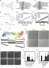
Mif1 binds to phosphoinositol lipids and possesses phospholipase activity in vitro. (A) Chemical structure of PtIns(3,5)P2. (B and C) Lipid-protein interaction assays were performed using purified Mif1 protein and Mif1 specific antibody on (B) a membrane lipid strip (Echelon Biosciences P-6002) or (C) a PIP strip membrane (Echelon Biosciences P-6001). (D) Chemical structure of 4-nitrophenyl phosphate (pNPP)-decanoic acid and Tween 20 esterase assay substrates. The chemical bond cleaved is indicated by a dotted red line. (E) Lipid cleavage assay with purified Mif1 protein, chaperone JF50_12605 (605) protein, or GFP protein incubated with Tween-20 in the presence of Ca2+ and liberated lauric acid was observed via turbidity. The average of four technical replicates is shown. (F) Esterase assay with purified Mif1, GFP, or chaperone JF50_12605(605) protein with decanoic acid-pNPP substrate. Cleavage and pNPP release occurs if acyl-ester linkage is hydrolyzed. Data are represented as the mean ± SD of n = 12 technical replicates across three independent biological replicates. (G) AlphaFold2 predicted Mif1 structural model and schematic representation of Mif1 fragments cloned for overexpression. Colors of the Mif1 model range from N- to C-terminus (blue-green-yellow-red) and are for orientation. (H) Escherichia coli expressing recombinant mif1 (Mif1), mif1 fragments, JF50_12605 (605), or gfp genes from an IPTG-inducible promoter in a pET15b vector in the presence or absence of 0.1 mM IPTG. Bacteria were grown overnight and then spotted by 1/5 serial dilutions starting at OD 1.0. (I and J) E. coli expressing recombinant full-length wild-type mif1 (Mif1 WT), mif1 fragments, or gfp genes from an IPTG-inducible promoter in a pET15b vector after induction with 0.1 mM IPTG for 2 hours. (I) Representative images of cells after 15 minutes of incubation with propidium iodide (PI). Scale bar is 5 µm. (J) Bar graph quantifying the number of cells (%) stained with PI indicating cell permeabilization. Permeabilization is significantly different between GFP vs Mif1 WT or A-frag WT and not significantly different between GFP vs A1-frag H55A, C1-frag H609A, or C1-frag (*P < 0.05; ns, not significant, one-way ANOVA, Dunnett’s multiple comparisons test). Results shown are the average of three independent biological replicates; error bars are SD. (K) Comparison of blank control, WT, ∆macB, ∆mif1, mif1H55A, and mif1H609A mutation on the stimulation of Hydroides metamorphosis (***P < 0.001; ns, not significant, one-way ANOVA, Dunnett’s multiple comparisons test).
Fig 3
Fig 3
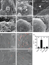
MACs bind to and permeabilize human tissue culture cells. (A–F) Scanning electron micrographs of human HEK293T cells exposed to MACs from (A–C) wild type, (D) ∆mif1, (E) ∆macB, or (F) buffer alone. MACs bound to HEK293T cells within 3 minutes of exposure (A: low magnification and B: high magnification). White box in panel A indicates the location of magnification for panel B. (C) MACs induce cell membrane degradation within 30 minutes. (D) HEK293T cells exposed to MACs from a ∆mif1 mutant. Cell membrane degradation is reduced in the absence of mif1. (B–D) White arrows indicate the location of MACs array. (G–J) Representative images of PI-stained HEK293T cells after exposure to (G) buffer, (H) wild-type, (I) ∆macB, or (J) ∆mif1 MACs. Scale bars are 50 µm. (K) Bar graph quantifying the number of cells (%) stained with PI indicating cell permeabilization. Permeabilization is significantly different between WT vs buffer, P = 0.0036; WT vs ∆macB; P = 0.0138; and WT vs ∆mif1, P = 0.0253; and not significantly different between buffer vs ∆macB, buffer vs ∆mif1, and ∆macB vs ∆mif1 (one-way ANOVA, Tukey’s multiple comparisons test, letters represent Tukey post hoc test results). Results shown are the average of three independent biological replicates; error bars are SD.
Fig 4
Fig 4
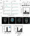
Mif1 activates MAPK activity and calcium flux. (A) Fluorescence intensity distribution of HEK293 cells transfected with a Ca2+ reporter after 10 minutes and 60 minutes of exposure to MACs’ extracts. GFP intensity values were transformed by log10. Mean intensity values were significantly different (P < 0.05, one-way ANOVA, Tukey post hoc test) between treatments after 10 minute exposure and were not significantly different after 1 hour exposure. (B) Western blot of HEK293T cells treated with MACs’ extracts for 5 minutes. Representative blots are shown for anti-phospho-p38 and total p38. (C) The ratio of phosphorylated p38 was compared to the total p38 (n = 3) (a one-way ANOVA with multiple comparisons showed WT vs ∆macB P-value = 0.80, WT vs buffer P-value = 0.79, and WT vs ∆mif1 P-value = 0.75). (D) Hybridizing chain reaction of p38 on larvae exposed to MACs’ extracts for 5 minutes from wild type, ∆mif1, ∆macB, or buffer alone. (E) Bar graph of percentage of larvae with ciliary band fluorescence (n = 10 per treatment averaged from two biological replicates). Letters represent one-way ANOVA and Tukey post hoc test results (P < 0.0001). (F) Graph of Hydroides percentage of metamorphosis in response to buffer, WT, ∆mif1, or MACs with Mif1-N200, Mif1-N200-RegIIIɑ, or Mif1-N200-MLKL. Letters represent one-way ANOVA and Tukey post hoc test results (Mif1-N200 vs Mif1-N200-RegIIIɑ =P < 0.03; Mif1-N200 vs Mif1-N200-MLKL = P < 0.001).
Fig 5
Fig 5
Model of Hydroides metamorphosis in response to MACs and Mif1. (A) P. luteoviolacea bacteria produce MACs loaded with the Mif1 payload, which are released via cell lysis. (B) Hydroides and MACs engage via the larvae’s ciliated band (blue). (C) Upon contact with Hydroides cilia, the MACs sheath contracts, driving the rigid inner tube through the cilia cell membrane. Subsequently, Mif1 (orange) located within the MACs inner tube lumen is delivered and promotes the number and size of pores. (D) Pore formation in the cilia, enhanced by Mif1, leads to calcium flux and phosphorylation of p38 MAPK, ultimately initiating the metamorphosis of the Hydroides larva to the juvenile form.

References

    1. Alegado RA, Brown LW, Cao S, Dermenjian RK, Zuzow R, Fairclough SR, Clardy J, King N. 2012. A bacterial sulfonolipid triggers multicellular development in the closest living relatives of animals. Elife 1:e00013. doi:10.7554/eLife.00013 - DOI - PMC - PubMed
    1. Coon KL, Valzania L, McKinney DA, Vogel KJ, Brown MR, Strand MR. 2017. Bacteria-mediated hypoxia functions as a signal for mosquito development. Proc Natl Acad Sci U S A 114:E5362–E5369. doi:10.1073/pnas.1702983114 - DOI - PMC - PubMed
    1. Fraune S, Bosch TCG. 2010. Why bacteria matter in animal development and evolution. Bioessays 32:571–580. doi:10.1002/bies.200900192 - DOI - PubMed
    1. Koropatnick TA, Engle JT, Apicella MA, Stabb EV, Goldman WE, McFall-Ngai MJ. 2004. Microbial factor-mediated development in a host-bacterial mutualism. Science 306:1186–1188. doi:10.1126/science.1102218 - DOI - PubMed
    1. Weiss BL, Wang J, Aksoy S. 2011. Tsetse immune system maturation requires the presence of obligate symbionts in larvae. PLoS Biol 9:e1000619. doi:10.1371/journal.pbio.1000619 - DOI - PMC - PubMed

Substances

LinkOut - more resources