

Accuracy of the Hounsfield Unit Values Measured by Implant Planning Software
- PMID: 39727470
- PMCID: PMC11674854
- DOI: 10.3390/dj12120413
Accuracy of the Hounsfield Unit Values Measured by Implant Planning Software
Abstract
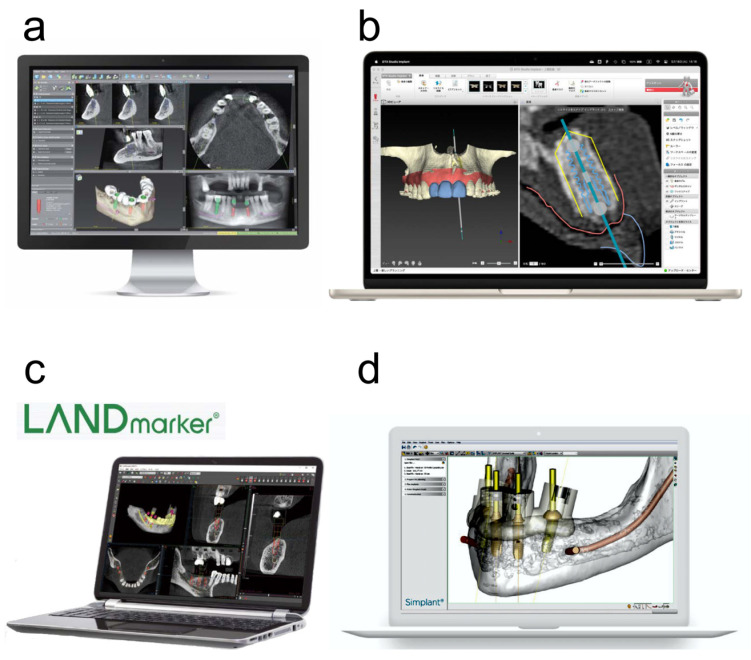
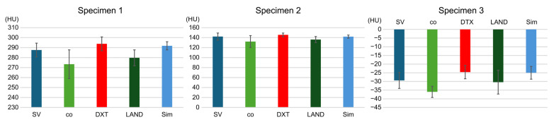
Background: The measurement of Hounsfield units (HU) during implant treatment planning is important. Currently, various manufacturers' implant planning software programs offer HU capabilities; however, their accuracy remains unverified. In this study, we aimed to validate the accuracy of HU values measured by implant planning software programs. Methods: This study used one type of multidetector computed tomography (MDCT), two types of cone-beam computed tomography (CBCT), and four implant planning software packages. Three specimens were prepared for the evaluation of HUs, and the standard values of the HUs were measured. Digital Imaging and Communications in Medicine (DICOM) data obtained from MDCT and CBCT were loaded into four implant planning software packages to measure the HU values. The HU and reference values of the four implant planning software programs obtained from MDCT and CBCT were compared. Additionally, the HU values between each implant planning software program were compared. Results: The HU values of the three specimens, as measured using the four implant planning software programs utilizing MDCT, did not exhibit a significant difference from the standard values. Conversely, those obtained from CBCT were significantly different. The measured HU values after the MDCT imaging of the specimens were not significantly different between the implant planning software programs; however, they differed after CBCT imaging. Conclusions: The results of this study indicate that it is not possible to measure HU values using CBCT with implant planning software programs. However, HU values can be measured by any implant planning software using MDCT.
Keywords: Hounsfield units (HUs); cone–beam computed tomography (CBCT); dental implant; implant planning software; multidetector row computed tomography (MDCT).
Conflict of interest statement
The authors declare no conflicts of interest.
Figures


Similar articles
-
Reliability and accuracy of cone beam computed tomography versus conventional multidetector computed tomography for image-guided craniofacial implant planning: An in vitro study.Int J Oral Maxillofac Implants. 2019 May/June;34(3):665–672. doi: 10.11607/jomi.6915. Epub 2019 Apr 1. Int J Oral Maxillofac Implants. 2019. PMID: 30934042
-
Improving Hounsfield Unit accuracy in dental CBCT through the integration of 2D anti-scatter grid.Oral Radiol. 2025 May 16. doi: 10.1007/s11282-025-00824-3. Online ahead of print. Oral Radiol. 2025. PMID: 40377825
-
Three-dimensional evaluation of lipiodol retention in HCC after chemoembolization: a quantitative comparison between CBCT and MDCT.Acad Radiol. 2014 Mar;21(3):393-9. doi: 10.1016/j.acra.2013.11.006. Acad Radiol. 2014. PMID: 24507426 Free PMC article. Clinical Trial.
-
Are multi-detector computed tomography and cone-beam computed tomography exams and software accurate to measure the upper airway? A systematic review.Eur J Orthod. 2023 Nov 30;45(6):818-831. doi: 10.1093/ejo/cjad060. Eur J Orthod. 2023. PMID: 37797294
-
Can gray values be converted to Hounsfield units? A systematic review.Dentomaxillofac Radiol. 2022 Jan 1;51(1):20210140. doi: 10.1259/dmfr.20210140. Epub 2021 Jun 19. Dentomaxillofac Radiol. 2022. PMID: 34148350 Free PMC article.
References
-
- Dings J.P., Verhamme L., Merkx M.A., Xi T., Meijer G.J., Maal T.J. Reliability and accuracy of cone beam computed tomography versus conventional multidetector computed tomography for image-guided craniofacial implant planning: An in vitro study. Int. J. Oral Maxillofac. Implants. 2019;34:665–672. doi: 10.11607/jomi.6915. - DOI - PubMed
LinkOut - more resources
Full Text Sources
