

Prevalence and Clinical Implications of Pulmonary Vein Stenosis in Bronchiectasis: A 3D Reconstruction CT Study
- PMID: 39727497
- PMCID: PMC11673734
- DOI: 10.3390/arm92060046
Prevalence and Clinical Implications of Pulmonary Vein Stenosis in Bronchiectasis: A 3D Reconstruction CT Study
Abstract
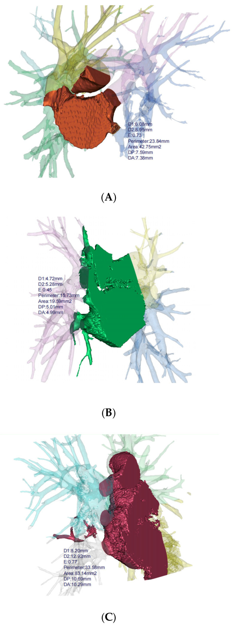
Background: Recent studies on bronchiectasis have revealed significant structural abnormalities and pathophysiological changes. However, there is limited research focused on pulmonary venous variability and congenital variation. Through our surgical observations, we noted that coarctation of pulmonary veins and atrophied lung volume are relatively common in bronchiectasis patients. Therefore, we conducted a retrospective study to explore pulmonary venous variation and secondary manifestations in bronchiectasis cases, utilizing 3D reconstruction software (Mimics Innovation Suite 21.0, Materialise Dental, Leuven, Belgium) to draw conclusions supported by statistical evidence.
Method: This retrospective study included patients with bronchiectasis and healthy individuals who underwent CT examinations at Beijing Chao-Yang Hospital between January 2017 and July 2023. Chest CT data were reconstructed using Materialise Mimics. Pulmonary veins and lung lobes were segmented from surrounding tissue based on an appropriate threshold determined by local grey values and image gradients. Subsequently, venous cross-sectional areas and lung volumes were measured for statistical analysis.
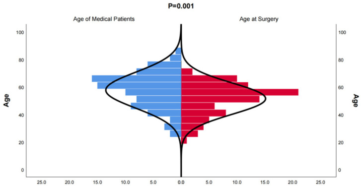
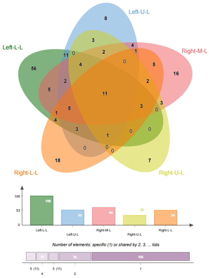
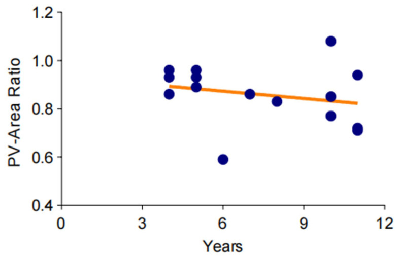
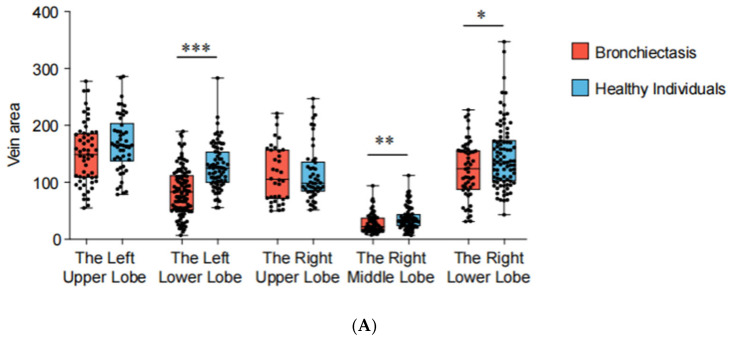
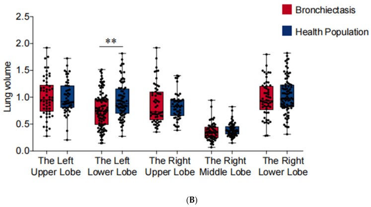
Result: CT data from 174 inpatients with bronchiectasis and 75 cases from the health examination center were included. Three-dimensional reconstruction data revealed a significant reduction in cross-sectional areas of pulmonary veins in the left lower lobe (p < 0.001), the right lower lobe (p = 0.030), and the right middle lobe (p = 0.009) of bronchiectasis patients. Subgroup analyses indicated that approximately 73.5% of localized cases of the left lower lobe exhibited pulmonary vein stenosis, while in the diffuse group, this proportion was only 52.6%. Furthermore, the cross-sectional area of pulmonary veins had a gradually decreasing trend, based on a small sample. Lung function tests showed significant reductions in FEV1, FVC, and FEV1% in bronchiectasis patients, attributed to the loss of lung volume in the left lower lobe, which accounted for 60.9% of the included sample.
Conclusions: Our recent findings suggest that pulmonary venous stenosis is a common variation in bronchiectasis and is often observed concurrently with reduced lung volume, particularly affecting the left lower lobe. Moreover, localized cases are more likely to suffer from pulmonary venous stenosis, with an ambiguous downtrend as the disease progresses. In conclusion, increased attention to pulmonary venous variation in bronchiectasis is warranted, and exploring new therapies to intervene in the early stages or alleviate obstruction may be beneficial.
Keywords: 3D reconstruction; bronchiectasis; lung function; lung volume; pulmonary venous stenosis.
Conflict of interest statement
The authors declare no conflicts of interest.
Figures
Similar articles
-
Computed tomographic parenchymal lung findings in premature infants with pulmonary vein stenosis.Pediatr Radiol. 2023 Aug;53(9):1874-1884. doi: 10.1007/s00247-023-05673-y. Epub 2023 Apr 28. Pediatr Radiol. 2023. PMID: 37106091
-
[Translobar Phenomenon of Pulmonary Veins and Its Clinical Significance in Lobectomy].Zhongguo Fei Ai Za Zhi. 2021 Feb 20;24(2):99-107. doi: 10.3779/j.issn.1009-3419.2021.104.01. Epub 2021 Jan 22. Zhongguo Fei Ai Za Zhi. 2021. PMID: 33478198 Free PMC article. Chinese.
-
Normative analysis of pulmonary vein drainage patterns on multidetector CT with measurements of pulmonary vein ostial diameter and distance to first bifurcation.Acad Radiol. 2007 Feb;14(2):178-88. doi: 10.1016/j.acra.2006.11.004. Acad Radiol. 2007. PMID: 17236990
-
The many faces and outcomes of pulmonary vein stenosis in early childhood.Pediatr Pulmonol. 2021 Mar;56(3):649-655. doi: 10.1002/ppul.24848. Epub 2020 Jun 7. Pediatr Pulmonol. 2021. PMID: 32506838 Review.
-
A review of the etiology and clinical presentation of non-cystic fibrosis bronchiectasis: A tertiary care experience.Respir Med. 2018 Apr;137:35-39. doi: 10.1016/j.rmed.2018.02.013. Epub 2018 Feb 24. Respir Med. 2018. PMID: 29605210 Review.
References
MeSH terms
Grants and funding
LinkOut - more resources
Full Text Sources
Medical
